

Radiomics MRI Phenotyping with Machine Learning to Predict the Grade of Lower-Grade Gliomas: A Study Focused on Nonenhancing Tumors
- PMID: 31464116
- PMCID: PMC6715562
- DOI: 10.3348/kjr.2018.0814
Radiomics MRI Phenotyping with Machine Learning to Predict the Grade of Lower-Grade Gliomas: A Study Focused on Nonenhancing Tumors
Abstract
Objective: To assess whether radiomics features derived from multiparametric MRI can predict the tumor grade of lower-grade gliomas (LGGs; World Health Organization grade II and grade III) and the nonenhancing LGG subgroup.
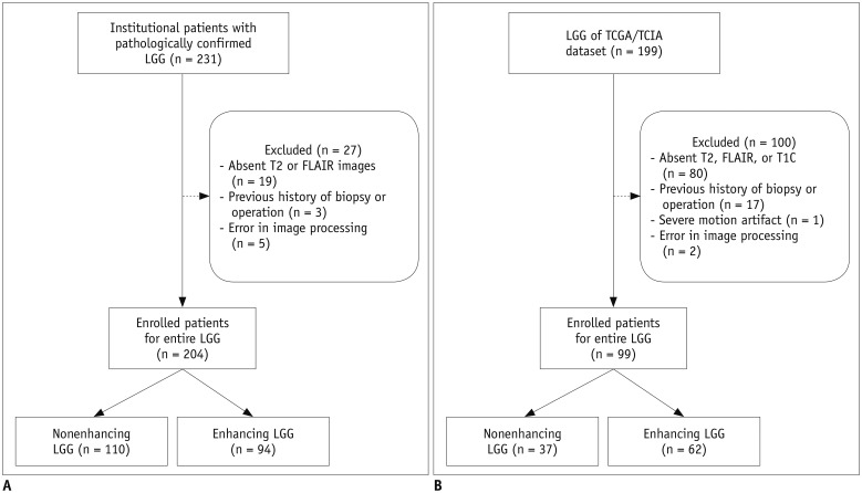
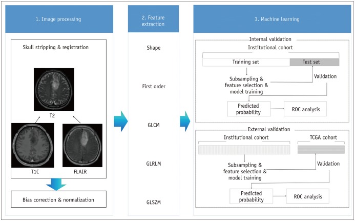
Materials and methods: Two-hundred four patients with LGGs from our institutional cohort were allocated to training (n = 136) and test (n = 68) sets. Postcontrast T1-weighted images, T2-weighted images, and fluid-attenuated inversion recovery images were analyzed to extract 250 radiomics features. Various machine learning classifiers were trained using the radiomics features to predict the glioma grade. The trained classifiers were internally validated on the institutional test set and externally validated on a separate cohort (n = 99) from The Cancer Genome Atlas (TCGA). Classifier performance was assessed by determining the area under the curve (AUC) from receiver operating characteristic curve analysis. An identical process was performed in the nonenhancing LGG subgroup (institutional training set, n = 73; institutional test set, n = 37; and TCGA cohort, n = 37) to predict the glioma grade.
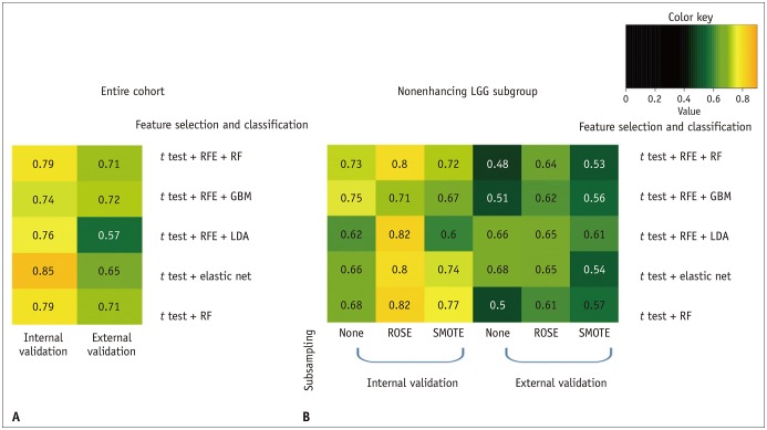
Results: The performance of the best classifier was good in the internal validation set (AUC, 0.85) and fair in the external validation set (AUC, 0.72) to predict the LGG grade. For the nonenhancing LGG subgroup, the performance of the best classifier was good in the internal validation set (AUC, 0.82), but poor in the external validation set (AUC, 0.68).
Conclusion: Radiomics feature-based classifiers may be useful to predict LGG grades. However, radiomics classifiers may have a limited value when applied to the nonenhancing LGG subgroup in a TCGA cohort.
Keywords: Grade; Lower-grade glioma; Magnetic resonance imaging; Radiomics; The Cancer Genome Atlas.
Copyright © 2019 The Korean Society of Radiology.
Conflict of interest statement
The authors have no potential conflicts of interest to disclose.
Figures
References
-
- Louis DN, Perry A, Reifenberger G, von Deimling A, Figarella-Branger D, Cavenee WK, et al. The 2016 World Health Organization classification of tumors of the central nervous system: a summary. Acta Neuropathol. 2016;131:803–820. - PubMed
-
- Rollin N, Guyotat J, Streichenberger N, Honnorat J, Tran Minh VA, Cotton F. Clinical relevance of diffusion and perfusion magnetic resonance imaging in assessing intra-axial brain tumors. Neuroradiology. 2006;48:150–159. - PubMed
Publication types
MeSH terms
LinkOut - more resources
Full Text Sources
Medical
Molecular Biology Databases
