. 2019 Oct;15(43):e1902971.
doi: 10.1002/smll.201902971. Epub 2019 Aug 29.

Deconstructed Microfluidic Bone Marrow On-A-Chip to Study Normal and Malignant Hemopoietic Cell-Niche Interactions

Affiliations

Deconstructed Microfluidic Bone Marrow On-A-Chip to Study Normal and Malignant Hemopoietic Cell-Niche Interactions

Julio Aleman et al. Small. 2019 Oct.

Abstract

Human hematopoietic niches are complex specialized microenvironments that maintain and regulate hematopoietic stem and progenitor cells (HSPC). Thus far, most of the studies performed investigating alterations of HSPC-niche dynamic interactions are conducted in animal models. Herein, organ microengineering with microfluidics is combined to develop a human bone marrow (BM)-on-a-chip with an integrated recirculating perfusion system that consolidates a variety of important parameters such as 3D architecture, cell-cell/cell-matrix interactions, and circulation, allowing a better mimicry of in vivo conditions. The complex BM environment is deconvoluted to 4 major distinct, but integrated, tissue-engineered 3D niche constructs housed within a single, closed, recirculating microfluidic device system, and equipped with cell tracking technology. It is shown that this technology successfully enables the identification and quantification of preferential interactions-homing and retention-of circulating normal and malignant HSPC with distinct niches.

Keywords: bone marrow niche; hematopoietic stem and progenitor cells; homing; microfluidics; tissue chip.


Conflict of interest statement

Conflict of Interest

The authors declare no conflict of interest.

Figures

Figure 1.
Figure 1.
Overall NOC experimental summary. a) Human bone marrow is b) separated into 3 niche populations (sinusoidal endothelial, arterial endothelial, and mesenchymal) by magnetic sorting. A subset of the MSC population is differentiated to the osteoblastic lineage. c) Using an extracellular matrix-mimicking hydrogel comprised of thiolated hyaluronic acid, thiolated gelatin, and a polyethylene glycol diacrylate (PEGDA) crosslinker, d) individual niche populations are encapsulated in 3D niche constructs inside the f) NOC microfluidic device. Homing and lodging/retention studies e) are initiated by infusing either healthy HSPC or malignant leukemia or lymphoma cells, after which homing and lodging/retention of infused cells that have traveled through circulation to each of the niche constructs is quantified in an unbiased manner.
Figure 2.
Figure 2.
Microfluidic chip device fabrication and niche construct integration. a) Microfluidic device fabrication is performed by bonding a molded PDMS layer in which channels and chamber features are defined by soft lithography to a glass slide. b) In situ 3D microconstruct formation workflow: Channels i) are filled with a mixture of photocurable hydrogel precursor, cells, and additional components (dark red, ii). A photomask (gray) is employed to define construct shape and location iii), and the remaining solution washed away with fresh PBS iv). c) Side and top view depictions of the niche constructs in the device chambers. d,e) Schematic and photograph, respectively, of operational recirculating multi-niche-on-a-chip systems. Scale bar: 1 cm. f) Four frames from a fluid dynamics computational model of the NOC using Flow3D software, in which a simulation of infusing HSPC into the device was performed by infusing 1000 roughly HSPC-sized particles. Time (T) is in seconds. Heatmap represents fluid velocity (m s−1).
Figure 3.
Figure 3.
Niche cell biomarkers in 3D niche constructs in NOC devices on Day 8 following construct biofabrication. a) Arterial niche constructs stained for Ephrin B2 (B2); b) Osteoblastic niche constructs stained with Alizarin Red for calcium deposits; c) Sinusoidal niche constructs stained for EphB4 (B4); and d) mesenchymal (MSC) niche constructs stained for CD44. Panels are organized as: i) DAPI, ii) indicated stain, and iii) merged image; e) LIVE/DEAD viability staining of each niche construct type at day 8. Green-calcein AM-stained viable cells; Red-ethidium homodimer-stained dead cells. Scale bars: 100 μm.
Figure 4.
Figure 4.
Tracking of fluorescently labeled normal/healthy, leukemic, or lymphoma HSPC using an onboard fluorescent camera system to visualize HSPC homing, lodging/retention, or passing by the 3D niche constructs. a) Working NOC device that is then monitored using b) a custom-built fluorescent camera system comprised of the NOC sandwiched between an LED, lens, filter, and camera, shown in operation in the right panel. This system allows (c,d) real-time visualization of infused labeled cells in the NOC system. c) A U937 cell is indicated (white arrows) moving toward an arterial niche construct i–iv) and remaining in place after contact v–vi). d) A different U937 cell is indicated (white arrows) that passes around the arterial construct i–iii), never making contact, while in panel iv), a second U937 cell is observed detaching from the construct and re-entering “circulation” v–vi). Both sequences occur in ≈5–10 s time.
Figure 5.
Figure 5.
Quantification of initial homing of HSPC to niches after 24 h. Cell counts at each niche for: a) normal/healthy CD34+ HSPC, b) U937 lymphoma cells, and c) MOLM13 leukemia cells.
Figure 6.
Figure 6.
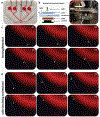
Niche-on-a-chip lodging/retention experiments using CD34+ cells from normal/healthy adult donors (CD34+ HSPC), CD34- cell line derived from a lymphoma patient (U937), CD34+ cells derived from an AML patient (MOLM13), which show distinct niche lodging/retention preferences between cell types. a–e) Normal/healthy CD34+ HSPC located preferentially within the ON, exhibited moderate lodging/retention within the MN and SN, and were only rarely found in the AN. f–j) The CD34- lymphoma cell line (U937) exhibited a marked predilection for the AN, followed by the ON. k–o) CD34+ cells derived from an acute monocytic leukemia patient (MOLM13) engrafted/lodged primarily in the ON and AN, with some lodging/retention in the MN and SN. a–n) show representative lodging/retention images with fluorescent images in the red channel highlighting the infused cell types overlaid on light microscopy images of each niche. Images were taken on day 1, day 3, and day 5 following infusion of the cells into each NOC device. e,j,o) provide quantified average cell numbers of lodged/retained CD34+ HSPC, U937 lymphoma cells, and MOLM13 leukemia cells, respectively, in each niche. Red highlighted borders indicate regions of common lodging/retention. One-way ANOVA (n = 10) **P < 0.005,***P < 0.001. Scale bar: 250 μm.

References

    1. Acar M, Kocherlakota KS, Murphy MM, Peyer JG, Oguro H, Inra CN, Jaiyeola C, Zhao Z, Luby-Phelps K, Morrison SJ, Nature 2015, 526, 126; - PMC - PubMed
    2. Ding L, Saunders TL, Enikolopov G, Morrison SJ, Nature 2012, 481, 457; - PMC - PubMed
    3. Kunisaki Y, Bruns I, Scheiermann C, Ahmed J, Pinho S, Zhang D, Mizoguchi T, Wei Q, Lucas D, Ito K, Mar JC, Bergman A, Frenette PS, Nature 2013, 502, 637; - PMC - PubMed
    4. Mendelson A, Frenette PS, Nat. Med 2014, 20, 833; - PMC - PubMed
    5. Morrison SJ, Scadden DT, Nature 2014, 505, 327. - PMC - PubMed
    1. Crane GM, Jeffery E, Morrison SJ, Nat. Rev. Immunol 2017, 17, 573. - PubMed
    1. Horn PA, Thomasson BM, Wood BL, Andrews RG, Morris JC, Kiem HP, Blood 2003, 102, 4329. - PubMed
    1. Ho WJ, Pham EA, Kim JW, Ng CW, Kim JH, Kamei DT, Wu BM, Cancer Sci 2010, 101, 2637. - PMC - PubMed
    1. Skardal A, Devarasetty M, Soker S, Hall AR, Biofabrication 2015, 7, 031001. - PMC - PubMed

Publication types