. 2019 Nov;33(11):12299-12310.
doi: 10.1096/fj.201900398RR. Epub 2019 Aug 28.

Enteric dysbiosis is associated with sepsis in patients

Affiliations

Enteric dysbiosis is associated with sepsis in patients

Zhanguo Liu et al. FASEB J. 2019 Nov.

Abstract

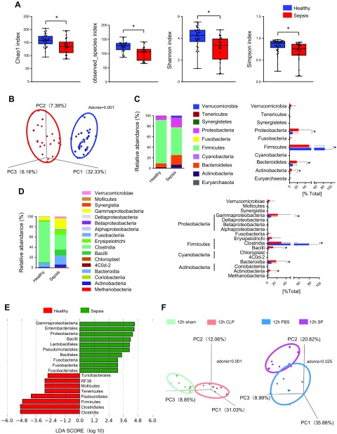
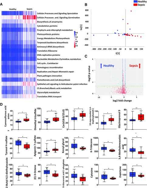
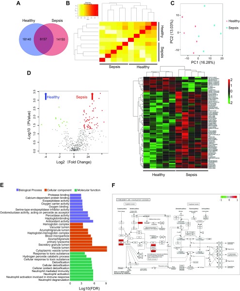
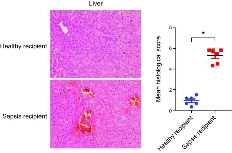
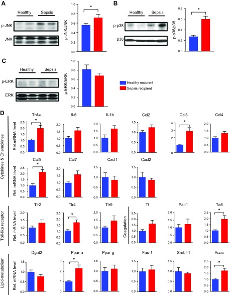
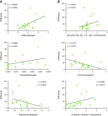
Sepsis is defined as a life-threatening organ dysfunction caused by a dysregulated host response to microbial infection. For decades, the potential role of gut microbiota in sepsis pathogenesis has been revealed. However, the systemic and functional link between gut microbiota and sepsis has remained unexplored. To address this gap in knowledge, we carried out systematic analyses on clinical stool samples from patients with sepsis, including 16S rDNA sequencing, metabolomics, and metaproteomics analyses. In addition, we performed fecal microbiota transplantation from human to mice to validate the roles of gut microbiota on sepsis progression. We found that the composition of gut microbiota was significantly disrupted in patients with sepsis compared with healthy individuals. Besides, the microbial functions were significantly altered in septic feces as identified by metabolomics and metaproteomics analyses. Interestingly, mice that received septic feces exhibited more severe hepatic inflammation and injury than mice that received healthy feces after cecal ligation and puncture. Finally, several strains of intestinal microbiota and microbial metabolites were corelated with serum total bilirubin levels in patients with sepsis. Taken together, our data indicated that sepsis development is associated with the disruption of gut microbiota at both compositional and functional levels, and such enteric dysbiosis could promote organ inflammation and injury during sepsis.-Liu, Z., Li, N., Fang, H., Chen, X., Guo, Y., Gong, S., Niu, M., Zhou, H., Jiang, Y., Chang, P., Chen, P. Enteric dysbiosis is associated with sepsis in patients.

Keywords: gut microbiota; metabolomics; metaproteomics.


Conflict of interest statement

This study was supported, in part, by the National Science Funds for Distinguished Young Scholars of the Guangdong Province (2016A030306043), the Young Pearl Scholar of the Guangdong Province Award and National Natural Science Foundation of China (NSFC) (81873926 to P.C.), the NSFC–Guangdong Joint Fund of China (Grant U1601225) and Key Science and Technology Projects Program of Guangzhou City (201607020016 to Y.J.), and the Clinical Research Startup Program of Southern Medical University by High-Level University Construction Funding of the Guangdong Provincial Department of Education (LC2019ZD014 to Z.L.). Z.L., N.L., H.F., and X.C. share co–first authorship. The authors declare no conflicts of interest.

Figures

Figure 1
Figure 1
Variations in the composition of gut microbiota caused by sepsis in humans. A) Composition of α-diversity (as accessed by the Chao1, Observed Species, Shannon, and Simpson indexes) in healthy people (n = 34) and patients with sepsis (n = 22). B) Scatter plots of PCoA (Bray-Curtis emperor) for gut microbiota composition to show β-diversity in healthy people (n = 34) and patients with sepsis (n = 22). C, D) The percentage of total bacteria presented at phylum and class levels (healthy, n = 34; sepsis, n = 22). E) The LEfSe analysis. F) Scatter plots of PCoA (Bray-Curtis emperor) for gut microbiota (cecal content) composition to show β-diversity in mice of the 12-h severe CLP model (n = 8) and SP-induced sepsis model (n = 8). *P < 0.05.
Figure 2
Figure 2
The variation in function and metabolism of gut microbiota caused by sepsis in humans. A) Heat map of phylogenetic investigation of communities by reconstruction of unobserved states for the function of gut microbiota in healthy people (n = 34) and patients with sepsis (n = 22). B) Scatter plots of OPLS-DA for the microbial metabolomics in the stool samples from healthy people (n = 26) and patients with sepsis (n = 22). C) A volcano plot for metabolomics analysis. D) The relative abundance of specific metabolites in the stool samples from healthy people (n = 26) and patients with sepsis (n = 22). *P < 0.05.
Figure 3
Figure 3
The variation in fecal proteins and functional pathways caused by sepsis in humans. A) Total protein Venn diagram for healthy and sepsis groups (n = 5). B) The correlation cluster analysis between samples in each group (n = 5). C) Principle component analysis for healthy and sepsis groups (n = 5). D) Total sum area normalization was performed for total protein to select 483 proteins (a missing ratio of <30%) and the volcano plot and heat map of differential protein (n = 5). E) Bar chart depicting the gene ontology classification of the differentially expressed proteins in terms of biologic processes, cellular components, and molecular functions (n = 5). F) KEGG pathway of complement and coagulation cascades; red represents enriched-in-sepsis group (n = 5).
Figure 4
Figure 4
Sepsis-linked gut dysbiosis promoted polymicrobial sepsis-induced liver injury in mice. Mice were intragastrically administered antibiotics once daily for 5 d to deplete the gut microbiota. Then, mice were randomly distributed into 2 groups (healthy and sepsis recipient groups). The stool samples from the participants of the 2 groups (healthy individuals and patients with sepsis) were collected and resuspended in PBS at 0.125 g/ml. Then, 0.15 ml was orally inoculated to mice of both groups. After 3 d, the mice were subjected to severe CLP and euthanized 8 h after CLP treatment. Hepatic hematoxylin and eosin staining and the histologic score, n = 6. *P < 0.05.
Figure 5
Figure 5
Sepsis-linked gut dysbiosis enhanced hepatic pathologic factors expression during polymicrobial sepsis in mice. Mice were intragastrically administered antibiotics once daily for 5 d to deplete the gut microbiota. Then mice were randomly distributed into 2 groups (healthy and sepsis recipient groups). The stool samples from the participants of the 2 groups (healthy individuals and patients with sepsis) were collected and resuspended in PBS at 0.125 g/ml. Then, 0.15 ml was orally inoculated to mice of both groups. After 3 d, the mice were subjected to severe CLP and euthanized 8 h after CLP treatment. A–C) Hepatic p-JNK (A), p-p38 (B), and p-ERK (C) levels (n = 3–6). D) mRNA level of key cytokines, chemokines, coagulation-related factors, and substances of lipid metabolism (n = 12). *P < 0.05.
Figure 6
Figure 6
The correlation between gut microbiota and STB. A) The correlation between specific bacterial strains and STB in patients with sepsis (n = 19; Spearman correlation; p under FDR correction). B) The correlation between specific bacterial metabolites and STB in patients with sepsis (n = 22; Spearman correlation; ρ under FDR correction).

References

    1. Haak B. W., Wiersinga W. J. (2017) The role of the gut microbiota in sepsis. Lancet Gastroenterol. Hepatol. 2, 135–143 - PubMed
    1. Vincent J. L., Marshall J. C., Namendys-Silva S. A., François B., Martin-Loeches I., Lipman J., Reinhart K., Antonelli M., Pickkers P., Njimi H., Jimenez E., Sakr Y.; ICON Investigators (2014) Assessment of the worldwide burden of critical illness: the intensive care over nations (ICON) audit. Lancet Respir. Med. 2, 380–386 - PubMed
    1. Gotts J. E., Matthay M. A. (2016) Sepsis: pathophysiology and clinical management. BMJ 353, i1585 - PubMed
    1. Singer M., Deutschman C. S., Seymour C. W., Shankar-Hari M., Annane D., Bauer M., Bellomo R., Bernard G. R., Chiche J. D., Coopersmith C. M., Hotchkiss R. S., Levy M. M., Marshall J. C., Martin G. S., Opal S. M., Rubenfeld G. D., van der Poll T., Vincent J. L., Angus D. C. (2016) The third international consensus definitions for sepsis and septic shock (Sepsis-3). JAMA 315, 801–810 - PMC - PubMed
    1. Vincent J. L., Opal S. M., Marshall J. C., Tracey K. J. (2013) Sepsis definitions: time for change. Lancet 381, 774–775 - PMC - PubMed

Publication types