. 2019 Oct 3;4(19):e126219.
doi: 10.1172/jci.insight.126219.

Siglec-8 antibody reduces eosinophils and mast cells in a transgenic mouse model of eosinophilic gastroenteritis

Affiliations

Siglec-8 antibody reduces eosinophils and mast cells in a transgenic mouse model of eosinophilic gastroenteritis

Bradford A Youngblood et al. JCI Insight. .

Abstract

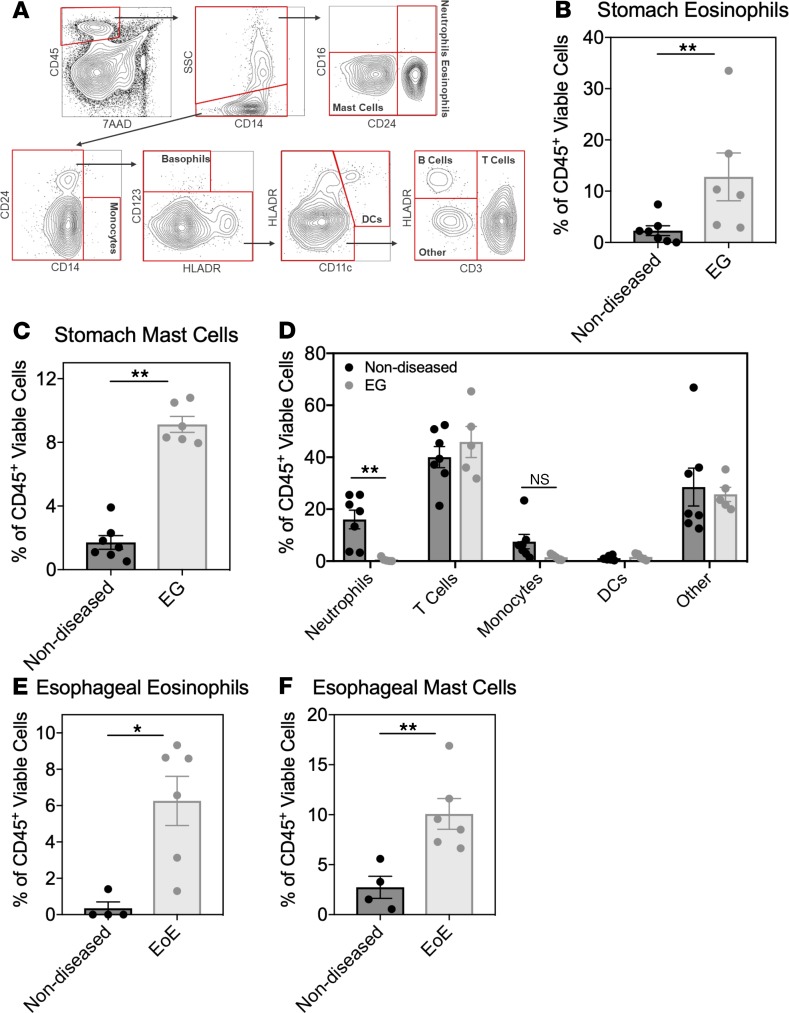
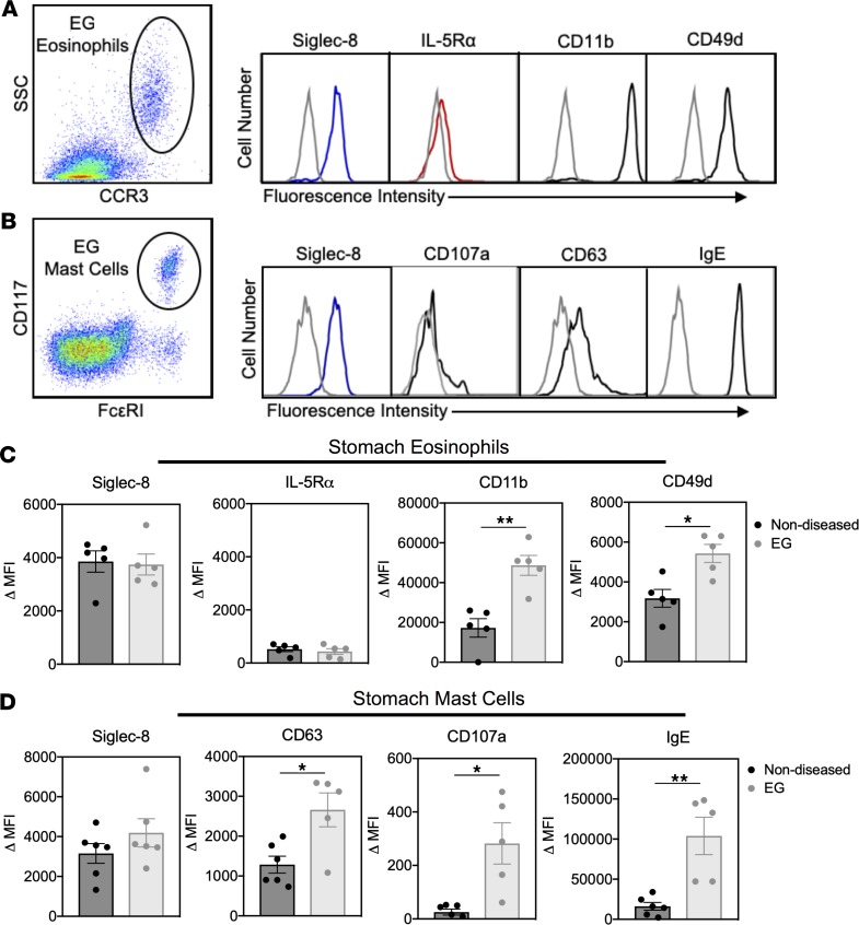
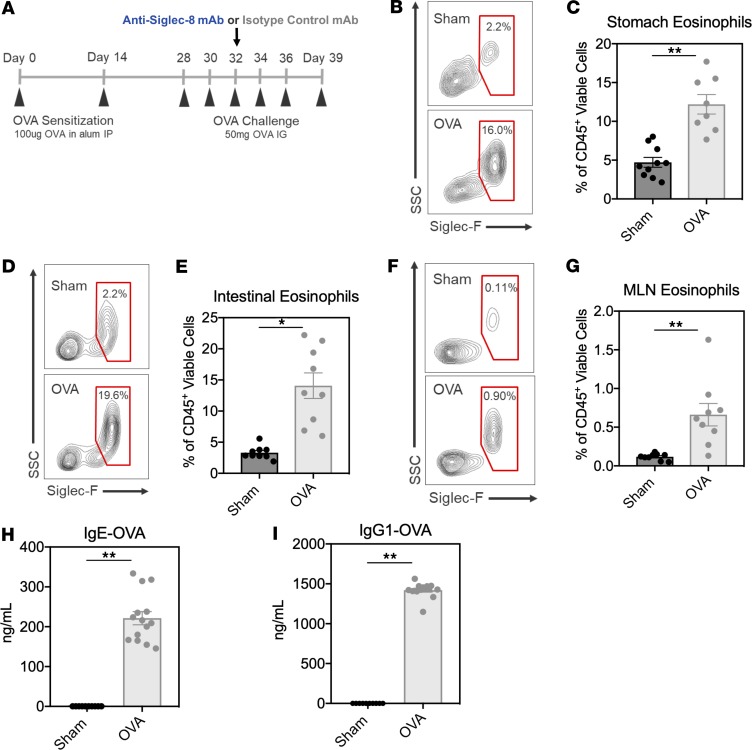
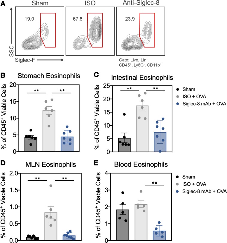
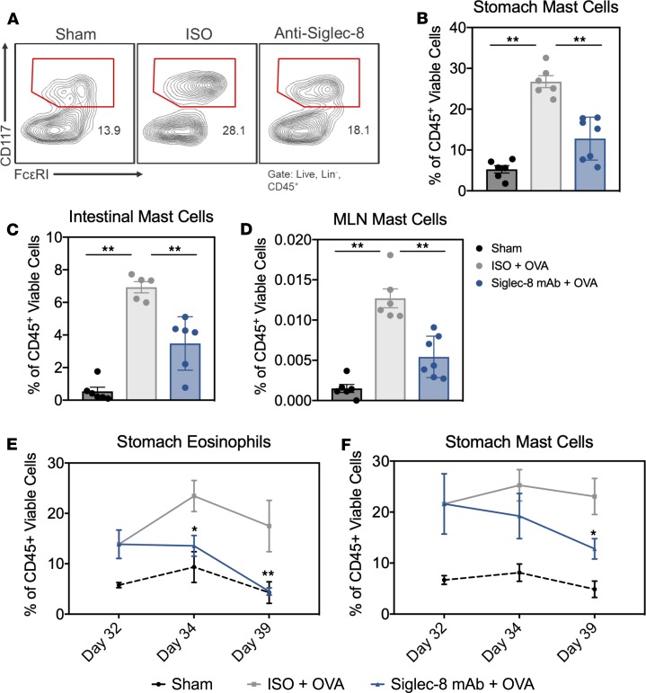
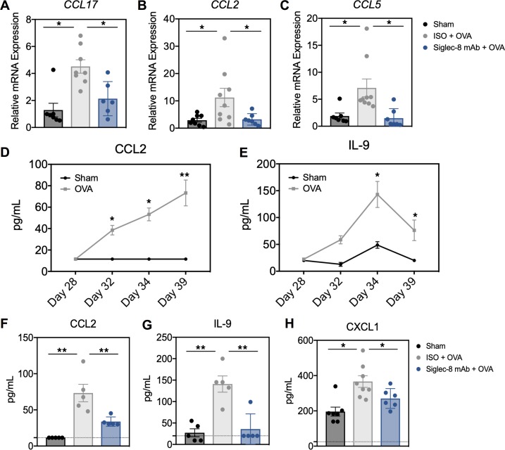
Aberrant accumulation and activation of eosinophils and potentially mast cells (MCs) contribute to the pathogenesis of eosinophilic gastrointestinal diseases (EGIDs), including eosinophilic esophagitis (EoE), gastritis (EG), and gastroenteritis (EGE). Current treatment options, such as diet restriction and corticosteroids, have limited efficacy and are often inappropriate for chronic use. One promising new approach is to deplete eosinophils and inhibit MCs with a monoclonal antibody (mAb) against sialic acid-binding immunoglobulin-like lectin 8 (Siglec-8), an inhibitory receptor selectively expressed on MCs and eosinophils. Here, we characterize MCs and eosinophils from human EG and EoE biopsies using flow cytometry and evaluate the effects of an anti-Siglec-8 mAb using a potentially novel Siglec-8-transgenic mouse model in which EG/EGE was induced by ovalbumin sensitization and intragastric challenge. MCs and eosinophils were significantly increased and activated in human EG and EoE biopsies compared with healthy controls. Similar observations were made in EG/EGE mice. In Siglec-8-transgenic mice, anti-Siglec-8 mAb administration significantly reduced eosinophils and MCs in the stomach, small intestine, and mesenteric lymph nodes and decreased levels of inflammatory mediators. In summary, these findings suggest a role for both MCs and eosinophils in EGID pathogenesis and support the evaluation of anti-Siglec-8 as a therapeutic approach that targets both eosinophils and MCs.

Keywords: Allergy; Gastroenterology; Mast cells; Mouse models; Therapeutics.


Conflict of interest statement

Conflict of interest: Research was funded by Alkalos, Inc. BAY, ECB, JL, RF, HSR, BSB, CB, and NT are or were employees of and/or own stock and/or stock options from Allakos, Inc. BSB is a paid consultant on the scientific advisory board of Allakos, Inc., and owns stock in Allakos. He is a coinventor on an existing Siglec-8–related patent (US20080213212A1) and thus may be entitled to a share of royalties received by Johns Hopkins University on the potential sales of such products. He is also a cofounder of Allakos, which makes him subject to certain restrictions under university policy. The terms of this arrangement are being managed by the Johns Hopkins University and Northwestern University in accordance with their conflict of interest policies.

Figures

Figure 1
Figure 1. EG and EoE patient tissues have significantly increased numbers of eosinophils and mast cells compared with nondiseased control tissue.
(A) Flow cytometry gating strategy used to identify immune cells, including eosinophils and mast cells, in human stomach tissue from patients with EG. Percentage of (B) eosinophils (CD45+7AADSSChiCD16CD24+) and (C) mast cells (CD45+7AADSSChiCD16CD24) present in nondiseased (black) or EG (gray) stomach tissue identified using the gating strategy in A. (D) Percentage of neutrophils, T cells, monocytes, DCs, and other (B cells, NK cells, macrophages, basophils) in nondiseased (black) or EG (gray) stomach tissue using the gating strategy shown in A. Percentage of (E) eosinophils and (F) mast cells present in nondiseased (black) or EoE (gray) esophageal tissue identified using the gating strategy in A. The percentage of cells was derived from the CD45+ viable population. Data are plotted as mean ± SEM for n = 7 nondiseased stomach tissue and n = 4 nondiseased esophageal tissue; n = 4 EG, n = 3 EG + EoE, and n = 3 EoE patients. *P < 0.05; **P < 0.01 by Mann-Whitney U test.
Figure 2
Figure 2. Mast cells and eosinophils from EG and EoE patient tissues are highly activated compared with nondiseased control cells.
(A) Dot plot of eosinophils in EG patient tissue identified by CD45+7AADCD117CD16CCR3+SSChi cells. Histogram of EG eosinophils labeled for analysis of surface expression of Siglec-8, IL-5Rα, CD11b, or CD49d or a fluorescence minus 1 (FMO) negative control (gray). (B) Dot plot of mast cells in EG patient tissue identified by CD45+7AADCD117+FcεRI+ cells. Histogram of EG mast cells labeled for analysis of surface expression of Siglec-8, CD107a, CD63, or IgE or an FMO negative control (gray). (C) Expression as shown by ΔMFI of Siglec-8, IL-5Rα, CD11b, and CD49d on stomach eosinophils from nondiseased controls (black) or patients with EG (gray). (D) Expression as shown by ΔMFI of the mast cell activation and degranulation markers, CD63, CD107a, and IgE, on stomach mast cells from nondiseased controls (black) or patients with EG (gray). Data are plotted as mean ± SD for n = 5–6 nondiseased stomach tissue; n = 2 EG, and n = 3 EG + EoE. *P < 0.05; **P < 0.01 by Mann-Whitney U test.
Figure 3
Figure 3. Systemic sensitization and intragastric challenge with OVA induces EG and EGE in Siglec-8–transgenic mice.
(A) Schematic of EG and EGE mouse model in Siglec-8–transgenic mice. Mice were systemically sensitized with OVA in aluminum hydroxide adjuvant (alum) on days 0 and 14, followed by 6 intragastric OVA challenges starting on day 28 until day 39. IP, intraperitoneal. (B) Representative flow cytometry contour plots of stomach eosinophils and (C) the percentage of eosinophils in the stomach in sham- or OVA-administered mice on day 39 quantified by flow cytometry. (D) Representative flow cytometry contour plots of duodenal eosinophils and (E) the percentage of eosinophils in the duodenum in sham- or OVA-administered mice on day 39 quantified by flow cytometry. (F) Representative flow cytometry contour plots of MLN eosinophils and (G) the percentage of eosinophils in the MLNs in sham- or OVA-administered mice on day 39 quantified by flow cytometry. (H and I) Serum levels of OVA-specific IgE or IgG1 in sham-treated mice (black) or mice sensitized and challenged with OVA (gray) on day 39. The percentage of eosinophils is derived from the CD45+ viable cell population. Data are plotted as mean ± SEM (n = 8–10 mice/group) and are representative of 3 experiments. *P < 0.05; **P < 0.01 by Mann-Whitney U test.
Figure 4
Figure 4. Administration of an anti–Siglec-8 mAb reduces eosinophils in GI tissues in mice with EG and EGE.
(A) Representative flow cytometry dot plots of stomach tissue eosinophils in mice treated with sham, OVA and isotype control mAb, or OVA and anti–Siglec-8 mAb. The percentage of eosinophils on day 39 in the (B) stomach, (C) duodenum, (D) MLNs, and (E) peripheral blood quantified by flow cytometry in sham-treated mice (black) or mice sensitized and challenged with OVA and dosed with either an isotype control mAb (gray) or anti–Siglec-8 mAb (blue). The percentage of eosinophils is derived from the CD45+ viable cell population. Data are plotted as mean ± SEM (n = 6–7 mice/group) and are representative of 3 experiments. **P < 0.01 by 1-way ANOVA with Tukey’s multiple-comparisons test.
Figure 5
Figure 5. Administration of an anti–Siglec-8 mAb reduces mast cells in GI tissues in mice with EG and EGE.
(A) Representative flow cytometry dot plots of stomach tissue mast cells in mice treated with sham, OVA and isotype control mAb, or OVA and anti–Siglec-8 mAb. The percentage of mast cells on day 39 in the (B) stomach, (C) duodenum, and (D) MLN quantified by flow cytometry in sham-treated mice (black) or mice sensitized and challenged with OVA and dosed with either an isotype control mAb (gray) or anti–Siglec-8 mAb (blue). The percentage of stomach (E) eosinophils or (F) mast cells on days 32, 34, and 39 in mice treated with sham (black), OVA and isotype control mAb (gray), or OVA and anti–Siglec-8 mAb (blue) quantified by flow cytometry. The percentage of mast cells is derived from the CD45+ viable cell population. Data are plotted as mean ± SEM (n = 6–7 mice/group for BD and n = 4–6 mice/group for E and F) and are representative of 3 experiments. *P < 0.05; **P < 0.01 by 1-way ANOVA with Tukey’s multiple-comparisons test (BD) or 2-tailed t test with Holm-Šídák’s posttest (E and F).
Figure 6
Figure 6. Mice treated with anti–Siglec-8 mAb display reduced expression of OVA-induced type 2 immune–associated inflammatory cytokines and chemokines in intestinal tissue and serum.
Quantitative PCR (qPCR) gene expression analysis of (A) CCL17, (B) CCL2, and (C) CCL5 in the duodenum at day 39 of study in sham-treated mice (black) or mice sensitized and challenged with OVA and dosed with either an isotype control mAb (gray) or anti–Siglec-8 mAb (blue). (D and E) CCL2 and IL-9 levels in serum in sham-treated (black) or OVA-treated (gray) mice on day 28 (before first OVA challenge) and days 32, 34, and 39. (FH) CCL2, IL-9, and CXCL1 levels in serum in mice treated with sham (black), OVA and isotype control mAb (gray), and OVA and anti–Siglec-8 mAb (blue) (n = 5 mice/group). Graphs are plotted as mean ± SEM (n = 6–8 mice/group) and are representative of 3 experiments. *P < 0.05; **P < 0.01 by 1-way ANOVA with Tukey’s multiple-comparisons test (AC) or 2-tailed t test with Holm-Šídák’s posttest (D and E).

References

    1. Uppal V, Kreiger P, Kutsch E. Eosinophilic gastroenteritis and colitis: a comprehensive review. Clin Rev Allergy Immunol. 2016;50(2):175–188. doi: 10.1007/s12016-015-8489-4. - DOI - PubMed
    1. Bochner BS, et al. Workshop report from the National Institutes of Health Taskforce on the Research Needs of Eosinophil-Associated Diseases (TREAD) J Allergy Clin Immunol. 2012;130(3):587–596. doi: 10.1016/j.jaci.2012.07.024. - DOI - PMC - PubMed
    1. Khoury P, et al. Revisiting the NIH Taskforce on the Research needs of Eosinophil-Associated Diseases (RE-TREAD) J Leukoc Biol. 2018;104(1):69–83. doi: 10.1002/JLB.5MR0118-028R. - DOI - PMC - PubMed
    1. Hsu Blatman KS, Gonsalves N, Hirano I, Bryce PJ. Expression of mast cell-associated genes is upregulated in adult eosinophilic esophagitis and responds to steroid or dietary therapy. J Allergy Clin Immunol. 2011;127(5):1307–1308. doi: 10.1016/j.jaci.2010.12.1118. - DOI - PMC - PubMed
    1. Abonia JP, et al. Involvement of mast cells in eosinophilic esophagitis. J Allergy Clin Immunol. 2010;126(1):140–149. doi: 10.1016/j.jaci.2010.04.009. - DOI - PMC - PubMed

MeSH terms

Supplementary concepts