. 2019 Aug 29;13(8):e0007056.
doi: 10.1371/journal.pntd.0007056. eCollection 2019 Aug.

The Accuracy of Histopathological and Cytopathological Techniques in the Identification of the Mycetoma Causative Agents

Affiliations

The Accuracy of Histopathological and Cytopathological Techniques in the Identification of the Mycetoma Causative Agents

Emmanuel Edwar Siddig et al. PLoS Negl Trop Dis. .

Abstract

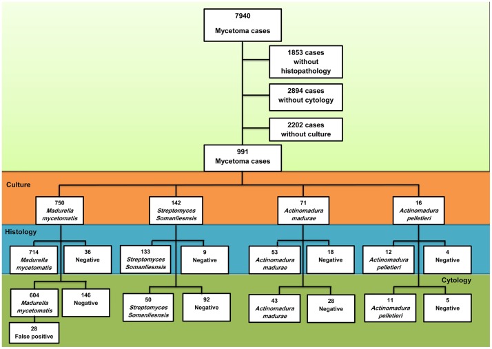
Mycetoma is a devastating neglected tropical disease, caused by various fungal and bacterial pathogens. Correct diagnosis to the species level is mandatory for proper treatment. In endemic areas, various diagnostic tests and techniques are in use to achieve that, and that includes grain culture, surgical biopsy histopathological examination, fine needle aspiration cytological (FNAC) examination and in certain centres molecular diagnosis such as PCR. In this retrospective study, the sensitivity, specificity and diagnostic accuracy of grain culture, surgical biopsy histopathological examination and FNAC to identify the mycetoma causative organisms were determined. The histopathological examination appeared to have better sensitivity and specificity. The histological examination results were correct in 714 (97.5%) out of 750 patients infected with Madurella mycetomatis, in 133 (93.6%) out of 142 patients infected with Streptomyces somaliensis, in 53 (74.6%) out of 71 patients infected with Actinomadura madurae and in 12 (75%) out of 16 patients infected with Actinomadura pelletierii. FNAC results were correct in 604 (80.5%) out of 750 patients with Madurella mycetomatis eumycetoma, in 50 (37.5%) out of 133 Streptomyces somaliensis patients, 43 (60.5%) out of 71 Actinomadura madurae patients and 11 (68.7%) out of 16 Actinomadura pelletierii. The mean time required to obtain the FNAC result was one day, and for the histopathological examinations results it was 3.5 days, and for grain it was a mean of 16 days. In conclusion, histopathological examination and FNAC are more practical techniques for rapid species identification than grain culture in many endemic regions.


Conflict of interest statement

The authors have declared that no competing interests exist.

Figures

Fig 1
Fig 1. identification of the causative agents: All culture, histopathological and cytological identification reports of patients seen between January 1991 and January 2018 in the Mycetoma Research Centre were retrieved from our electronic database.
Of these patients in 2202 cases no microbiological report was available, of 1853 cases no histopathological report was available and in 2894 cases no cytological report was available leaving 991 mycetoma cases which met our inclusion criteria. The culture identification was considered the golden standard. Of these patients 750 cases were caused by M. mycetomatis, 142 cases by S. somaliensis, 71 were caused by A. madurae and 12 by A. pelletieri. From these cases the histological and cytological reports were compared to the culture results. A histological report or cytological report was either identical to the culture identification or negative. Results for both histology and cytology reports are shown.
Fig 2
Fig 2. Cytology of mycetoma causative agents.
(A): The cytological appearance of M. mycetomatis, (B): A. pelletierii, (C): S. somaliensis in fine needle aspirates stained with HE (magnification 10 times).
Fig 3
Fig 3. Histopathological appearance of mycetoma causative agents.
The histopathological appearance of A. madurae (A), S. somaliensis (B), A. pelletierii (C) and M. mycetomatis (D) in HE stained sections (Magnification 10x).
Fig 4
Fig 4. Histopathological section from patient diagnosed as M. mycetomatis based on the culture results; Histopathology report was been negative for fungus and when we retrieved the slide we can see adherence of histiocytes at the center with sparse amount of the grains (H and E, X 10).
Fig 5
Fig 5. False positive identifications in cytology.
Showing cytological smears of (A) M. mycetomatis hyphae stained with Giemsa stain, (B) Synthetic Fibers stained Wright-Giemsa stain. (Giemsa stain, X40). (C) Smear showing hyphae of M. mycetomatis. (D) Smear showing elongated structures of the Oedogoniales order. The chloroplasts form a chain interrupted by clear zones (X40). (E) Smear showing M. mycetomatis grains after being crushed on the smear. (F) smear with abundant calcific debris without intact cells taken from patient with tumoral calcinosis (Diff Quick, X10).

References

    1. Reis CMS, Reis-Filho EGM. Mycetomas: an epidemiological, etiological, clinical, laboratory and therapeutic review. An Bras Dermatol. 2018; 93 (1):8–18. 10.1590/abd1806-4841.20187075 - DOI - PMC - PubMed
    1. Fahal AH. Mycetoma: A global medical and socio-economic dilemma. PLoS Negl Trop Dis. 2017; 11(4): e0005509 10.1371/journal.pntd.0005509 - DOI - PMC - PubMed
    1. Zijlstra EE, van de Sande WWJ, Fahal AH. Mycetoma: A Long Journey from Neglect. PLoS Negl Trop Dis. 2016; 10(1): e0004244 10.1371/journal.pntd.0004244 - DOI - PMC - PubMed
    1. Suleiman SH, Wadaella ES, Fahal AH. The Surgical Treatment of Mycetoma. PLoS Negl Trop Dis. 2016; 10(6): e0004690 10.1371/journal.pntd.0004690 - DOI - PMC - PubMed
    1. van de Sande Wendy, Fahal Ahmed, Ahmed Sarah Abdalla, Serrano Julian Alberto, Bonifaz Alexandro, Zijlstra Ed, on behalf of the eumycetoma working group; Closing the mycetoma knowledge gap, Medical Mycology, 2018; 56(supp 1):153–164. - PubMed

MeSH terms

Supplementary concepts