. 2019 Nov 6;27(11):1906-1918.
doi: 10.1016/j.ymthe.2019.07.019. Epub 2019 Aug 5.

Recombinant Adenovirus Expressing a Soluble Fusion Protein PD-1/CD137L Subverts the Suppression of CD8+ T Cells in HCC

Affiliations

Recombinant Adenovirus Expressing a Soluble Fusion Protein PD-1/CD137L Subverts the Suppression of CD8+ T Cells in HCC

Yonghui Zhang et al. Mol Ther. .

Abstract

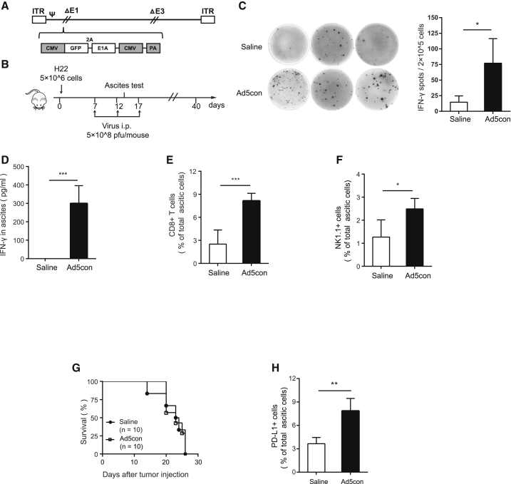
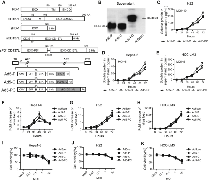
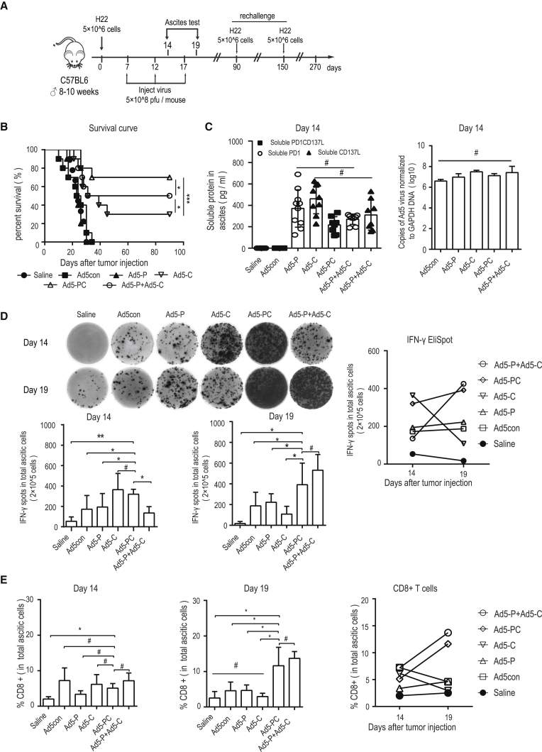
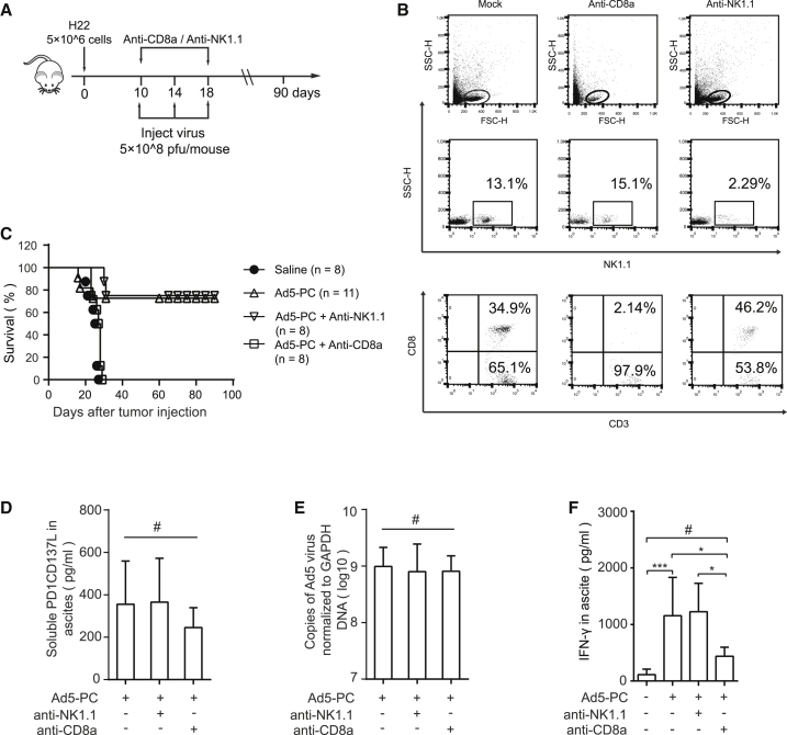
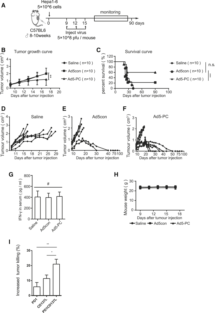
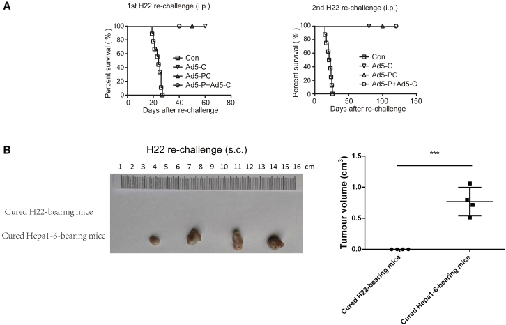
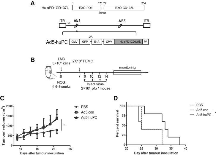
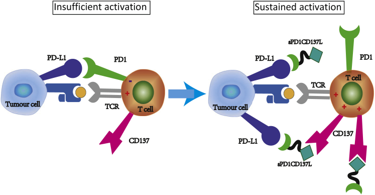
Oncolytic viruses are an excellent platform for developing effective strategies in cancer immunotherapy. Several challenges remain in the use of viro-immunotherapy for cancer, such as the lack of costimulatory signals and negative regulation of immune checkpoints. In this study, we designed a novel adenovirus expressing a soluble fusion protein, programmed cell death protein 1 (PD-1)/CD137L, which contains the extracellular domains of PD-1 and CD137L at each terminus (Ad5-PC). Ad5-PC preserved the costimulatory activity of CD137L and facilitated the persistence of activated CD8+ T cells. Ad5-PC induced strikingly increased antitumor activity in both ascitic and subcutaneous hepatocellular carcinoma (HCC) tumor models, with 70% and 60% long-term cure rates, respectively. The improved antitumor effect of Ad5-PC was attributed to the sustained high-level lymphocyte activation and interferon (IFN)-γ production in the tumor microenvironment, and was essentially dependent on CD8+ T cells rather than natural killer (NK) cells. Moreover, Ad5-huPC-expressing human soluble PD-1/CD137L fusion protein was effective in suppressing tumor growth and improving survival in a humanized mouse model. We confirmed that Ad5-PC induced tumor-specific and systematic protection against tumor rechallenges at both in situ and distant sites. Thus, Ad5-PC harnesses several distinct functions to efficiently overcome several major hurdles of viro-immunotherapy.

Keywords: adenovirus; hepatocellular carcinoma; immune checkpoints.


Figures

Figure 1
Figure 1
Replication-Competent Adenovirus Improves Immune Responses in TME but Fails to Prolong Survival in an Ascitic HCC Murine Model Male C57BL/6 mice were injected peritoneally with 5 × 106 H22 cells. On days 7, 12, and 17, mice were treated i.p. with Ad5con (5 × 108 PFUs), with saline used as a control (n = 10 for each group). (A) Construction of the replication-competent adenovirus. (B) Schematic diagram of the experimental setup for adenovirus therapy. (C) Mouse ascites were collected on day 14, and the IFN-γ-producing lymphocytes were determined by ELISpot. The representative results for ELISpot are shown in the left panel, and the associated plot counts are shown in the right panel. (D) IFN-γ concentrations in the ascites were determined by ELISA. (E and F) Frequencies of (E) CD8+ T cells and (F) NK cells were determined by flow cytometry. (G) Survival curves of mice with or without Ad5con treatment. (H) PD-L1-positive cells in ascites were detected by flow cytometry. Data shown are the means ± SD. Data are representative of at least three independent experiments. *p < 0.05, **p < 0.01, ***p < 0.001.
Figure 2
Figure 2
Generation of Recombinant Adenoviruses Regulating PD-L1/PD-1 Negative Feedback Signaling and CD137L/CD137 Costimulatory Signaling Recombinant adenoviruses were generated to secrete soluble PD-1 (sPD-1), soluble CD137L (sCD137L), or a fusion PD-1/CD137L (sPD-1CD137L). (A) Recombinant adenovirus constructs. (B) H22 cells were infected with recombinant adenovirus (MOI = 20) for 72 h, and the levels of sPD-1, sCD137L, and sPD-1CD137L in the supernatant were detected by western blot. The molecular weights were as expected. H22 (C), Hepa1-6 (D), and HCC-LM3 cells (E) were infected with recombinant adenoviruses at MOIs of 20 or 5. The supernatants were collected at the indicated time points, and the concentrations of soluble proteins were determined by ELISA. H22 cells (F), Hepa1-6 cells (G), and HCC-LM3 cells (H) were infected with recombinant adenoviruses at an MOI of 10 and harvested at varying time points. DNA was extracted, and the viral copy number was determined by qPCR. The fold changes were calculated by dividing the copy number at 6 h. H22 cells (I), Hepa1-6 cells (J), and HCC-LM3 cells (K) were infected with recombinant adenoviruses at the indicated MOI for 72 h, and cell viability was determined by MTT. Data shown are the means ± SD. Ad5-C, recombinant adenovirus encoding sCD137L; Ad5-P, recombinant adenovirus encoding sPD-1; Ad5-PC, recombinant adenovirus encoding sPD-1CD137L; ENDO, endocellular domain; EXO, extracellular domain; TM, transmembrane region.
Figure 3
Figure 3
Recombinant Ad5-PD-1/CD137L Activates Immune Responses and Increases Durable Cure Rate Male C57BL/6 mice were injected peritoneally with 5 × 106 H22 cells. On days 7, 12, and 17, mice were treated i.p. with recombinant adenoviruses (5 × 108 PFUs), and ascites were collected on days 14 and 19 (n = 10 for each group). (A) Schematic diagram of the experimental setup for adenovirus therapy. (B) Survival curves of mice treated with recombinant adenoviruses. (C) On day 14, the concentrations of soluble proteins in ascites were detected by ELISA, and viral copies were determined by qPCR. Data shown are the means ± SD. (D) On days 14 and 19, the IFN-γ-producing lymphocytes in ascites were determined by ELISpot. The representative results are shown in the left panel, and the associated plot counts are shown in the right panel. (E) CD8+ T cell frequencies in ascites were determined by flow cytometry. Data shown are the means ± SD. #p value was not significant; *p < 0.05, **p < 0.01, ***p < 0.001.
Figure 4
Figure 4
CD8+ T Cells Mediate the Antitumor Activity of Ad5-PC Male C57BL/6 mice were injected peritoneally with 5 × 106 H22 cells. Mice were treated i.p. with recombinant adenoviruses (5 × 108 PFUs/mouse), combined with or without anti-CD8a or anti-NK1.1. (A) Schematic diagram of the experimental setup for CD8+ T cell or NK cell depletion. (B) Mouse blood was collected 5 days after the administration of blocking antibody, and the depletion effects of anti-CD8a and anti-NK1.1 were confirmed via flow cytometry. (C) Survival curves of mice treated with recombinant adenoviruses in the presence or absence of anti-CD8a or anti-NK1.1 antibodies. The concentrations of soluble proteins (D) and IFN-γ (F) in ascites were determined by ELISA. (E) Viral copies in ascites were detected by qPCR. Data shown are the means ± SD. #Not significant; *p < 0.05, ***p < 0.001.
Figure 5
Figure 5
Ad5-PC Attenuates Tumor Growth in a Subcutaneous HCC Murine Model Male C57BL/6 mice were subcutaneously inoculated with 5 × 106 Hepa1-6. Recombinant adenovirus (5 × 108 PFUs/mouse) was locally injected when the tumor volume reached 0.4 cm3. The tumor volume and mouse weights were monitored, and the mice were considered to have died when the tumor volume reached 2 cm3. (A) Schematic diagram of the experimental setup for adenovirus therapy in solid tumors. Tumor volumes (B) and survival curves (C) of mice with or without treatment of recombinant adenovirus were determined. (D–F) Volume for each tumor in the group of mice treated with (D) saline, (E) Ad5con, or (F) Ad5-PC. (G) On day 14, the concentrations of sPD-1CD137 and IFN-γ in plasma were determined by ELISA. (H) Mouse weights were determined at the indicated time points. (I) Splenocytes and Hepa1-6 were mixed at a ratio of 5:1, in the presence of purified PD1, CD137L, or fusion protein PD1/CD137L, respectively. The number of Hepa1-6 cells was reflected by the bioluminescence intensity. Data shown are the means ± SD. #Not significant; *p < 0.05, **p < 0.01, ***p < 0.001.
Figure 6
Figure 6
Ad5-PC-Cured Mice Exhibit a Tumor-Specific and Long-Term Immune Memory (A) Mice that had been cured were re-challenged with 5 × 106 H22 cells on days 90 and 150 post-inoculation. Naive mice were used as a control. The day that mice received the re-challenge was regarded as day 0 in the survival curves. (B) Cured mice from H22 ascetic tumor or hepa1-6 solid HCC were subcutaneously injected with 5 × 106 H22 cells. Mice were sacrificed for tumor measure 16 days after re-challenge with H22 cells. i.p., injected peritoneally; s.c., injected subcutaneously. ***p < 0.001.
Figure 7
Figure 7
Ad5-huPC Significantly Inhibits HCC Growth and Prolongs Survival in a Humanized Mouse Model (A) The recombinant adenovirus coding for human PD1/CD137L. (B) Schematic diagram of the experimental setup for Ad5-PC therapy in the NCG mouse model. Male NCG mice were injected subcutaneously with 5 × 106 LM3 cells. Human PBMC was administrated on day 7, followed by the Ad5-huPC treatment. Tumor volumes (C) and mice survival (D) were monitored. *p < 0.05.
Figure 8
Figure 8
Ad5-PC Harnesses Multiple Distinct Functions to Improve Antitumor Immune Responses Recombinant adenovirus producing the soluble fusion protein PD1/CD137L harnesses multiple distinct functions to promote antitumor immune responses. The infiltrated T cells induced by adenovirus are suppressed because of the inhibitory molecule PD-L1 and the paucity of costimulatory molecules (left panel). The soluble fusion protein PD1/CD137L provides a costimulatory signal to the virus-recruited CD8+ T cells and sustains the immune activity of CD8+ T cells by blocking PD-L1. Furthermore, sPD1/CD137L may bridge the gap of effecter T cells and malignant cells to enhance cytotoxicity and convert PD-L1-mediated suppressive signal to activating signal (right panel).

Comment in

References

    1. O’Donnell J.S., Teng M.W.L., Smyth M.J. Cancer immunoediting and resistance to T cell-based immunotherapy. Nat. Rev. Clin. Oncol. 2019;16:151–167. - PubMed
    1. Dyer A., Baugh R., Chia S.L., Frost S., Iris S., Jacobus E.J., Khalique H., Pokrovska T.D., Scott E.M., Taverner W.K. Turning cold tumours hot: oncolytic virotherapy gets up close and personal with other therapeutics at the 11th Oncolytic Virus Conference. Cancer Gene Ther. 2019;26:59–73. - PubMed
    1. Bischoff J.R., Kirn D.H., Williams A., Heise C., Horn S., Muna M., Ng L., Nye J.A., Sampson-Johannes A., Fattaey A., McCormick F. An adenovirus mutant that replicates selectively in p53-deficient human tumor cells. Science. 1996;274:373–376. - PubMed
    1. Nemunaitis J., Ganly I., Khuri F., Arseneau J., Kuhn J., McCarty T., Landers S., Maples P., Romel L., Randlev B. Selective replication and oncolysis in p53 mutant tumors with ONYX-015, an E1B-55kD gene-deleted adenovirus, in patients with advanced head and neck cancer: a phase II trial. Cancer Res. 2000;60:6359–6366. - PubMed
    1. Ribas A., Dummer R., Puzanov I., VanderWalde A., Andtbacka R.H.I., Michielin O., Olszanski A.J., Malvehy J., Cebon J., Fernandez E. Oncolytic Virotherapy Promotes Intratumoral T Cell Infiltration and Improves Anti-PD-1 Immunotherapy. Cell. 2018;174:1031–1032. - PubMed

Publication types

MeSH terms