Review
. 2019 Oct 11;294(41):15137-15157.
doi: 10.1074/jbc.REV119.006901. Epub 2019 Aug 29.

Multifaceted HIV integrase functionalities and therapeutic strategies for their inhibition

Affiliations
Review

Multifaceted HIV integrase functionalities and therapeutic strategies for their inhibition

Alan N Engelman. J Biol Chem. .

Abstract

Antiretroviral inhibitors that are used to manage HIV infection/AIDS predominantly target three enzymes required for virus replication: reverse transcriptase, protease, and integrase. Although integrase inhibitors were the last among this group to be approved for treating people living with HIV, they have since risen to the forefront of treatment options. Integrase strand transfer inhibitors (INSTIs) are now recommended components of frontline and drug-switch antiretroviral therapy formulations. Integrase catalyzes two successive magnesium-dependent polynucleotidyl transferase reactions, 3' processing and strand transfer, and INSTIs tightly bind the divalent metal ions and viral DNA end after 3' processing, displacing from the integrase active site the DNA 3'-hydroxyl group that is required for strand transfer activity. Although second-generation INSTIs present higher barriers to the development of viral drug resistance than first-generation compounds, the mechanisms underlying these superior barrier profiles are incompletely understood. A separate class of HIV-1 integrase inhibitors, the allosteric integrase inhibitors (ALLINIs), engage integrase distal from the enzyme active site, namely at the binding site for the cellular cofactor lens epithelium-derived growth factor (LEDGF)/p75 that helps to guide integration into host genes. ALLINIs inhibit HIV-1 replication by inducing integrase hypermultimerization, which precludes integrase binding to genomic RNA and perturbs the morphogenesis of new viral particles. Although not yet approved for human use, ALLINIs provide important probes that can be used to investigate the link between HIV-1 integrase and viral particle morphogenesis. Herein, I review the mechanisms of retroviral integration as well as the promises and challenges of using integrase inhibitors for HIV/AIDS management.

Keywords: AIDS; HIV/AIDS; antiretroviral therapy; intasome; integrase; integrase strand transfer inhibitor; integration; microbiology; polynucleotidyl transferase; retrovirus; viral DNA; virology; virus structure.


Conflict of interest statement

The author has received fees from ViiV Healthcare Co. within the past 12 months

Figures

Figure 1.
Figure 1.
HIV replication cycle. After entry into a susceptible target cell, RT converts genomic RNA into linear DNA within the confines of the reverse transcription complex (RTC) (272). Processing of the viral DNA ends by IN yields the PIC (30), which can integrate the endogenous DNA made by reverse transcription into recombinant target DNA in vitro (5). Following nuclear import and integration, the provirus (flanked by composite cyan/yellow/magenta LTRs) serves as a transcriptional template to produce viral mRNAs for translation of viral proteins as well as nascent viral genomes that co-assemble with viral proteins to form immature virions that bud out from the infected cell (2). Shown is a generalized scheme that depicts the major steps of HIV-1 replication, although it is important to note that deviations from this plan exist throughout Retroviridae. Most notably, spumavirus reverse transcription occurs during the second half of the infectious cycle (after integration), and spumaviral particles accordingly predominantly contain dsDNA (104, 273). The primary steps in the HIV-1 replication cycle that are inhibited by the two major classes of IN inhibitors discussed herein are indicated.
Figure 2.
Figure 2.
DNA cutting and joining steps of retroviral integration. The linear viral reverse transcript (lavender lines; plus-strands shaded more darkly than same-colored minus-strands throughout the cartoon) contains a copy of the LTR at each end composed of cyan U3, yellow R repeat, and magenta U5 sequences. The upstream LTR is abutted by the primer-binding site (PBS; purple box), whereas the downstream element is abutted by the polypurine tract (PPT; lavender box). During 3′ processing, IN hydrolyzes the DNA adjacent to invariant CA dinucleotides, which for HIV-1 liberates the pGTOH dinucleotide from each end. After nuclear localization, the intasome interacts with host target DNA (gray lines with targeted green sequence) to promote DNA strand transfer. The DNA gaps that persist after strand transfer are repaired by host cell machinery to yield a target site duplication (thin green lines) flanking the integrated provirus.
Figure 3.
Figure 3.
Retroviral intasome structures. A–C, representative intasomes from the spumavirus PFV (A; protein database (PDB) accession code 3OY9), β-retrovirus MMTV (B; PDB code 3JCA), and lentivirus MVV (C; PDB code 5M0Q) are color-coded to highlight the CIC. Green and blue, catalytically active IN protomers; cyan, supporting IN CCDs; black, DNA strands. Whereas four PFV IN molecules suffice to form the CIC, both MMTV and MVV require six IN protomers. For MMTV, critical CTDs (magenta) are donated by flanking IN dimers, leading to an overall IN octamer. In MVV, flanking IN tetramers provide the critical CTDs, resulting in an overall IN hexadecamer. Gray coloring in B and C deemphasizes IN elements that do not compose the CIC. D–F, resected CCD and CTD domains from above green IN protomers, oriented to highlight the different CCD-CTD linker regions (dark gray). Associated magenta CTDs from separate IN protomers in E and F assume similar positions as the green CTD in D. Red sticks, DDE catalytic triad residues.
Figure 4.
Figure 4.
INSTI structures and ALLINI chemotypes. A, diagrams of the four FDA-licensed INSTIs as well as investigational second-generation compound 6p (113, 119). B, representative ALLINI chemotypes. Asterisks mark common positions of t-butoxyacid moieties.
Figure 5.
Figure 5.
Mechanisms of INSTI action. A, close-up view of one PFV IN active site in the CSC intasome structure (PDB code 3OY9) with IN secondary elements labeled. Additional labels highlight the conserved CA dinucleotide of the transferred DNA strand (magenta sticks) and associated G nucleotide of the nontransferred strand (orange), the 3′-OH nucleophile of the terminal deoxyadenylate used by IN to cut chromosomal DNA, as well as IN residues (in sticks) that compose the catalytic triad (Asp-128, Asp-185, and Glu-221) and that when changed confer INSTI resistance (Tyr-212 and Asn-224). Blue and red stick colors denote nitrogen and oxygen atoms, respectively. Gray spheres, divalent metal ions; α, β, and η denote α-helix, β-strand, and 310-helix, respectively. B, RAL (cyan sticks)-bound PFV intasome structure (PDB code 3OYA) oriented as in A to highlight the mode of INSTI binding. RAL binding results in a greater than 6-Å displacement of the 3′-OH of the terminal deoxyadenylate from the IN active site. The position of the RAL methyl-oxadiazole group is highlighted in the cartoon on the left as well as the chemical diagram on the right, which was reconfigured from Fig. 4A to accentuate the position within the crystal structure. Other colors and labeling are the same as in A or as described under “INSTIs.” C, same as in B, except with DTG bound (PDB code 3S3M). The position of the IN β4-α2 connector is noted; other labeling is the same as in A and B. D, RAL (magenta) from the drug-bound PFV CSC structure (PDB code 3OYA) is overlaid with the PFV SSC structure (green; PDB code 4E7I) to highlight mimicry between drug oxygen atoms and oxygen atoms critical for IN 3′ processing activity (red sphere, nucleophilic water (W) molecule; red bridge in viral DNA (vDNA), scissile phosphodiester bond). E, similar to D; RAL is superimposed onto the PFV TCC structure (IN and target DNA in cyan and vDNA in blue; PDB code 4E7K) to highlight similarly positioned drug oxygen atoms with the vDNA 3′-oxygen and scissile phosphodiester bond in target DNA (tDNA) critical for strand transfer activity. Other labeling is the same as in A and B.
Figure 6.
Figure 6.
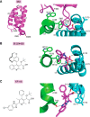
ALLINI mimicry of LEDGF/p75 binding to the HIV-1 IN CCD dimer. A, solution structure of the LEDGF/p75 IBD (left; PDB code 1Z9E) and IBD-CCD co-crystal structure (right; PDB code 2B4J) highlight the locations of hotspot-interacting residues Ile-365 and Asp-366 in the hairpin that connects α-helices 1 and 2 and Phe-406 in the α4-α5 hairpin (left). Whereas Asp-366 interacts with the backbone amide groups of IN residues Glu-170 and His-171 of the cyan IN monomer (dashed lines), Ile-365 occupies a hydrophobic pocket composed of IN residues from each monomer (i.e. Trp-132 of the green IN monomer and Met-178 of the cyan monomer; right). Other colorings denote atoms of interacting amino acid residues: nitrogen (blue), sulfur (yellow), and oxygen (red). B, quinoline ALLINI BI 224436 (left, chemical diagram) bound to the IN CCD dimer (right, PDB code 6NUJ). The interactions between the compound carboxylic acid and backbone amides within the IN cyan monomer are analogous to those shown in A for LEDGF/p75 residue Asp-366. Thr-174 of the IN cyan monomer additionally interacts with the t-butoxy moiety of the drug. Other labeling is the same as in A. C, binding of pyridine ALLINI KF116 (left, chemical structure) to the IN CCD dimer (right, PDB code 4O55). This view, rotated down ∼90° from A and B, is shown to accentuate the drug-binding pocket. In addition to the contacts described in B, Thr-125 of the green IN monomer interacts with the benzimidazole moiety of KF116. Other labeling is as defined in A and B.

References

    1. Arts E. J., and Hazuda D. J. (2012) HIV-1 antiretroviral drug therapy. Cold Spring Harb. Perspect. Med. 2, a007161 10.1101/cshperspect.a007161 - DOI - PMC - PubMed
    1. Sundquist W. I., and Kräusslich H.-G. (2012) HIV-1 assembly, budding, and maturation. Cold Spring Harb. Perspect. Med. 2, a006924 - PMC - PubMed
    1. Yamashita M., and Engelman A. N. (2017) Capsid-dependent host factors in HIV-1 infection. Trends Microbiol. 25, 741–755 10.1016/j.tim.2017.04.004 - DOI - PMC - PubMed
    1. Engelman A. (2010) Reverse transcription and integration. In Retroviruses: Molecular Biology, Genomics and Pathogenesis (Kurth R., and Bannert N., eds) pp. 129–159, Caister Academic Press, Norfolk, UK
    1. Brown P. O., Bowerman B., Varmus H. E., and Bishop J. M. (1987) Correct integration of retroviral DNA in vitro. Cell 49, 347–356 10.1016/0092-8674(87)90287-X - DOI - PubMed

Publication types

MeSH terms