. 2019 Sep 13;39(9):BSR20191433.
doi: 10.1042/BSR20191433. Print 2019 Sep 30.

ERK1/2 signaling regulates the immune microenvironment and macrophage recruitment in glioblastoma

Affiliations

ERK1/2 signaling regulates the immune microenvironment and macrophage recruitment in glioblastoma

Claire Lailler et al. Biosci Rep. .

Abstract

The tumor microenvironment is an important determinant of glioblastoma (GBM) progression and response to treatment. How oncogenic signaling in GBM cells modulates the composition of the tumor microenvironment and its activation is unclear. We aimed to explore the potential local immunoregulatory function of ERK1/2 signaling in GBM. Using proteomic and transcriptomic data (RNA seq) available for GBM tumors from The Cancer Genome Atlas (TCGA), we show that GBM with high levels of phosphorylated ERK1/2 have increased infiltration of tumor-associated macrophages (TAM) with a non-inflammatory M2 polarization. Using three human GBM cell lines in culture, we confirmed the existence of ERK1/2-dependent regulation of the production of the macrophage chemoattractant CCL2/MCP1. In contrast with this positive regulation of TAM recruitment, we found no evidence of a direct effect of ERK1/2 signaling on two other important aspects of TAM regulation by GBM cells: (1) the expression of the immune checkpoint ligands PD-L1 and PD-L2, expressed at high mRNA levels in GBM compared with other solid tumors; (2) the production of the tumor metabolite lactate recently reported to dampen tumor immunity by interacting with the receptor GPR65 present on the surface of TAM. Taken together, our observations suggest that ERK1/2 signaling regulates the recruitment of TAM in the GBM microenvironment. These findings highlight some potentially important particularities of the immune microenvironment in GBM and could provide an explanation for the recent observation that GBM with activated ERK1/2 signaling may respond better to anti-PD1 therapeutics.

Keywords: CCL2/MCP1; ERK1/2; Glioblastoma; Immune checkpoints; The Cancer Genome Atlas; Tumour-associated macrophages.


Conflict of interest statement

The authors declare that there are no competing interests associated with the manuscript.

Figures

Figure 1
Figure 1. Identification of a subgroup of GBM with high phosphorylation levels of the ERK1/2 kinases
(A) Hierarchical clustering of RPPA data (40 phosphoproteins including phosphorylated forms of ERK1/2, EGFR, and MET, highlighted in red) in GBM tumors from TCGA (n=244 samples). (B) Clustered heatmap of RPPA data for GBM with samples (columns) and phosphoproteins (rows). High protein expression is shown in red, low protein expression in blue. The following clusters are labeled at the bottom of the heatmap: MET, EGFR, PKC, ERK1/2 and MTOR. (C) Immunoblot analysis of EGFR phosphorylation in the human GBM cell line A172, carrying an amplified EGFR gene. Phosphorylation of EGFR on Y1068 (its major autophosphorylation site) reflects the activation of EGFR. Note the complete inhibition of phosphorylation upon exposure of A172 cells to afatinib (1 μM, 18 h). (D) A kinetic assessment of oncogenic signaling upon exposure of A172 cells to afatinib, MK2206 (2.5 μM, Akt/PKB inhibitor) or trametinib (1 μM, MEK inhibitor) both applied to A172 cells for the indicated time.
Figure 2
Figure 2. GBM tumors with high levels of phosphorylated ERK1/2 are characterized by a dense monocyte-macrophage infiltrate
(A) Box plot analysis showing the relative levels of tumor infiltration for eight cell types based on transcript level analysis (MCP-counter), for tumors from the indicated GBM clusters: EGFR (n=12), ERK (n=18), MET (n=6), MTOR (n=23), and PKC (n=12). P values obtained with Kruskal–Wallis analysis; ns: nonsignificant. (B) M1/M2 polarization is based on the comparison of GBM tumors with high levels of ERK1/2 phosphorylation with all other GBM. *P<0.05
Figure 3
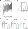
Figure 3. ERK1/2 signaling regulates CCL2/MCP1 expression in GBM
(A) Tumor type ranking according to the expression levels of the CCL2/MCP1 mRNA. Data were retrieved from TCGA, with n=23 tumor types and a total number of n=8823 tumors. GBM is indicated with the red arrow. (B) Box plot comparison of CCL2/MCP1 mRNA in GBM tumors with high ERK1/2 phosphorylation levels vs others (P=0.039 using Wilcoxon–Mann–Whitney test). (C) Quantitative PCR analysis of CCL2/MCP1 mRNA levels in the indicated GBM cell lines (trametinib at 1 μM for 24 h). *P<0.05
Figure 4
Figure 4. ERK1/2 signaling regulates the production of the cytokines CCL2/MCP1 and CSF1/MCSF in GBM
(A) Protein concentration of CCL2/MCP1 and (B) CSF1/MCSF measured in the cell culture supernatant of GBM cell lines. Trametinib was applied as indicated at a concentration of 1 μM for 24 h. Each measurement was performed in triplicate. ***P<0.001 compared with control conditions, **P<0.01 (Student’s t test).
Figure 5
Figure 5. Lack of evidence of a direct regulation of the immune checkpoint ligands PD-L1 / PD-L2 by ERK1/2 signaling in GBM
(A) Pan-cancer analysis of the expression of the immune checkpoint ligands PD-L1/PD-L2. Tumor types are ranked according to their mRNA expression levels of the immune checkpoint ligands PD-L1 (CD274) and PDL2 (PDCD1LG2). Data are from n=23 tumor types and a total number of n=8823 tumors. GBM is indicated with the red arrow. (B) Box plot comparison of PDL1 and PDL2 mRNA expression levels in GBM tumors with high ERK1/2 phosphorylation levels vs others (ns : nonsignificant; Wilcoxon–Mann–Whitney test). (C) Immunoblot analysis of PD-L1 and PD-L2 expression in GBM cell lines. Protein extracts were prepared from the indicated cell lines, exposed to trametinib (1 μM, 24 h) as indicated.
Figure 6
Figure 6. Lack of evidence of modulation of the metabolic inhibitory signaling mediated by lactate and its macrophage receptor GPR65 by ERK1/2 in GBM
(A) Tumor ranking according to the expression levels of GPR65 mRNA. Data are from n=23 tumor types and a total number of n=8823 tumors. GBM is indicated with the red arrow. (B) Lactate concentrations measured in the cell supernatant of GBM cells vs the melanoma cell line SK-MEL3. Lactate concentrations are expressed as pmol/μg prot/24 h. (C) Box plot comparison of GBM tumors with high levels of ERK1/2 phosphorylation vs other GBM (ns: not significant, Wilcoxon–Mann–Whitney test). (D) Inhibition of tumor glycolysis upon ERK1/2 inhibition by trametinib. Data are expressed as percent of lactate production upon exposure to trametinib (1 μM, 24 h), of the indicated GBM cell lines, taking control conditions as 100%. The cell line SK-MEL-3, carrying an activated BRAF V600E oncogene, is given here as positive control. Note the significantly greater inhibition of lactate production in SK-MEL-3 compared with GBM cells. **P<0.01, ***P<0.001 compared with SK-MEL3.

References

    1. Thakkar J.P., Dolecek T.A., Horbinski C.. et al. (2014) Epidemiologic and molecular prognostic review of glioblastoma. Cancer Epidemiol. Biomarkers Prev. 23, 1985–1996 10.1158/1055-9965.EPI-14-0275 - DOI - PMC - PubMed
    1. Stupp R., Mason W.P., van den Bent M.J.. et al. (2005) Radiotherapy plus concomitant and adjuvant temozolomide for glioblastoma. N. Engl. J. Med. 352, 987–996 10.1056/NEJMoa043330 - DOI - PubMed
    1. Brennan C.W., Verhaak R.G., McKenna A.. et al. (2013) The somatic genomic landscape of glioblastoma. Cell 155, 462–477 10.1016/j.cell.2013.09.034 - DOI - PMC - PubMed
    1. Hannen R., Hauswald M. and Bartsch J.W. (2017) A rationale for targeting extracellular regulated kinases ERK1 and ERK2 in glioblastoma. J. Neuropathol. Exp. Neurol. 76, 838–847 10.1093/jnen/nlx076 - DOI - PubMed
    1. Reardon D.A., Ligon K.L., Chiocca E.A. and Wen P.Y. (2015) One size should not fit all: advancing toward personalized glioblastoma therapy. Discov. Med 19, 471–477 - PubMed

Publication types

MeSH terms