. 2019 Aug 29;9(1):12510.
doi: 10.1038/s41598-019-48878-8.

Biosynthesis of Hyaluronic acid polymer: Dissecting the role of sub structural elements of hyaluronan synthase

Affiliations

Biosynthesis of Hyaluronic acid polymer: Dissecting the role of sub structural elements of hyaluronan synthase

Garima Agarwal et al. Sci Rep. .

Abstract

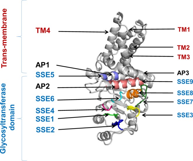
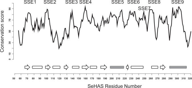
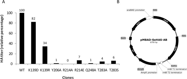
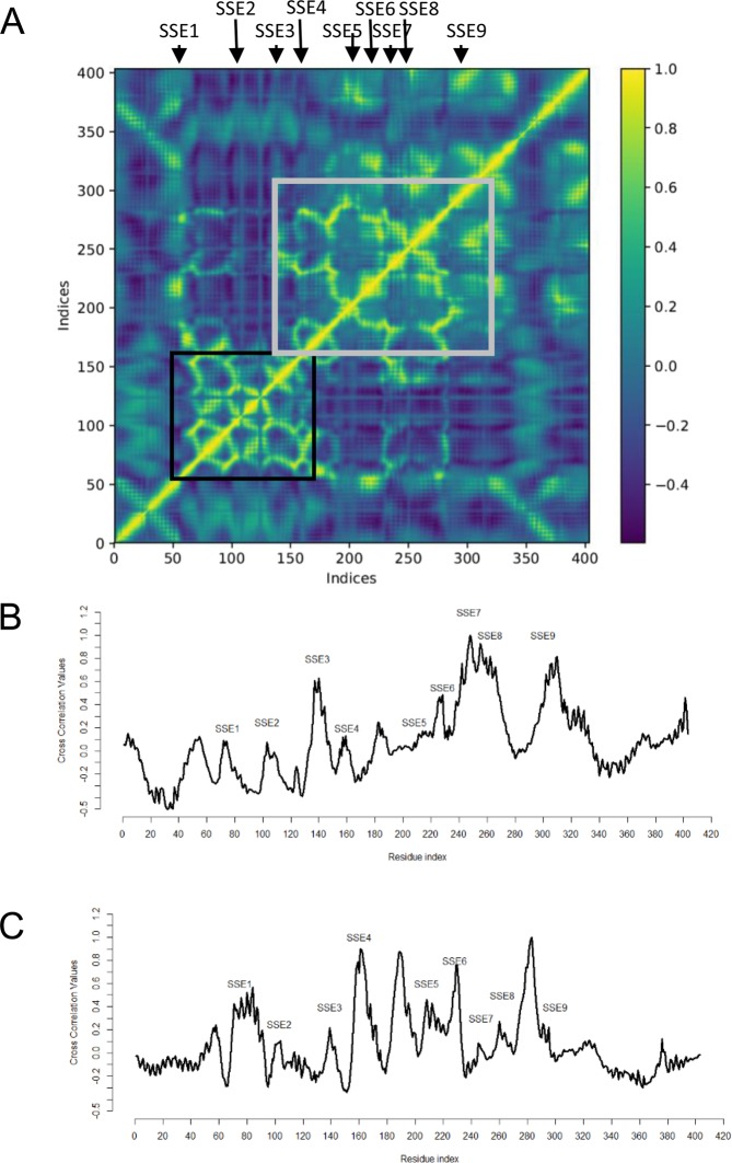
Hyaluronic acid (HA) based biomaterials have several biomedical applications. HA biosynthesis is catalysed by hyaluronan synthase (HAS). The unavailability of 3-D structure of HAS and gaps in molecular understanding of HA biosynthesis process pose challenges in rational engineering of HAS to control HA molecular weight and titer. Using in-silico approaches integrated with mutation studies, we define a dictionary of sub-structural elements (SSE) of the Class I Streptococcal HAS (SeHAS) to guide rational engineering. Our study identifies 9 SSE in HAS and elucidates their role in substrate and polymer binding and polymer biosynthesis. Molecular modelling and docking assessment indicate a single binding site for two UDP-substrates implying conformationally-driven alternating substrate specificities for this class of enzymes. This is the first report hypothesizing the involvement of sites from SSE5 in polymer binding. Mutation at these sites influence HA production, indicating a tight coupling of polymer binding and synthase functions. Mutation studies show dispensable role of Lys-139 in substrate binding and a key role of Gln-248 and Thr-283 in HA biosynthesis. Based on the functional architecture in SeHAS, we propose a plausible three-step polymer extension model from its reducing end. Together, these results open new avenues for rational engineering of Class I HAS to study and regulate its functional properties and enhanced understanding of glycosyltransferases and processive enzymes.


Conflict of interest statement

The authors declare no competing interests.

Figures

Figure 1
Figure 1
Structural model and features of SeHAS. SSE: Substructural element, AP: Amphipathic helix, TM: Trans-membrane helix.
Figure 2
Figure 2
A plot of conservation score with respect to SeHAS sequence number. SSE indicated on top. The secondary structure projection of sequence is indicated at the bottom. The strands are marked as arrows and helices represented as rectangles. Filled rectangles correspond to amphipathic helices. Refer text for details.
Figure 3
Figure 3
(A) Frequency of ligand contacting residues for energetically favourable conformers of UDP-N Acetylglucosamine (UDP-GlcNAc) and UDP- D Glucuronic acid (UDP-GlcA). Frequency is plotted on the X-axis and Sites on Y-axis. (B) Role of SSE1–4, SSE8 and SSE9 in UDP-sugar substrate binding. (C) Role of SSE6 in ligand binding. (D) Role of SSEs in polymer binding. (E) Role of SSE7. (i) SSE7 loop in hyaluronan synthase, (ii) Equivalent loop in cellulose synthase, (iii) Equivalent loop in non-processive glycosyltransferase. (F) Energy minimized HAS structure showing UDP-N-Acetylglucosamine and disaccharide of glucuronic acid and N-Acetylglucosamine moeities.
Figure 4
Figure 4
Mutation studies on SeHAS. (A) Relative activity of SeHAS mutants conducted in this study. Experiments were conducted in triplicates and the standard error for HA titer was in the range of ±0.01 to ±0.05. (B) pMBAD vector construct. hasA and hasB genes from Streptococcus equi subsp. zooepidemicus were sequentially cloned. For mutational studies, hasA wildtype was replaced with corresponding mutant(s).
Figure 5
Figure 5
Assessment of global dynamics in SeHAS. (A) Average correlation coefficient values for SeHAS across residue pairs. Average Correlation coefficient values for Gln-248 (B) and Thr-283 (C) with other SeHAS residues.
Figure 6
Figure 6
Proposed molecular mechanism in SeHAS. (A) Mechanism in a prototypic glycosyltransferase with inversion chemistry. (B) Proposed mechanism in Class I HAS. P: Polymeric sugar, S: Substrate sugar.UDP is indicated as a circle. B1, B2 and B3 are the catalytic bases participating in the reaction mechanism.

References

    1. Weissmann B, Meyer K. The Structure of Hyalobiuronic Acid and of Hyaluronic Acid from Umbilical Cord1,2. J. Am. Chem. Soc. 1954;76:1753–1757. doi: 10.1021/ja01636a010. - DOI
    1. Parker NP, Bailey SS, Walner DL. Effects of basic fibroblast growth factor-2 and hyaluronic acid on tracheal wound healing. Laryngoscope. 2009;119:734–739. doi: 10.1002/lary.20131. - DOI - PubMed
    1. Evanko SP, Potter-Perigo S, Johnson PY, Wight TN. Organization of Hyaluronan and Versican in the Extracellular Matrix of Human Fibroblasts Treated With the Viral Mimetic Poly I:C. J. Histochem. Cytochem. 2009;57:1041–1060. doi: 10.1369/jhc.2009.953802. - DOI - PMC - PubMed
    1. Nishida Y, Knudson CB, Nietfeld JJ, Margulis A, Knudson W. Antisense inhibition of hyaluronan synthase-2 in human articular chondrocytes inhibits proteoglycan retention and matrix assembly. J. Biol. Chem. 2014;274:21893–21899. doi: 10.1074/jbc.274.31.21893. - DOI - PubMed
    1. Y Lee J, Spicer A. Hyaluronan: a multifunctional, megaDalton, stealth molecule. Curr Opin Cell Biol. 2000;12:581–6. Curr. Opin. Cell Biol. 2000;12:581–586. doi: 10.1016/S0955-0674(00)00135-6. - DOI - PubMed

Publication types

MeSH terms