. 2019 Aug 29;9(1):12551.
doi: 10.1038/s41598-019-49108-x.

Mobility of pectin methylesterase in pectin/cellulose gels is enhanced by the presence of cellulose and by its catalytic capacity

Affiliations

Mobility of pectin methylesterase in pectin/cellulose gels is enhanced by the presence of cellulose and by its catalytic capacity

Estelle Bonnin et al. Sci Rep. .

Abstract

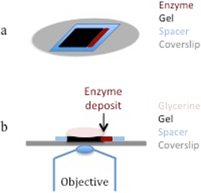
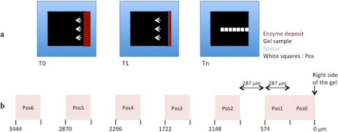
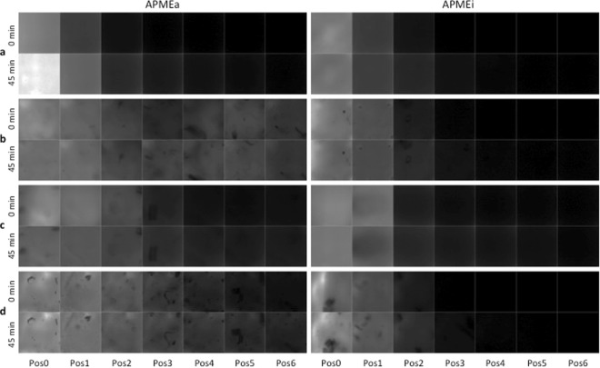
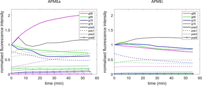
The pectin methylesterase action is usually studied in a homogeneous aqueous medium in the presence of a large excess of soluble substrate and water. However in the cell wall, the water content is much lower, the substrate is cross-linked with itself or with other polymers, and the enzyme has to diffuse through the solid matrix before catalysing the linkage breakdown. As plant primary cell walls can be considered as cellulose-reinforced hydrogels, this study investigated the diffusion of a fungal pectin methylesterase in pectin/cellulose gels used as cell wall-mimicking matrix to understand the impact of this matrix and its (micro) structure on the enzyme's diffusion within it. The enzyme mobility was followed by synchrotron microscopy thanks to its auto-fluorescence after deep-UV excitation. Time-lapse imaging and quantification of intensity signal by image analysis revealed that the diffusion of the enzyme was impacted by at least two criteria: (i) only the active enzyme was able to diffuse, showing that the mobility was related to the catalytic ability, and (ii) the diffusion was improved by the presence of cellulose in the gel.


Conflict of interest statement

The authors declare no competing interests.

Figures

Figure 1
Figure 1
Sample presentation. (a) Schematic perspective view of the sample presentation: the gel (black) was poured in a 10 × 10 mm spacer (blue) on the coverslip (gray). The enzyme (maroon) was deposited on the right side of the gel. The sample was covered with glycerine before observation (not represented). (b) Schematic side view of the gel (black) poured in a spacer (blue) on the coverslip (gray), and then covered with glycerine (light pink) and observed on the inverted microscope. The enzyme (maroon) was deposited on the right side of the gel
Figure 2
Figure 2
Definition of the regions of interest. (a) Schematic top view of enzyme diffusion (maroon) inside the gel sample (black) and definition of the regions of interest, referred to as Pos for Position (white squares). Three time points are represented: at T0, the channel is full of enzyme (maroon); at T1, the enzyme diffuses into the gel according to the white arrows; at Tn, the channel is empty. (b) From the deposit point of the enzyme (right side of the gel), the 2 first Pos corresponded to the two first fields at magnification x40, the next ones corresponded to 1 field over 2. The 7 positions were numbered from Pos0 to Pos6. Values below the positions are the distance from the deposit point.
Figure 3
Figure 3
Raw fluorescence images of Pos0 at time T0 of two kinetics carried out on each gel type: (a), 0% cellulose; (b), 0.5% cellulose; (c), 1.0% cellulose; (d), 1.5% cellulose. To allow comparison, brightness and contrast were similarly adjusted for all images. The full field of view was 287 × 287 µm2.
Figure 4
Figure 4
Treated fluorescence images acquired at 0 min and 45 min on all positions (Pos0 to Pos6) for the native (APMEa) and the heat-inactivated (APMEi) enzyme. Gels were prepared by mixing 1.5% LM pectin with (a), 0% cellulose; (b), 0.5% cellulose; (c), 1.0% cellulose; (d), 1.5% cellulose. The full field of view was 287 × 287 µm2.
Figure 5
Figure 5
Evolution of fluorescence intensity as a function of time. The fluorescent intensities were extracted from images acquired during 55 min on positions 0 (Pos0), position 1 (Pos1), position 3 (Pos3) and position 6 (Pos6) for the native (APMEa) and the heat-inactivated (APMEi) enzyme. The intensity values were normalized on the values at Pos0 and T0 for each kinetic experiment. The gels were prepared by mixing 1.5% LM pectin with no cellulose (g00, pink), 0.5% cellulose (g05, green), 1.0% cellulose (g10, blue), 1.5% cellulose (g15, black).
Figure 6
Figure 6
Evolution of fluorescence intensity as a function of Position. The fluorescent intensities were extracted from images acquired on all positions at 0, 15, 30 and 45 min for the native (APMEa) and the heat-inactivated (APMEi) enzyme. The intensity values were normalized on the values at Pos0 and T0 for each kinetic experiment. The gels were prepared by mixing 1.5% LM pectin with no cellulose (g00, pink), 0.5% cellulose (g05, green), 1.0% cellulose (g10, blue), 1.5% cellulose (g15, black).

References

    1. Chen EMW, Mort AJ. Nature of sites hydrolyzable by endopolygalacturonase in partially-esterified homogalacturonans. Carbohydr. Polym. 1996;29:129–136. doi: 10.1016/0144-8617(96)00005-7. - DOI
    1. Benen JAE, Kester HCM, Visser J. Kinetic characterization of Aspergillus niger N400 endopolygalacturonases I, II and C. Eur. J. Biochem. 1999;259:577–585. doi: 10.1046/j.1432-1327.1999.00080.x. - DOI - PubMed
    1. Thibault J-F, Rinaudo M. Interactions of mono- and divalent counterions with alkali- and enzyme-deesterified pectins in salt-free solutions. Biopolymers. 1985;24:2131–2143. doi: 10.1002/bip.360241109. - DOI
    1. Ralet M-C, Dronnet V, Buchholt HC, Thibault J-F. Enzymatically and chemically de-esterified lime pectins: characterisation, polyelectrolyte behaviour and calcium binding properties. Carbohydr. Res. 2001;336:117–125. doi: 10.1016/S0008-6215(01)00248-8. - DOI - PubMed
    1. Ström A, et al. Influence of pectin fine structure on the mechanical properties of Calcium-pectin and acid-pectin gels. Biomacromolecules. 2007;8:2668–2674. doi: 10.1021/bm070192r. - DOI - PubMed