

Concurrent pulmonary and hepatic hydatid cysts managed with single stage surgery
- PMID: 31467627
- PMCID: PMC6710642
- DOI: 10.1016/j.radcr.2019.08.006
Concurrent pulmonary and hepatic hydatid cysts managed with single stage surgery
Abstract
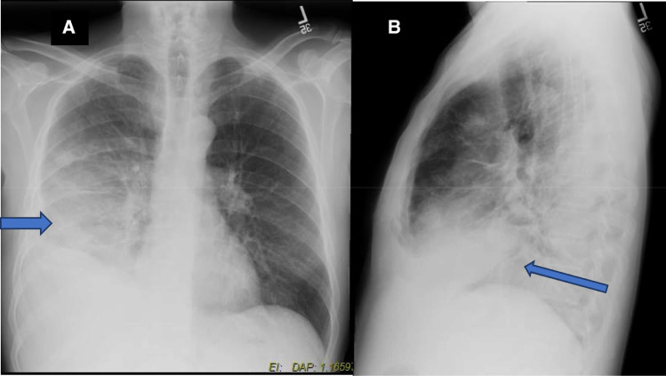
Most of the case reports on hydatid cyst have documented its diagnosis and treatment involving either lungs or liver. This case report is unique as it documents the standard diagnostic and treatment approach followed for curing concurrent multiple hydatid cysts involving liver and right lung simultaneously via single stage surgery. A 52-year-old male presented with symptoms of simple pneumonia along with mild pain in the right upper quadrant. Hydatid cyst was considered as the differential diagnosis after physical examination. Both CT scan and MRI confirmed the presence of multiple cysts both in the liver as well as right lung. A single stage radical removal of cysts from both the organs was performed. The patient was hemodynamically stable, and no complications were reported postoperatively. This case report highlights the importance of considering hydatid cyst as a differential diagnosis in the light of vague presenting symptoms. Also, it emphasizes on the benefits of single stage surgery for removing cysts from both the organs simultaneously.
Keywords: Hydatid cyst; Liver; Lungs; Single stage surgery; Water lily appearance.
Conflict of interest statement
I declare that i have no significant competing financial, professional, or personal interests that might have influence the performance or presentation of the work described in this manuscript.
Figures


References
-
- Global Health – Division of Parasitic Diseases . Centers for Disease Control and Prevention; Atlanta, United States of America: 2012. Echinococcosis Biology.https://www.cdc.gov/parasites/echinococcosis/biology.html Available from.
-
- Brown R.A., Millar A.I.W., Steiner Z., Krige J.E.J., Burkimsher D., Cywes S. Hydatid cyst of the pancreas: a case report in a child. Eur J Pediatr Surg. 1995;5:121–124. - PubMed
-
- Bartholomot G., Vuitton D.A., Harraga S., Shi D.Z., Giraudoux P., Barnish G. Combined ultrasound and serologic screening for hepatic alveolar echinococcosis in central China. Am J Trop Med Hyg. 2002;66:23–29. - PubMed
-
- Filippou D., Tselepis D., Filippou G., Papadopoulos V. Advances in liver echinococcosis: diagnosis and treatment. Clin Gastroenterol Hepatol. 2007;5:152–159. - PubMed
Publication types
LinkOut - more resources
Full Text Sources
