. 2019 Aug 21:11:576-587.
doi: 10.1016/j.dadm.2019.06.003. eCollection 2019 Dec.

White matter alterations in early-stage Alzheimer's disease: A tract-specific study

Affiliations

White matter alterations in early-stage Alzheimer's disease: A tract-specific study

Qiuting Wen et al. Alzheimers Dement (Amst). .

Abstract

Introduction: Diffusion magnetic resonance imaging may allow for microscopic characterization of white matter degeneration in early stages of Alzheimer's disease.

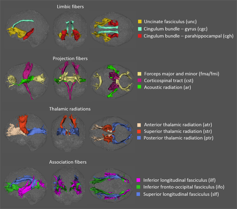
Methods: Multishell Diffusion magnetic resonance imaging data were acquired from 100 participants (40 cognitively normal, 38 with subjective cognitive decline, and 22 with mild cognitive impairment [MCI]). White matter microscopic degeneration in 27 major tracts of interest was assessed using diffusion tensor imaging (DTI), neurite orientation dispersion and density imaging, and q-space imaging.

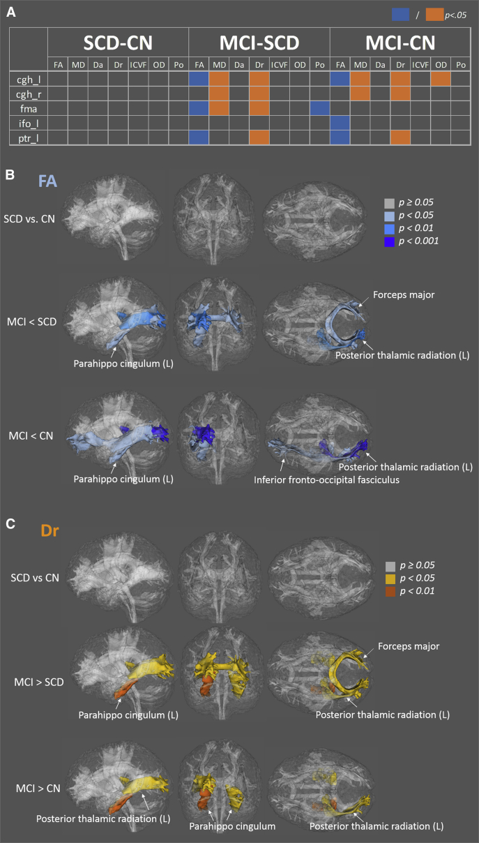
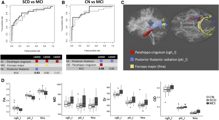
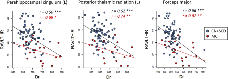
Results: Lower DTI fractional anisotropy and higher radial diffusivity were observed in the cingulum, thalamic radiation, and forceps major of participants with MCI. These tracts of interest also had the highest predictive power to discriminate groups. Diffusion metrics were associated with cognitive performance, particularly Rey Auditory Verbal Learning Test immediate recall, with the highest association observed in participants with MCI.

Discussion: While DTI was the most sensitive, neurite orientation dispersion and density imaging and q-space imaging complementarily characterized reduced axonal density accompanied with dispersed and less restricted white matter microstructures.

Keywords: Alzheimer's disease; Diffusion imaging; MCI; Magnetic resonance imaging; NODDI; SCD; Tract; Tractography; White matter; diffusion tensor imaging.


Figures

Fig. 1
Fig. 1
White matter tracts of interest in one cognitively normal subject generated using probabilistic tractography rendered in sagittal (left), coronal (center), and axial (right) views. The 27 tracts of interest were grouped into five categories: the brainstem (not shown here), limbic fibers connecting the limbic system (first row), projection fibers (second row), thalamic radiations (third row), and association fibers (fourth row).
Fig. 2
Fig. 2
Tracts exhibiting significant differences after the Bonferroni correction. (A) Tests showing significant differences are highlighted in orange (higher in disease group) or blue (lower in disease group). (B) Tracts exhibiting significant differences in FA, with color labeling indicating different significance levels. Rendered in sagittal (left), coronal (center), and axial (right) views. (C) Tracts showing significant differences in Dr. Abbreviations: SCD, subjective cognitive decline; CN, cognitively normal; MCI, mild cognitive impairment; FA, fractional anisotropy; MD, mean diffusivity; Da, axial diffusivity; Dr, radial diffusivity; ICVF, intra-axonal volume fraction; OD, orientation dispersion; P0, zero-displacement probability; cgh_l, left parahippocampal cingulum; cgh_r, right parahippocampal cingulum; fma, forceps major; ifo_l, inferior fronto-occipital fasciculi left; ptr_l, left posterior thalamic radiation.
Fig. 3
Fig. 3
Best predictors identified by penalized logistic regression (LASSO). (A) The receiver operating characteristic (ROC) curves for each combination of predictors (i.e., diffusion and tract of interest pairs) selected by LASSO for discriminating between SCD and MCI. The best predictors for SCD and MCI were Dr in the left parahippocampal cingulum (cgh_l), MD in the forceps major (fma), and FA in the left posterior thalamic radiation (ptr_l). The area under the ROC curve (AUC) was 0.83 with all three predictors (bold black line), 0.81 with Dr-cgh_l and MD-fma (gray line), and 0.80 with only Dr-cgh_l (light gray line). (B) The ROC curves for each combination of predictors selected by LASSO for discriminating between CN and MCI. The best predictors for CN and MCI were FA-ptr_l and OD-cgh_l. The areas under the ROC curve were 0.88 using both predictors (bold black line) and 0.82 using only FA-ptr_l (gray line). (C) Anatomical locations of the three most sensitive tracts of interest: left parahippocampal cingulum (cgh_l), left posterior thalamic radiation (ptr_l), and forceps major (fma). (D) Boxplots of LASSO-selected diffusion parameters (vertical axes) in the three most sensitive tracts of interest (horizontal axes) for all groups. Abbreviations: LASSO, least absolute shrinkage and selection operator; SCD, subjective cognitive decline; CN, cognitively normal; MCI, mild cognitive impairment; FA, fractional anisotropy; MD, mean diffusivity; Dr, radial diffusivity; OD, orientation dispersion.
Fig. 4
Fig. 4
Linear regression analyses between Rey Auditory Verbal Learning Test immediate recall (RAVLT-IR) and radial diffusivity (Dr) in the left parahippocampal cingulum, posterior thalamic radiation, and forceps major. The correlation coefficient as in goodness of fit (r) is labeled in black for all subjects combined (n = 100, black regression line) and with red for MCI (n = 22, red regression line). Significance level is labeled as: * for P < .05, ** for P < .01, and *** for P < .001. Abbreviations: SCD, subjective cognitive decline; CN, cognitively normal; MCI, mild cognitive impairment.

References

    1. Jack C.R., Jr., Lowe V.J., Weigand S.D., Wiste H.J., Senjem M.L., Knopman D.S., Alzheimer's Disease Neuroimaging Initiative Serial PIB and MRI in normal, mild cognitive impairment and Alzheimer's disease: implications for sequence of pathological events in Alzheimer's disease. Brain. 2009;132:1355–1365. - PMC - PubMed
    1. Sperling R.A., Aisen P.S., Beckett L.A., Bennett D.A., Craft S., Fagan A.M. Toward defining the preclinical stages of Alzheimer's disease: recommendations from the National Institute on Aging-Alzheimer's Association workgroups on diagnostic guidelines for Alzheimer's disease. Alzheimers Dement. 2011;7:280–292. - PMC - PubMed
    1. Braak H., Braak E. Neuropathological stageing of Alzheimer-related changes. Acta Neuropathol. 1991;82:239–259. - PubMed
    1. Brun A., Englund E. A white matter disorder in dementia of the Alzheimer type: a pathoanatomical study. Ann Neurol. 1986;19:253–262. - PubMed
    1. Bartzokis G. Alzheimer's disease as homeostatic responses to age-related myelin breakdown. Neurobiol Aging. 2011;32:1341–1371. - PMC - PubMed