. 2019 Aug 26;4(4):527-541.
doi: 10.1016/j.jacbts.2019.04.002. eCollection 2019 Aug.

Adaptive Reductions in Left Ventricular Diastolic Compliance Protect the Heart From Stretch-Induced Stunning

Affiliations

Adaptive Reductions in Left Ventricular Diastolic Compliance Protect the Heart From Stretch-Induced Stunning

Brian R Weil et al. JACC Basic Transl Sci. .

Abstract

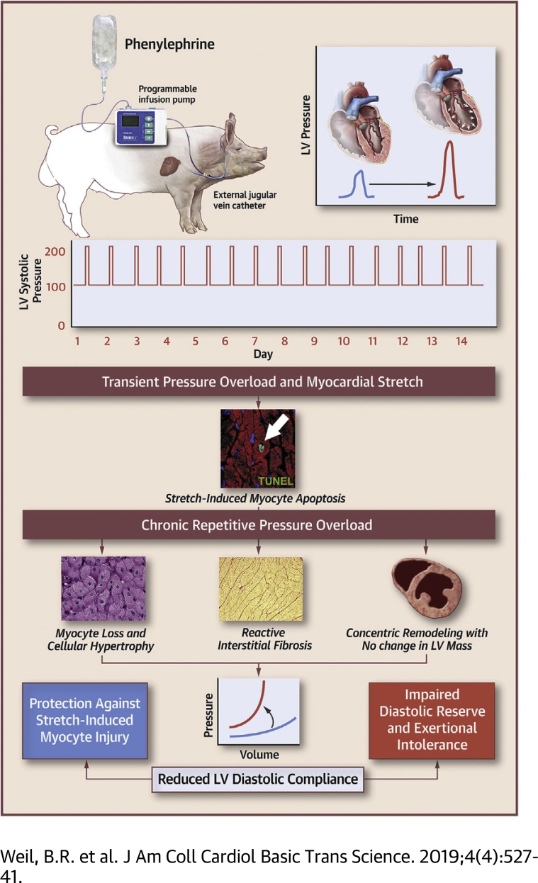
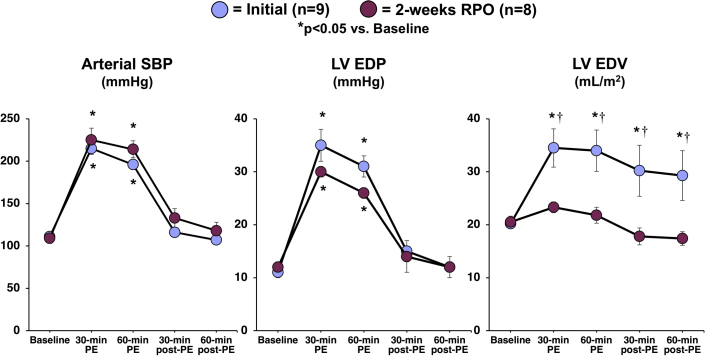
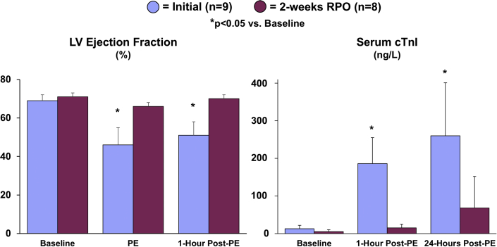
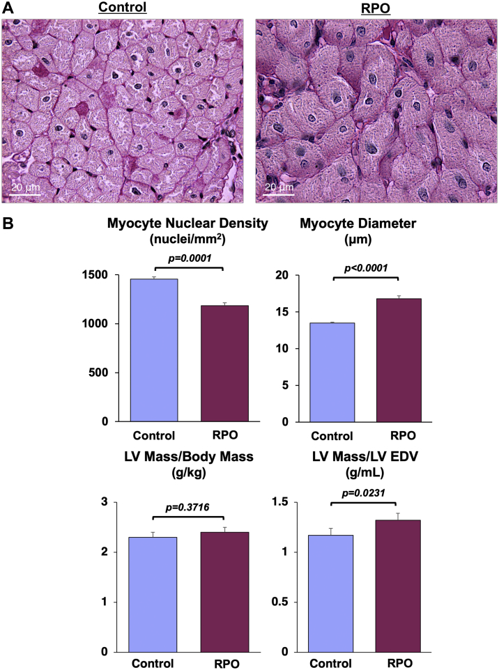
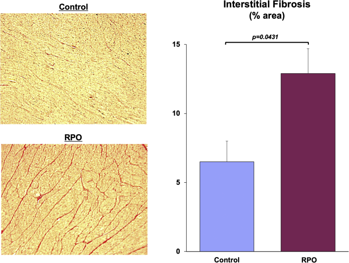
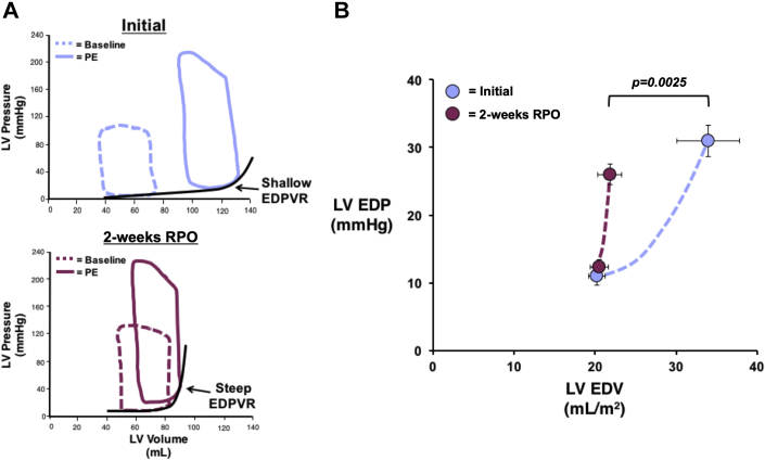
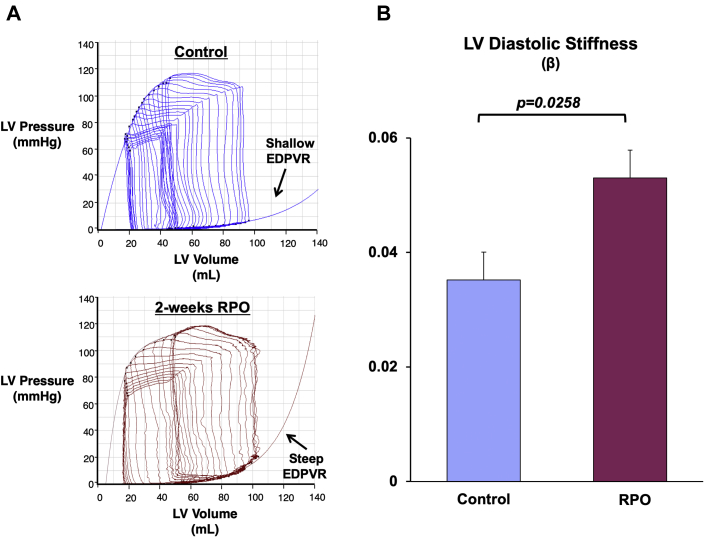
Swine subjected to 2 weeks of repetitive pressure overload (RPO) exhibited significant myocyte loss, but left ventricular (LV) systolic function was preserved, and chamber dilatation did not occur. Instead, myocardial remodeling characterized by myocyte hypertrophy and interstitial fibrosis led to a marked reduction in LV diastolic compliance, which protected the heart from stretch-induced myocyte injury and preserved LV ejection fraction without anatomic LV hypertrophy. These results support a novel paradigm that links cardiac adaptations to RPO with the pathogenesis of reduced LV diastolic compliance and may explain how LV stiffening can occur in the absence of sustained hypertension or anatomic hypertrophy.

Keywords: BP, blood pressure; EDPVR, end-diastolic pressure−volume relationship; HFpEF, heart failure with preserved ejection fraction; LV, left ventricular; LVEDP, left ventricular end-diastolic pressure; LVEDV, left ventricular end-diastolic volume; PE, phenylephrine; PV, pressure−volume; RPO, repetitive pressure overload; TUNEL, terminal deoxynucleotidyl transferase-mediated dUTP nick end labeling; cTnI, cardiac troponin I; diastolic dysfunction; fibrosis; heart failure; myocardial stunning; stretch; ΔEDP/ΔEDV, changes in end-diastolic pressure/end-diastolic volume.


Figures

None
Graphical abstract
Figure 1
Figure 1
RPO Elicits a Reduction in LV Compliance That Prevents Chamber Distension During an Acute Elevation in Afterload A 60-min intravenous infusion of phenylephrine (PE) elicited a similar increase in arterial systolic blood pressure (SBP) and left ventricular end-diastolic pressure (LVEDP) at the initial study (blue) and after 2 weeks of repetitive pressure overload (RPO) (purple). At the initial study, this was associated with an increase in left ventricular end-diastolic volume (LVEDV) that persisted after cessation of PE despite normalization of SBP and LVEDP. However, transient PE infusion no longer produced a significant increase in LVEDV when the study was repeated after 2 weeks of RPO, which was indicative of reduced LV compliance after RPO. Values are mean ± SEM. *p < 0.05 versus baseline; †p < 0.05 versus initial.
Figure 2
Figure 2
Cardiac Adaptations to RPO Prevent Acute Stretch-Induced Stunning and Myocyte Injury During a Transient Elevation in Afterload The attenuation of LV distension during PE infusion after RPO was associated with a preserved LV ejection fraction and the absence of a significant elevation in serum cardiac troponin I (cTnI) concentrations, indicating that RPO led to an adaptive reduction in LV compliance that protected the heart from stretch-induced stunning and myocyte injury during PE-mediated pressure overload. Values are mean ± SEM. *p < 0.05 versus baseline. Representative videos of short-axis echocardiograms that illustrate the LV response to PE at the initial study (Supplemental Videos 1A, 1B, 1C) and after 2-weeks of RPO (Supplemental Videos 2A, 2B, 2C) are included in the Supplemental Appendix. Abbreviations as in Figure 1.
None
Video Files 1A-1C: Example Left Ventricular Short-Axis Echocardiograms at the Initial Study Short-axis echocardiograms from the initial study demonstrate normal left ventricular systolic function at rest (arterial blood pressure (BP): 124/78 mmHg; Video 1A), followed by marked dilatation and systolic dysfunction during phenylephrine (PE) infusion (arterial BP: 216/155 mmHg; Video 1B) that persists following cessation of PE despite normalization of arterial BP (arterial BP: 125/86 mmHg; Video 1C).
None
Video Files 2A-2C: Example Left Ventricular Short-Axis Echocardiograms After 2-weeks of Repetitive Pressure Overload Short-axis echocardiograms from the same animal shown in Video Files 1A-1C obtained after 2-weeks of RPO demonstrate normal left ventricular systolic function at rest (arterial blood pressure (BP): 102/61 mmHg; Video 2A) as well as during (arterial BP: 230/154 mmHg; Video 2B) and after (arterial BP: 132/82 mmHg; Video 2C) phenylephrine (PE) infusion despite a similar arterial BP response to PE at both studies. *Note: All example echocardiograms were collected from the same animal subject.
Figure 3
Figure 3
RPO Produces Significant Cardiomyocyte Loss and Compensatory Cellular Hypertrophy of Remaining Myocytes Examples of periodic acid-Schiff−stained myocardial tissue sections from a normal control animal (left) and an animal subjected to RPO (right) illustrate the reduction in myocyte nuclear density and increase in myocyte diameter elicited by RPO. Although RPO did not elicit a change in total LV mass, the LV mass/LVEDV was increased compared with normal control animals (n = 20), which was indicative of concentric LV remodeling in the absence of overt anatomic hypertrophy in swine subjected to RPO. Values are mean ± SEM. Abbreviations as in Figure 1.
Figure 4
Figure 4
Swine Subjected to RPO Exhibit Increased Interstitial Fibrosis Picrosirius red−stained tissue sections demonstrate a marked increase in interstitial collagen deposition (red areas) in myocardial samples from animals subjected to RPO compared with normal control swine. Values are mean ± SEM. Abbreviations as in Figure 1.
Figure 5
Figure 5
RPO Reduces LV Diastolic Compliance (A) Example admittance catheter-derived LV pressure−volume loops at rest and during PE infusion demonstrate that swine subjected to 2 weeks of RPO exhibited a steeper end-diastolic pressure-volume relationship during elevations in afterload. (B) This was visualized via construction of 2-point diastolic pressure−volume curves and quantified via calculation of the ΔLVEDV/ΔLVEDP ratio during acute episodes of PE-induced pressure overload, which was significantly lower after RPO. Values are mean ± SEM. EDPVR = end-diastolic pressure-volume relationship; other abbreviations as in Figure 1.
Figure 6
Figure 6
RPO Elicits an Upward and Leftward Shift in the LV EDPVR (A) Example admittance catheter-derived LV pressure−volume loops during transient inferior vena cava occlusion show a steeper EDPVR after 2 weeks of RPO compared with normal control animals. (B) The upward and leftward shift of the EDPVR led to a higher LV diastolic chamber stiffness constant (β) in animals subjected to RPO compared with normal control swine. Values are mean ± SEM; n = 5/group. Abbreviations as in Figures 1 and 5.

References

    1. Whelan R.S., Kaplinskiy V., Kitsis R.N. Cell death in the pathogenesis of heart disease: mechanisms and significance. Ann Rev Physiol. 2010;72:19–44. - PubMed
    1. Braunwald E. Heart failure. J Am Coll Cardiol HF. 2013;1:1–20. - PubMed
    1. Thygesen K., Alpert J.S., Jaffe A.S. Fourth universal definition of myocardial infarction (2018) J Am Coll Cardiol. 2018;72:2231–2264. - PubMed
    1. Giannitsis E., Katus H.A. Cardiac troponin level elevations not related to acute coronary syndromes. Nat Rev Cardiol. 2013;10:623–634. - PubMed
    1. Weil B.R., Suzuki G., Young R.F., Iyer V., Canty J.M., Jr. Troponin release and reversible left ventricular dysfunction after transient pressure overload. J Am Coll Cardiol. 2018;71:2906–2916. - PMC - PubMed