

Case Reports
doi: 10.1016/j.case.2019.04.005.
eCollection 2019 Aug.
A Vicious Circle: Heyde Syndrome in Mild Aortic Stenosis
Affiliations
- PMID: 31468021
- PMCID: PMC6710854
- DOI: 10.1016/j.case.2019.04.005
Item in Clipboard
Case Reports
A Vicious Circle: Heyde Syndrome in Mild Aortic Stenosis
CASE (Phila).
.
No abstract available
Keywords: Aortic stenosis; Heyde Syndrome; High-output cardiac failure; von Willebrand factor.
Figures


Echocardiogram demonstrating significant but proportional dilatation of all cardiac chambers. (A) The left ventricular end-diastolic volume was 230 mL (indexed 85 mL). (B) The tricuspid anulus was 4.78 cm, and the basal right ventricular diameter was 5.89 cm. (C) The left atrial area was 38 cm² (indexed 14 cm²). (D) The right atrial area was 33.2 cm² (indexed 12 cm²).

Pulsed-wave Doppler across mitral valve demonstrating near normal filling of left ventricle.

Pulsed-wave Doppler right upper pulmonary vein demonstrating near normal filling of left atrium.

M-Mode aortic valve demonstrating good opening of aortic valve.
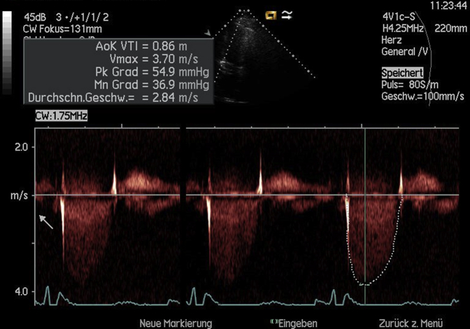
Continuous-wave Doppler signal across aortic valve demonstrating a maximum velocity of 3.7 m/sec and a mean gradient of 36.9 mm Hg.

Planimetric measurement of aortic valve orifice (2.4 cm2) during transesophageal echocardiography.

Angiodysplastic vessels in the small intestines seen in capsule endoscopy.

Multimeric analysis of plasma demonstrating a deficiency of large von Willebrand multimers at presentation, which completely resolved after aortic valve replacement.
References
-
- Vincentelli A., Susen S., Le T.T., Six I., Fabre O., Juthier F. Acquired von Willebrand syndrome in aortic stenosis. N Engl J Med. 2003;349:343–349. - PubMed
-
- Undas A., Natorska J. Bleeding in patients with severe aortic stenosis in the era of transcatheter aortic valve replacement. JACC Cardiovasc Interv. 2015;8:701–703. - PubMed
-
- Natorska J., Mazur P., Undas A. Increased bleeding risk in patients with aortic valvular stenosis: from new mechanisms to new therapies. Thromb Res. 2016;139:85–89. - PubMed
-
- Blackshear J.L., Wysokinska E.M., Safford R.E., Thomas C.S., Shapiro B.P., Ung S. Shear stress-associated acquired von Willebrand syndrome in patients with mitral regurgitation. J Thromb Haemost. 2014;12:1966–1974. - PubMed
Publication types
LinkOut - more resources
Full Text Sources
