Review
. 2019 Nov;61(11):801-815.
doi: 10.1007/s12033-019-00207-0.

Advances in the Production and Batch Reformatting of Phage Antibody Libraries

Affiliations
Review

Advances in the Production and Batch Reformatting of Phage Antibody Libraries

Rose H Reader et al. Mol Biotechnol. 2019 Nov.

Abstract

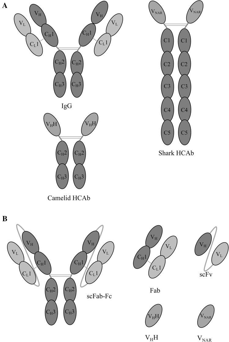
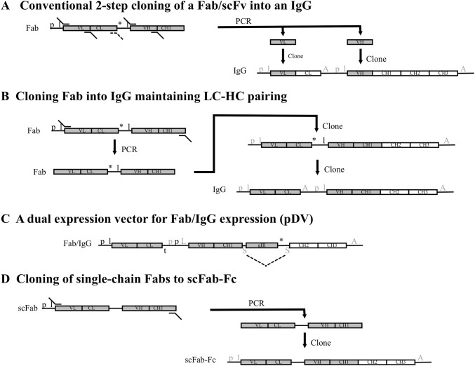
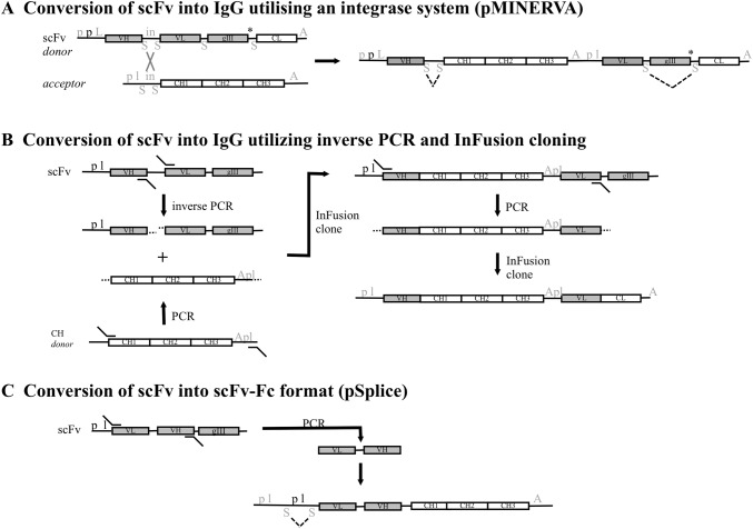
Phage display antibody libraries have proven an invaluable resource for the isolation of diagnostic and potentially therapeutic antibodies, the latter usually being antibody fragments converted into IgG formats. Recent advances in the production of highly diverse and functional antibody libraries are considered here, including for Fabs, scFvs and nanobodies. These advances include codon optimisation during generation of CDR diversity, improved display levels using novel signal sequences, molecular chaperones and isomerases and the use of highly stable scaffolds with relatively high expression levels. In addition, novel strategies for the batch reformatting of scFv and Fab phagemid libraries, derived from phage panning, into IgG formats are described. These strategies allow the screening of antibodies in the end-use format, facilitating more efficient selection of potential therapeutics.

Keywords: Fab; Nanobody; Phage display; Recombinant antibody; VHH; VNAR; scFv.


Figures

Fig. 1
Fig. 1
Representations of the different structures of antibodies and antibody fragments for phage display. The multi-domain structure of conventional IgG and heavy chain antibody (camelid and shark) (a). Antibody fragments can be displayed on phage as Fabs, scFvs or nanobodies (VHHs or VNAR, derived from camelid or shark, respectively). The synthetic scFab-Fc dimer is also shown (b). V variable domain, C constant domain, H heavy chain, L light chain
Fig. 2
Fig. 2
A summary of key elements within different strategies to produce full-length IgG or Fc-fusion constructs from Fab phage display vectors. Conventional cloning of Fab usually includes the amplification of the VH and VL domains and cloning into appropriate sites within an ‘IgG cassette’ that contains the CL and CH1, CH2 and CH3 domains within a mammalian expression vector (a). For Fabs, an analogous 2 step cloning method can be used to clone the entire light chain along with the VH domain (A, dashed primer would be used). The phagemid (left hand side) will contain E. coli promoter (p) and leader (l) sequences for the expression of the Fab. The antibody fragment gene is expressed as a fusion with a downstream gIII phage coat protein gene. Once the VL (or light chain) and VH domains are cloned into the IgG expression plasmid, the light and heavy chains are expressed separately with their own eukaryotic promoter (p) and leader (l) sequences and poly(A) tail (A) [mammalian regulatory elements are indicated in grey font]. Fabs have also been cloned from a phagemid vector in two steps into a mammalian expression vector to retain the pairing of the light chain and VH-CH1 domains, allowing batch cloning of polyclonal phagemid [21] (b). In the first step the Fab is amplified and cloned into the vector and then in a second step the mammalian regulatory elements are introduced between the light chain and heavy chain genes. With a Fab library that only contain heavy chain diversity, this can be expressed within Fab or full-length IgG formats from the same vector (c). The pDV system [24] expresses the Fab or IgG when in E. coli or mammalian cells, respectively, without any requirement for sub-cloning steps. The construct uses a bacterial p and l for the light chain and has a bacterial transcription terminator (t) after the light chain. The sequence upstream of the VH-CH1 genes contains bacterial and eukaryotic promoters along with a leader sequence that functions in either host cell (L). Downstream of the gIII gene is a stop codon (*) followed by a splice domain (S). A second S is present between the CH1 and gIII domains. In bacteria, Fab-gIII is expressed and in mammalian cells, intron splicing (dashed lines) removes the gIII region and the heavy chain is expressed and paired with an invariant light chain derived from a different vector. Alternatively, single chain Fabs expressed as fusions with pIII can be sub-cloned in a single step into a mammalian expression vector containing the Fc domain to make scFab-Fc [25] (d)
Fig. 3
Fig. 3
A summary of key elements within different strategies to produce full-length IgG or Fc-fusion constructs from scFv-phage display vectors. A conventional 2-step cloning strategy for scFvs is analogous to that for Fabs shown in Fig. 2a. Alternatively, the pMINERVA system [58] utilises both bacterial integrases and also intron splicing in mammalian cells (a). A phagemid ‘donor’ vector contains mammalian (grey font) and bacterial (black font) promoters and a leader sequence compatible with both cell types (symbols are the same as in Fig. 2), in addition the scFv has a linker that is also the substrate for phiC31 integrase (in). The construct also contains splice sites either side of the integrase substrate region and either side of gIII. The gIII has an ochre stop at its 5’ end followed by the CL domain. The construct can be transferred to an E. coli strain that expresses the integrase and where a second acceptor plasmid is present that contains the CH domain (CH1-CH2-CH3) and regulatory elements (p and l) for mammalian expression of the light chain. Integrase mediated recombination results in an IgG expression vector where, upon transfer to mammalian cells, intron splicing (dashed lines) results in the removal of the gIII region and expression of light chain and heavy chain. Alternatively, InFusion technology, allowing the cloning of overlapping 15 bp sequences, has been used in a multi-step cloning strategy to produce IgG from scFv [59] (b). The phagemid was amplified by inverse PCR to produce a linear version of the whole vector. This step removed the scFv linker and added an overlapping sequence (indicated by …). This same overlapping sequence was added in a separate PCR reaction to a DNA fragment containing CH domains and mammalian promoter and leader sequences (amplified from a ‘donor’ vector). InFusion cloning produced the heavy chain and VL region, the latter with mammalian regulatory elements, that are then amplified by PCR and subsequently cloned using InFusion into a mammalian expression vector containing the regulatory elements for the heavy chain as well as the CL domain. ScFv from phagemid can potentially be batch cloned into a vector (pSplice) that can express scFv-Fc in both bacterial and mammalian cells [56] (c). The scFv could be amplified by PCR and cloned into the pSplice vector that contains a mammalian promoter and leader sequence in addition to E. coli promoter and leader sequences, all upstream of the scFv-Fc coding region. The mammalian leader sequence is incomplete with the 3′ region contained after the prokaryotic p l sequences that are flanked by splice domains. The scFv-Fc is expressed from the cassette in E. coli and in mammalian cells the intron containing the bacterial regulation elements is excised (dashed lines) and the mammalian leader sequence spliced together allowing expression of the scFv-Fc

References

    1. Smith GP, Petrenko VA. Phage display. Chemical Reviews. 1997;97:391–410. - PubMed
    1. Carmen S, Lutz J. Concepts in antibody phage display. Briefings in Functional Genomics Proteomics. 2002;1:189–203. - PubMed
    1. Paschke M. Phage display systems and their applications. Applied Microbiology and Biotechnology. 2006;70:2–11. - PubMed
    1. Zhang H, Torkmani A, Jones TM, Ruiz DI, Pons J, Lerner RA. Phenotype-information-phenotype cycle for deconvolution of combinatorial antibody libraries selected against complex systems. Proceedings of the National Academy of Sciences USA. 2011;108:13456–13461. - PMC - PubMed
    1. D’Angelo S, Kumar S, Naranjo L, Ferrara F, Kiss C, Bradbury ARM. From deep sequencing to actual clones. Protein Engineering, Design & Selection. 2014;27:301–307. - PMC - PubMed

LinkOut - more resources