

Eosinophilic bronchitis, eosinophilic granuloma, and eosinophilic bronchopneumopathy in 75 dogs (2006-2016)
- PMID: 31468629
- PMCID: PMC6766508
- DOI: 10.1111/jvim.15605
Eosinophilic bronchitis, eosinophilic granuloma, and eosinophilic bronchopneumopathy in 75 dogs (2006-2016)
Abstract
Background: Eosinophilic lung disease is a poorly understood inflammatory airway disease that results in substantial morbidity.
Objective: To describe clinical findings in dogs with eosinophilic lung disease defined on the basis of radiographic, bronchoscopic, and bronchoalveolar lavage fluid (BAL) analysis. Categories included eosinophilic bronchitis (EB), eosinophilic granuloma (EG), and eosinophilic bronchopneumopathy (EBP).
Animals: Seventy-five client owned dogs.
Methods: Medical records were retrospectively reviewed for dogs with idiopathic BAL fluid eosinophilia. Information abstracted included duration and nature of clinical signs, bronchoscopic findings, and laboratory data. Thoracic radiographs were evaluated for the pattern of infiltrate, bronchiectasis, and lymphadenomegaly.
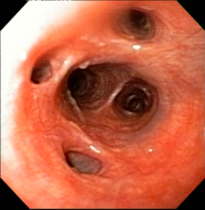
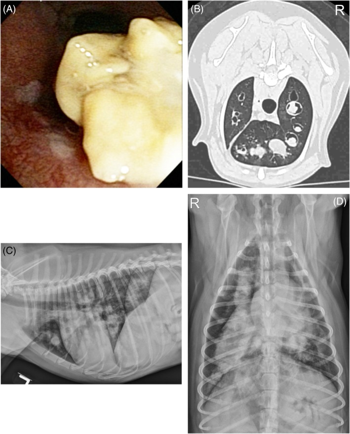
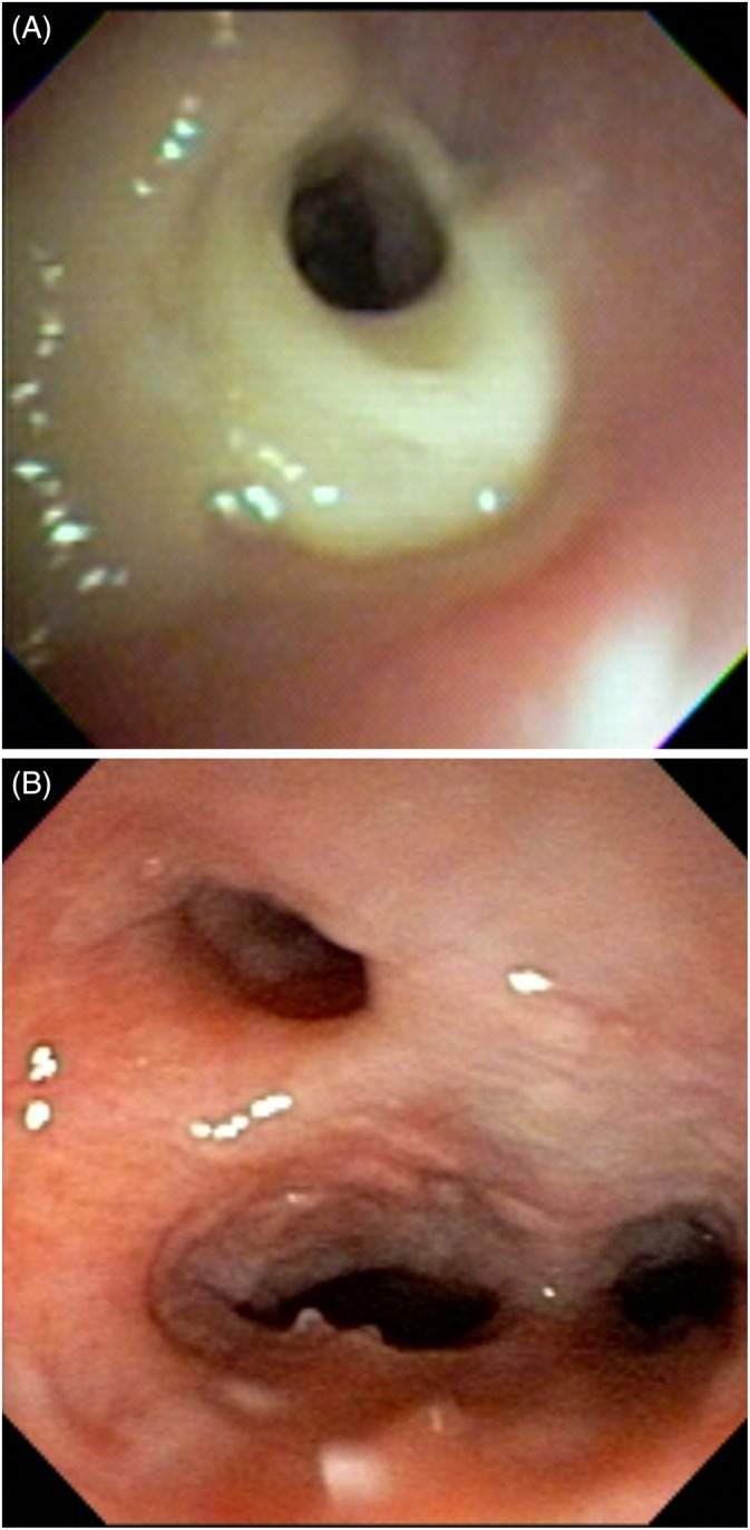
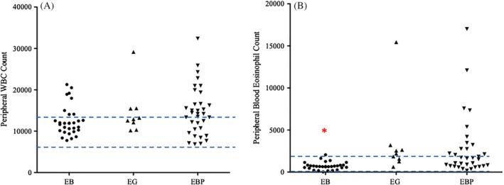
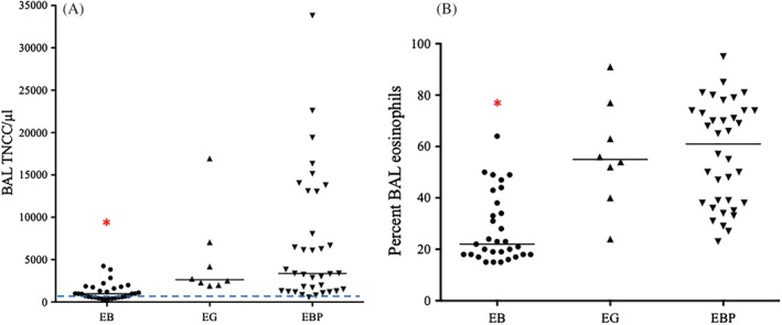
Results: Thoracic radiographs were normal or demonstrated a bronchial pattern in 31 dogs assigned a diagnosis of EB. Nine dogs had intraluminal mass lesions and were bronchoscopically diagnosed with EG. The remaining 35 dogs were categorized as having EBP based on radiographic changes, yellow green mucus in the airways, mucosal changes, and airway collapse. Age and duration of cough did not differ among groups. Dogs with EB were less likely to have bronchiectasis or peripheral eosinophilia, had lower total nucleated cell count in BAL fluid, and lower percentage of eosinophils in BAL fluid compared to dogs in the other 2 groups. In contrast to previous reports, prolonged survival (>55 months) was documented in dogs with EG.
Conclusions and clinical importance: Dogs with eosinophilic lung disease can be categorized based on imaging, bronchoscopic and BAL fluid cytologic findings. Further studies are needed to establish response to treatment in these groups.
Keywords: bronchitis; bronchomalacia; bronchopneumopathy; granuloma; infection.
© 2019 The Authors. Journal of Veterinary Internal Medicine published by Wiley Periodicals, Inc. on behalf of the American College of Veterinary Internal Medicine.
Conflict of interest statement
Authors declare no conflict of interest.
Figures
References
-
- Corcoran BM, Thoday KL, Henfrey JI, Simpson JW, Burnie AG, Mooney CT. Pulmonary infiltration with eosinophils in 14 dogs. J Small Anim Pract. 1991;32(10):494‐502.
-
- Clercx C, Peeters D, Snaps F, et al. Eosinophilic bronchopneumopathy in dogs. J Vet Intern Med. 2000;14(3):282‐291. - PubMed
-
- Calvert CA, Mahaffey MB, Lappin MR, et al. Pulmonary and disseminated eosinophilic granulomatosis in dogs. J Am Anim Hosp Assoc. 1988;24:311‐320.
-
- Katajavuori P, Melamies M, Rajamäki MM. Eosinophilic pulmonary granulomatosis in a young dog with prolonged remission after treatment. J Small Anim Pract. 2013;54(1):40‐43. - PubMed
-
- Fina C, Vignoli M, Terragni R, Rossi F, Wisner E, Saunders JH. Computed tomographic characteristics of eosinophilic pulmonary granulomatosis in five dogs. Vet Radiol Ultrasound. 2014;55(1):16‐22. - PubMed