Review
. 2019 Nov;52(6):e12659.
doi: 10.1111/cpr.12659. Epub 2019 Aug 30.

Extracellular vesicles in urologic malignancies-Implementations for future cancer care

Affiliations
Review

Extracellular vesicles in urologic malignancies-Implementations for future cancer care

Zhangsong Wu et al. Cell Prolif. 2019 Nov.

Abstract

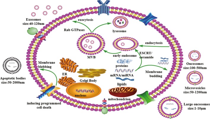
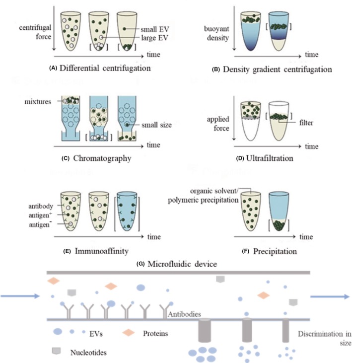
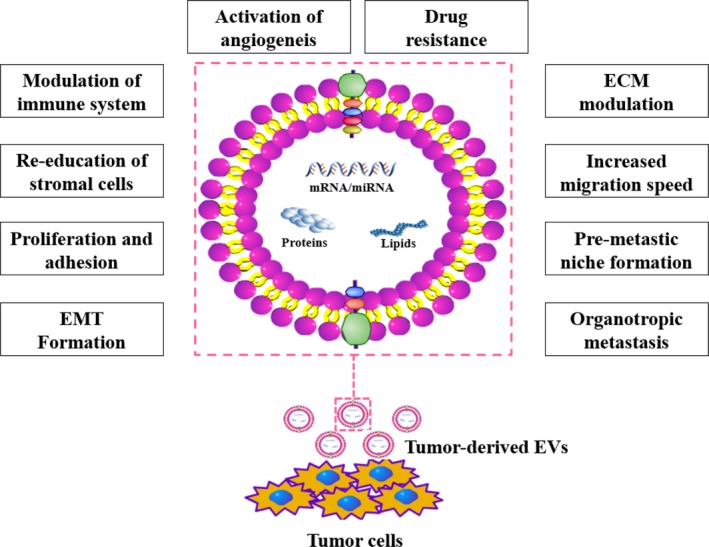
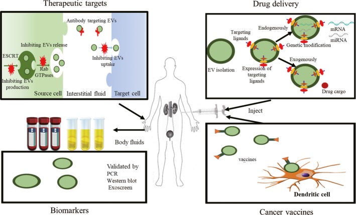
Extracellular vesicles (EVs), a heterogeneous group of vesicles differing in size and shape, cargo content and function, are membrane-bound and nano-sized vesicles that could be released by nearly all variations of cells. EVs have gained considerable attention in the past decades for their functions in modulating intercellular signalling and roles as potential pools for the novel diagnostic and prognostic biomarkers, as well as therapeutic targets in several cancers including urological neoplasms. In general, human and animal cells both can release distinct types of EVs, including exosomes, microvesicles, oncosomes and large oncosomes, and apoptotic bodies, while the content of EVs can be divided into proteins, lipids and nucleic acids. However, the lack of standard methods for isolation and detection platforms rein the widespread usage in clinical applications warranted furthermore investigations in the development of reliable, specific and sensitive isolation techniques. Whether and how the EVs work has become pertinent issues. With the aid of high-throughput proteomics or genomics methods, a fully understanding of contents contained in EVs from urogenital tumours, beyond all doubt, will improve our ability to identify the complex genomic alterations in the process of cancer and, in turn, contribute to detect potential therapeutic target and then provide personalization strategy for patient.

Keywords: bladder cancer; exosomes; extracellular vesicles; kidney cancer; microvesicles; prostate cancer.


Conflict of interest statement

The authors declare that they have no competing interests.

Figures

Figure 1
Figure 1
Release and uptake mechanisms of extracellular vesicles. Extracellular vesicles can be classed as exosomes, microvesicles and apoptotic bodies, based on the mechanism by which they are released from cells and differentiated based on their size and content. MVs, and oncosomes or large oncosomes are directly shed or bud from the plasma membrane. Apoptotic bodies are released from the cell undergoing programmed cell death. Exosomes are formed by inward budding of multivesicular bodies
Figure 2
Figure 2
The common methods to isolate EVs. Straight brackets: isolated EVs; yellow: soluble components; and blue: buffer. A, In differential centrifugation, separation is based on sedimentation velocity, largely depended by size; B, in density gradient centrifugation, separation is relied on buoyant and density; C, size exclusion chromatography uses a porous matrix (dotted circles) that separates on size; D, in ultrafiltration, separation is based on size; E, in immunocapture assays, EVs are captured based on the presence of specific EVs surface molecules. F, in precipitation, EVs isolation via adding some water‐excluding polymers to sample to force the precipitation of small EVs out. G, In microfluidic device, EVs isolation via combining several methods such as immunoaffinity and filtration systems. Copyright 2017, University of Helsinki, Frank AW Coumans31
Figure 3
Figure 3
Physiological processes influenced by EVs. Extracellular vesicles are involved in most physiological processes that are associated with intercellular communication, and the content of extracellular vesicles, including mRNAs, microRNAs (miRNAs), lipids and proteins, is depicted
Figure 4
Figure 4
Future implements for EVs in urological cancer. EVs impact the multistep process of cancer; therefore, EVs should be a novel treatment strategy by inhibiting intercellular crosstalk. EVs could serve as promising diagnostic and prognostic biomarkers to dynamically trace the changes in cancer due to their high specificity and sensitivity. In addition, EVs have the potential functions to stably deliver substantial therapeutic cargoes liking miRNAs and siRNAs with stability, few side effects and organ specificity. Furthermore, several studies have reported the potential of EVs derived from dendritic cells used as vaccine vesicles. Copyright 2018, The Jikei University School of Medicine, Fumihiko Urabe19

References

    1. Siegel L, Miller KD, Jemal A. Cancer statistics, 2018. CA Cancer J Clin. 2018;68:7‐30. - PubMed
    1. Dy GW, Gore JL, Forouzanfar MH, Naghavi M, Fitzmaurice C. Global burden of urologic cancers, 1990–2013. Eur Urol. 2017;71(3):437‐446. - PubMed
    1. Pitt JM, Kroemer G, Zitvogel L. Extracellular vesicles: masters of intercellular communication and potential clinical interventions. J Clin Invest. 2016;126(4):1139. - PMC - PubMed
    1. Colombo M, Raposo G, Théry C. Biogenesis, secretion, and intercellular interactions of exosomes and other extracellular vesicles. Annu Rev Cell Dev Biol. 2014;30(1):255‐289. - PubMed
    1. Xu R, Greening DW, Zhu H‐J, Takahashi N, Simpson RJ. Extracellular vesicle isolation and characterization: toward clinical application. J Clin Invest. 2016;126(4):1152‐1162. - PMC - PubMed

LinkOut - more resources