. 2019 Aug 30;10(1):279.
doi: 10.1186/s13287-019-1389-4.

Autograft microskin combined with adipose-derived stem cell enhances wound healing in a full-thickness skin defect mouse model

Affiliations

Autograft microskin combined with adipose-derived stem cell enhances wound healing in a full-thickness skin defect mouse model

Yuansen Luo et al. Stem Cell Res Ther. .

Abstract

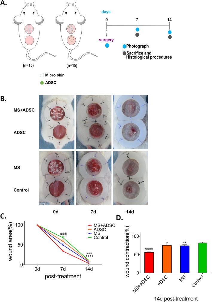
Objective: Autograft microskin transplantation has been widely used as a skin graft therapy in full-thickness skin defect. However, skin grafting failure can lead to a pathological delay wound healing due to a poor vascularization bed. Considering the active role of adipose-derived stem cell (ADSC) in promoting angiogenesis, we intend to investigate the efficacy of autograft microskin combined with ADSC transplantation for facilitating wound healing in a full-thickness skin defect mouse model.

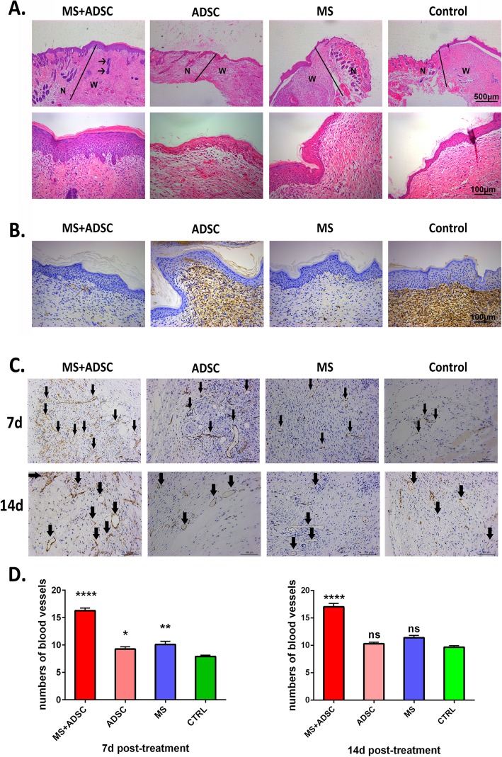
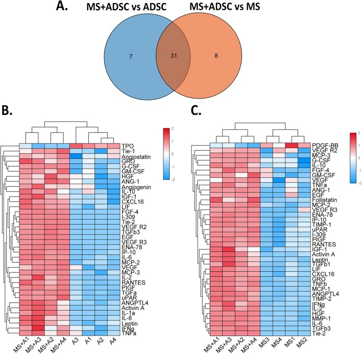
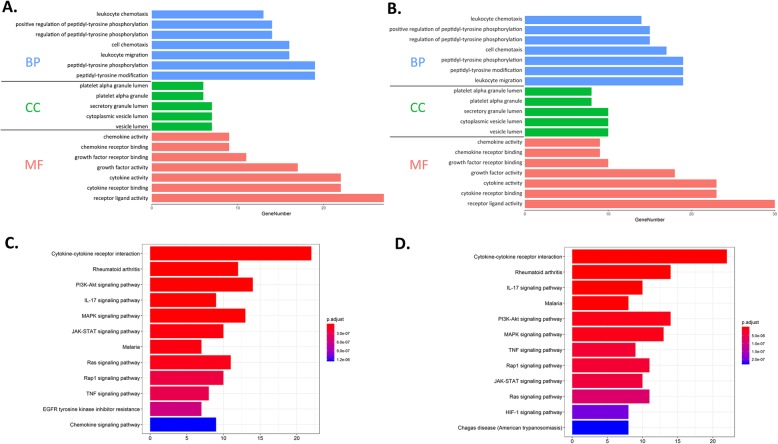
Material and methods: An in vivo full-thickness skin defect mouse model was used to evaluate the contribution of transplantation microskin and ADSC in wound healing. The angiogenesis was detected by immunohistochemistry staining. In vitro paracrine signaling pathway was evaluated by protein array and Gene Ontology, Kyoto Encyclopedia of Genes and Genomes pathway, and protein-protein interaction network analysis.

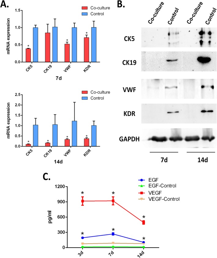
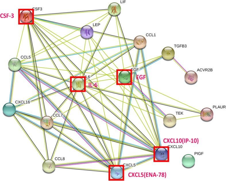
Results: Co-transplantation of microskin and ADSC potentiated the wound healing with better epithelization, smaller scar thickness, and higher angiogenesis (CD31) in the subcutaneous layer. We found both EGF and VEGF cytokines were secreted by microskin in vitro. Additionally, secretome proteomic analysis in a co-culture system of microskin and ADSC revealed that ADSC could secrete a wide range of important molecules to form a reacting network with microskin, including VEGF, IL-6, EGF, uPAR, MCP-3, G-CSF, and Tie-2, which most likely supported the angiogenesis effect as observed.

Conclusion: Overall, we concluded that the use of ADSC partially modulates microskin function and enhances wound healing by promoting angiogenesis in a full-thickness skin defect mouse model.

Keywords: Adipose-derived stem cell; Full-thickness skin defect; Microskin; Secretome; Wound healing.


Conflict of interest statement

The authors declare that they have no competing interests.

Figures

Fig. 1
Fig. 1
Treatment of ADSC+MS promoted wound healing in a full-thickness skin defect mouse model. a Mice were randomized into two parts (n = 15 each part). One group accepted the treatment of microskin or PBS, the other accepted microskin plus ADSCs or ADSCs after surgery. Photograph of the wound area was performed at days 0, 7, and 14, and mice were sacrificed to have IHC procedure at days 7 and 14. b Representative of a full-thickness wound of each group at day 0, and wound closure can be observed at days 7 and 14 which the MS+ADSC group presented the most remarkable effect of wound healing. c Quantitative evaluation of the wound area on days 0, 7, and 14 post-treatment. ###p < .001, the MS+ADSC group compared to all other groups; ****p < .0001, the MS+ADSC group compared to the ADSC group and control group; ***p < .001, the MS+ADSC group compared to the MS group. d Quantitative evaluation of wound contraction at day 14 post-treatment. ****p < .0001; **p < .01; *p < .05, compared to the control group
Fig. 2
Fig. 2
Microscopic appearance of wound beds post-surgery. a Hematoxylin and eosin staining of wound beds at day 14 post-surgery. Wounds treated with MS+ADSC showed a newly formed, hyperplastic epithelium that covered the wound area. The black arrows indicate the microskin grafts had become appendages of the newly formed skin. The asterisks indicated normal adnexal structures. “N” is represented for normal skin, and “w” is represented for wound area. b α-SMA staining of wound beds at day 14 post-treatment. At day 14, the expression of α-SMA in wound tissue was decreased in the MS+ADSC group compared to others. Scale bars are 100 and 500 μm. c CD31 staining of the wound area at days 7 and 14 after treatment. Black arrows indicate CD31-positive vessels. d the number of CD31-positive (+) blood vessels per high-power fields (HPFs) (× 20) were quantified to a particular time point. The data expressed are the average means ± SEM, n = 5. ****p < .0001, compared to the ADSC group, MS group, and control group; **p < .01; *p < .05; ns, not significant, compared to the control group
Fig. 3
Fig. 3
MS+ADSC may enhance wound healing through paracrine function rather than the differentiation of ADSCs. a mRNA expression of CK5, CK19, KDR, and VWF are shown in each group. b Representative Western blot bands for CK5, CK19, VWF, and KDR expression are shown in each group including at days 7 and 14 post-treatment. In co-cultured system, ADSCs were all downregulated compared to the control group. c The concentration of EGF and VEGF secreted by microskin kept stabilizing at 7 days and decreased at 14 days. Both at 7 and 14 days, EGF and VEGF secreted by microskin were higher than the control group (FBS) which demonstrated the microskin could release biological factor. *p < .05, compared with the control group
Fig. 4
Fig. 4
The differentially expressed proteins (DEPs) among the MS+ADSC group, the ADSC group, and the MS group. a Venn diagram showed there were 31 common DEPs detected in the MS+ADSC group compared to the ADSC group and MS group. R Bioconductor package and golots were performed to obtain heat map of DEPs in the MS+ADSC group compared with the ADSC group (b) and MS group (c), with the cluster analysis results
Fig. 5
Fig. 5
GO/KEGG enrichment analysis of DEPs in the ADSC+MS group compared to the ADSC group and MS group by DAVID online tool and R package clusterProfiler. a GO enrichment results of the ADSC+MS group versus ADSC group. b GO enrichment results of the ADSC+MS group versus MS group. c KEGG pathway enrichment results of the ADSC+MS group versus ADSC group. d KEGG pathway enrichment results of the ADSC+MS group versus ADSC group. GO, Gene Ontology; KEGG, Kyoto Encyclopedia of Genes and Genomes
Fig. 6
Fig. 6
Intermolecular interactions of vital associated differentially expressed proteins (DEPs). IL-6, G-CSF, EGF, IP-10, and ENA-78 held a large number of interactions. The interaction relationship of DEPs was shown as the lines. Network edges: line color represents the type of interaction evidence from the interaction sources, line shape represents the predicted mode of action, and line thickness represents the strength of data support. Colored nodes: query proteins and first shell of interactors; empty nodes: proteins of unknown 3D structure; filled nodes: proteins of some known or predicted 3D structure

References

    1. Richardson R, Slanchev K, Kraus C, et al. Adult zebrafish as a model system for cutaneous wound-healing research. J Invest Dermatol. 2013;133(6):1655–1665. - PMC - PubMed
    1. Gabarro P. A new method of grafting. Br Med J. 1943;1(4301):723–724. - PMC - PubMed
    1. Wang WZ, Baynosa RC, Zamboni WA. Update on ischemia-reperfusion injury for the plastic surgeon: 2011. Plast Reconstr Surg. 2011;128(6):685e–692e. - PubMed
    1. Li P, Guo X. A review: therapeutic potential of adipose-derived stem cells in cutaneous wound healing and regeneration. Stem Cell Res Ther. 2018;9(1):302. - PMC - PubMed
    1. Lee DE, Ayoub N, Agrawal DK. Mesenchymal stem cells and cutaneous wound healing: novel methods to increase cell delivery and therapeutic efficacy. Stem Cell Res Ther. 2016;7:37. - PMC - PubMed

Publication types