Review
. 2019 Oct 11;294(41):15121-15136.
doi: 10.1074/jbc.REV119.006129. Epub 2019 Aug 30.

How structural subtleties lead to molecular diversity for the type III polyketide synthases

Affiliations
Review

How structural subtleties lead to molecular diversity for the type III polyketide synthases

Hiroyuki Morita et al. J Biol Chem. .

Abstract

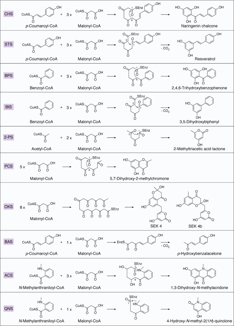
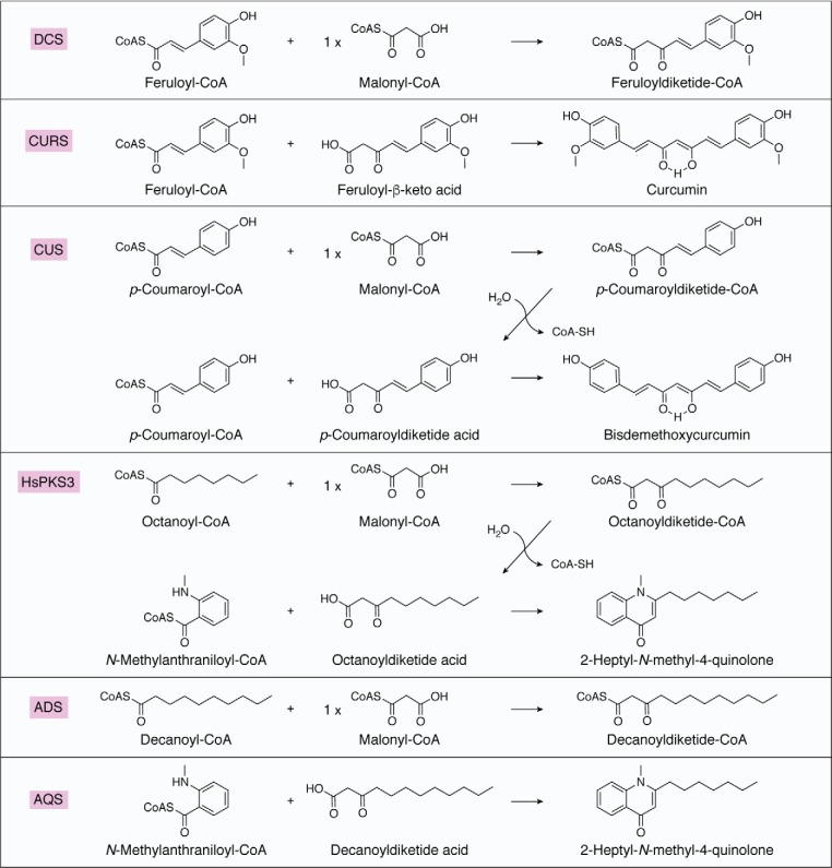
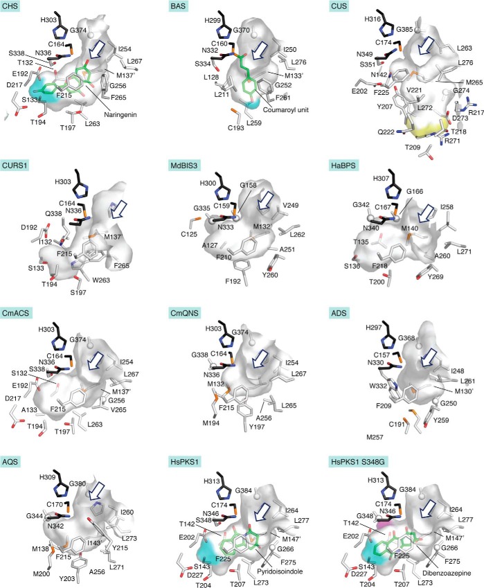
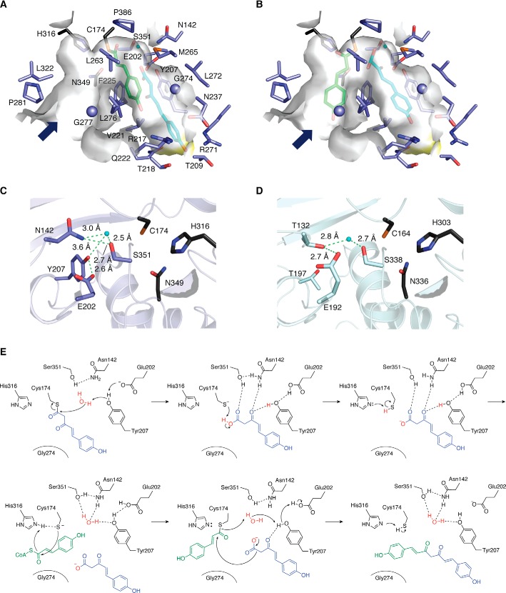
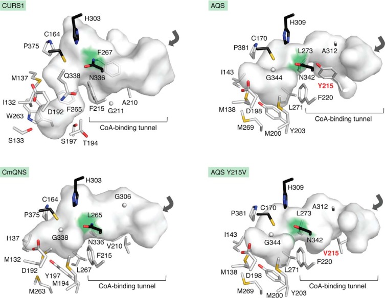
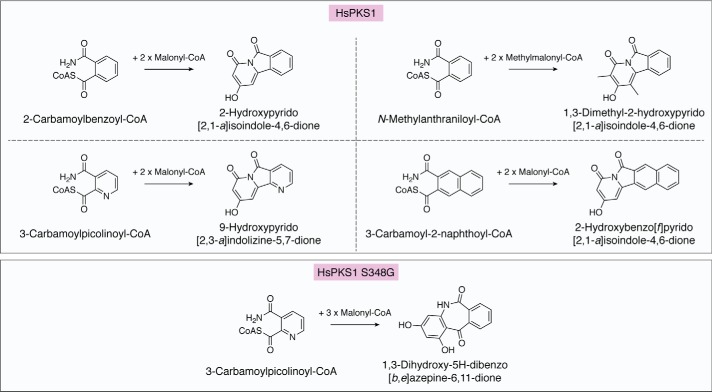
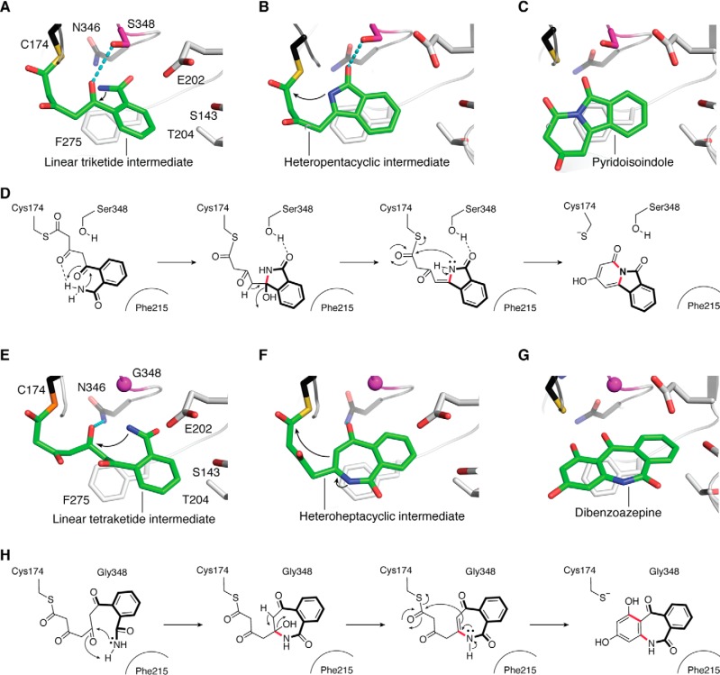
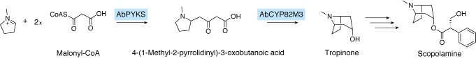
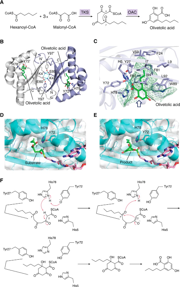
Type III polyketide synthases (PKSs) produce an incredibly diverse group of plant specialized metabolites with medical importance despite their structural simplicity compared with the modular type I and II PKS systems. The type III PKSs use homodimeric proteins to construct the molecular scaffolds of plant polyketides by iterative condensations of starter and extender CoA thioesters. Ever since the structure of chalcone synthase (CHS) was disclosed in 1999, crystallographic and mutational studies of the type III PKSs have explored the intimate structural features of these enzyme reactions, revealing that seemingly minor alterations in the active site can drastically change the catalytic functions and product profiles. New structures described in this review further build on this knowledge, elucidating the detailed catalytic mechanism of enzymes that make curcuminoids, use extender substrates without the canonical CoA activator, and use noncanonical starter substrates, among others. These insights have been critical in identifying structural features that can serve as a platform for enzyme engineering via structure-guided and precursor-directed engineered biosynthesis of plant polyketides. In addition, we describe the unique properties of the recently discovered "second-generation" type III PKSs that catalyzes the one-pot formation of complex molecular scaffolds from three distinct CoA thioesters or from "CoA-free" substrates, which are also providing exciting new opportunities for synthetic biology approaches. Finally, we consider post-type III PKS tailoring enzymes, which can also serve as useful tools for combinatorial biosynthesis of further unnatural novel molecules. Recent progress in the field has led to an exciting time of understanding and manipulating these fascinating enzymes.

Keywords: biosynthesis; enzyme; enzyme mechanism; enzyme structure; natural product biosynthesis.


Conflict of interest statement

The authors declare that they have no conflicts of interest with the contents of this article

Figures

Figure 1.
Figure 1.
Typical reactions catalyzed by plant type III PKSs. 2-PS, 2-pyrone synthase; PCS, pentaketide synthase; OKS, octaketide synthase.
Figure 2.
Figure 2.
Formations of curcumin by DCS and CURS, bisdemethoxycurcumin by CUS, 2-heptyl-N-methyl-4-quinolone by HsPKS3, and 2-heptyl-N-methyl-4-quinolone by ADS and AQS.
Figure 3.
Figure 3.
Crystal structure of M. sativa CHS. A, overall structure. Each monomer is colored white or blue. The catalytic Cys164 is represented by a black sphere. The Met137 residues, which form a partial wall of the active-site cavity in the opposing monomer, are represented by cyan stick models. Black arrows denote the entrance of the CoA-binding tunnel of each monomer. B, close-up view of the catalytic triad. Cys164, His303, and Asn338 are represented by black sticks. The CoA-SH molecules are depicted by green stick models.
Figure 4.
Figure 4.
Active-site cavities observed in the crystal structures of M. sativa CHS (PDB entry 1BI5), Rheum palmatum BAS (PDB entry 3A5Q), O. sativa CUS (PDB entry 3OIT), C. longa CURS1 (PDB entry 3OV2), MdBIS3 (PDB entry 5W8Q), HaBPS (PDB entry 5UCO), CmACS (PDB entry 3WD7), CmQNS (PDB entry 3WD8), E. rutaecarpa ADS (PDB entry 5WX3), E. rutaecarpa AQS (PDB entry 5WX4), and HsPKS1 (PDB entry 3AWK) and in the model structure of the HsPKS1 S348G mutant enzyme. The sizes and shapes of the active-site cavities are represented by surface models. The catalytic triads are represented by black stick models. The bottoms of the coumaroyl-binding pocket in M. sativa CHS, the corresponding pockets in HsPKS1 and its mutant, and the alternative coumaroyl-binding pocket in R. palmatum BAS are highlighted by cyan surfaces. The bottom of the downward-expanding pocket in CUS is indicated by a yellow surface. The expanded wall of the HsPKS1 S348G mutant is highlighted by a pink surface. The entrances to the active-site cavities are shown by blue arrows. In the M. sativa CHS and R. palmatum BAS crystal structures, the complexed naringenin and coumaroyl unit, respectively, are shown with green stick models. Three-dimensional structures of pyridoisoindole (2-hydroxybenzo[f]pyrido[2,1-a]isoindole-4,6-dione) and dibenzoazepine (1,3-dihydroxy-5H-dibenzo[b,e]azepine-6,11-dione), shown as green stick models, are docked into the active-site cavities of the WT and S348G mutant HsPKS1 enzymes, respectively. All active-site cavities in this figures were rendered using PyMOL.
Figure 5.
Figure 5.
Close-up views of the active-site cavity and the CoA-binding tunnel of O. sativa CUS. A and B, three-dimensional models of the coumaroyl monoketide covalently bound to the catalytic Cys174 (green stick model) and the p-coumaroyldiketide acid (cyan stick model) within the active-site cavity (A) and those within the CoA-binding tunnel and the active-site cavity (B), respectively. C and D, the hydrogen bond networks of O. sativa CUS (C) and P. sylvestris STS (D). The catalytic triads are represented by black stick models. The water molecules and hydrogen bonds are represented by cyan spheres and green dotted lines, respectively. E, proposed catalytic mechanism for the formation of bisdemethoxycurcumin by CUS.
Figure 6.
Figure 6.
Comparison of the active sites of C. longa CURS1, E. rutaecarpa AQS, E. rutaecarpa QNS, and the AQS Y215V mutant. The catalytic triads are represented by black stick models. The junctions between the CoA-binding tunnel and the active-site cavity are highlighted with green surfaces. The entrances to the CoA-binding tunnels are shown with gray arrows. Tyr215 and the mutated Val215 are highlighted with red designations.
Figure 7.
Figure 7.
Enzymatic formation of pyridoisoindole and benzoazepine alkaloids by WT HsPKS1 and its S348G mutant.
Figure 8.
Figure 8.
Proposed mechanism for the formation of 2-hydroxybenzo[f]pyrido[2,1-a]isoindole-4,6-dione by WT HsPKS1 and 1,3-dihydroxy-5H-dibenzo[b,e]azepine-6,11-dione by the HsPKS1 S348G mutant. A–C, model structures of HsPKS1 complexed with the linear triketide intermediate (A), the heteropentacyclic intermediate (B), and 2-hydroxybenzo[f]pyrido[2,1-a]isoindole-4,6-dione (pyridoisoindole) (C). The catalytic Cys174 and Asn346 are depicted by black stick models. Hydrogen bonds between Ser348 (purple stick model) and the intermediate are represented by cyan dotted lines. D, schematic representation of the proposed mechanism for the formation of pyridoisoindole. E–G, model structures of HsPKS1 S348G complexed with the linear tetraketide intermediate (E), the heteroheptacyclic intermediate (F), and 1,3-dihydroxy-5H-dibenzo[b,e]azepine-6,11-dione (dibenzoazepine) (G). H, schematic representation of the proposed mechanism for the formation of dibenzoazepine. The catalytic Cys174 and Asn346 are depicted by black stick models. Ser348 and Gly348 are highlighted by purple stick models. Hydrogen bonds between Ser348 and the intermediate are represented by cyan dotted lines.
Figure 9.
Figure 9.
Proposed biosynthetic pathway for the formation of tropinone and scopolamine, catalyzed by AbPYKS.
Figure 10.
Figure 10.
Proposed biosynthetic pathway for the formation of olivetolic acid and structure of OAC. A, the formation of olivetolic acid by tetraketide synthase (TKS) and OAC. B, overall binary structure of OAC complexed with olivetolic acid. Each monomer is colored white or purple. Olivetolic acid molecules are shown as green stick models. Hydrogen bonds forming the dimer interface between two monomers are represented with cyan dotted lines. C, close-up view of the active-site cavity. The size and shape of the active-site cavity are represented with green mesh. D and E, docking model of pentyltetra-β-ketide-CoA (substrate) (D) and product (E) in OAC. F, the proposed mechanism catalyzed by OAC to generate olivetolic acid.

References

    1. Smith S., and Tsai S. C. (2007) The type I fatty acid and polyketide synthases: a tale of two megasynthases. Nat. Prod. Rep. 24, 1041–1072 10.1039/b603600g - DOI - PMC - PubMed
    1. Hertweck C., Luzhetskyy A., Rebets Y., and Bechthold A. (2007) Type II polyketide synthases: gaining a deeper insight into enzymatic teamwork. Nat. Prod. Rep. 24, 162–190 10.1039/B507395M - DOI - PubMed
    1. Schröder J. (1999) The chalcone-stilbene synthase-type family of condensing enzymes, in Comprehensive Natural Products Chemistry (Sankawa U., ed) pp. 749–771, Elsevier, Oxford, UK
    1. Austin M. B., and Noel J. P. (2003) The chalcone synthase superfamily of type III polyketide synthases. Nat. Prod. Rep. 20, 79–110 10.1039/b100917f - DOI - PubMed
    1. Abe I., and Morita H. (2010) Structure and function of the chalcone synthase superfamily of plant type III polyketide syntheses. Nat. Prod. Rep. 27, 809–838 10.1039/b909988n - DOI - PubMed

Publication types

MeSH terms

Substances