. 2019 Oct 25;294(43):15698-15710.
doi: 10.1074/jbc.RA119.009980. Epub 2019 Aug 30.

The Vibrio cholerae minor pilin TcpB mediates uptake of the cholera toxin phage CTXφ

Affiliations

The Vibrio cholerae minor pilin TcpB mediates uptake of the cholera toxin phage CTXφ

Miguel Gutierrez-Rodarte et al. J Biol Chem. .

Abstract

Virulent strains of the bacterial pathogen Vibrio cholerae cause the diarrheal disease cholera by releasing cholera toxin into the small intestine. V. cholerae acquired its cholera toxin genes by lysogenic infection with the filamentous bacteriophage CTXφ. CTXφ uses its minor coat protein pIII, located in multiple copies at the phage tip, to bind to the V. cholerae toxin-coregulated pilus (TCP). However, the molecular details of this interaction and the mechanism of phage internalization are not well-understood. The TCP filament is a polymer of major pilins, TcpA, and one or more minor pilin, TcpB. TCP are retractile, with both retraction and assembly initiated by TcpB. Consistent with these roles in pilus dynamics, we hypothesized that TcpB controls both binding and internalization of CTXφ. To test this hypothesis, we determined the crystal structure of the C-terminal half of TcpB and characterized its interactions with CTXφ pIII. We show that TcpB is a homotrimer in its crystallographic form as well as in solution and is present in multiple copies at the pilus tip, which likely facilitates polyvalent binding to pIII proteins at the phage tip. We further show that recombinant forms of TcpB and pIII interact in vitro, and both TcpB and anti-TcpB antibodies block CTXφ infection of V. cholerae Finally, we show that CTXφ uptake requires TcpB-mediated retraction. Our data support a model whereby CTXφ and TCP bind in a tip-to-tip orientation, allowing the phage to be drawn into the V. cholerae periplasm as an extension of the pilus filament.

Keywords: CTXφ; TcpB; Vibrio cholerae; X-ray crystallography; bacteriophage; minor pilins; pIII; protein structure; toxin coregulated pilus; type IV pili.


Conflict of interest statement

The authors declare that they have no conflicts of interest with the contents of this article

Figures

Figure 1.
Figure 1.
Structure of the C-terminal half of V. cholerae TcpB and comparison with ETEC CofB. A, domain organization of TcpB and CofB and their recombinantly expressed forms. Domain boundaries and cysteines are indicated. B, amino acid sequences of TcpB and CofB aligned based on their structures and colored as in A. Identical residues are shown in boldface text. TcpB and CofB are 20% identical in their C-terminal halves (β-repeat and effector domain) and 16% identical overall. Disulfide bonds seen in the crystal structures are indicated with solid lines. The disulfide bond predicted between TcpB Cys-250 and Cys-261 is shown with a dashed line. The peptide used to generate anti-TcpB(64–78) antibodies is indicated. C, crystal structure of TcpB-C (residues 243–423) shown in ribbon representation, colored and labeled as in A and B. Residues 243–259 are not resolved in the crystal structure and are illustrated as the first strand of the three-stranded βR based on their alignment with βR2 of CofB. D, crystal structure of ΔN-CofB (22). Only the C-terminal half of ΔN-CofB (residues 261–518) is shown, omitting the pilin domain.
Figure 2.
Figure 2.
TcpB-C forms a homotrimer. A, TcpB-C and ΔN-CofB crystallographic trimers, with each subunit, A, B, and C, colored differently. Only the β-repeats and effector domains of the ΔN-CofB structure are shown. B, chain C of TcpB-C, shown in the same orientation as in A, at left in cartoon representation and at right with electrostatic surface potential, calculated using DelPhi (54). Red represents negative charge, blue is positive charge, and white is neutral (scale from −5 to +5 kT). The trimerization surface is mostly uncharged. C, Coomassie-stained nonreducing SDS-PAGE of recombinant TcpB forms in the absence and presence of cross-linker DSP. Fragments containing the C-terminal half of TcpB (residues 243–423) appear at approximately triple their mass in the presence of DSP, indicating the presence of trimers, whereas the TcpB fragment comprising the pilin domain remains as a monomer. The theoretical masses of monomeric and trimeric forms are shown in the table on the right. MM, mass markers (kDa).
Figure 3.
Figure 3.
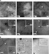
Immunogold labeling of TcpB at pilus ends. Purified TCP were adhered to carbon-coated copper grids, treated with chicken anti-TcpB antibody raised against ΔN-TcpB (Table 2) and gold-labeled secondary antibody, then stained with uranyl acetate, and imaged by transmission EM. Single (A) and multiple (B) gold labels are seen at ends of TCP. Magnification of insets is indicated.
Figure 4.
Figure 4.
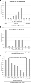
V. cholerae TcpB binds to CTXφ pIII in ELISAs. A, CTXφ pIII protein was immobilized on ELISA microplate wells, and V. cholerae TcpB was added in increasing amounts in solution. Captured TcpB was detected with rabbit anti-TcpB(64–78) antibody and enzyme-linked secondary antibody. TcpB was immobilized as a positive control; negative controls were blocked wells lacking immobilized antigen (−) and immobilized antigens TcpA and BSA to show the specificity of the TcpB:pIII interaction. B, TcpB was immobilized on ELISA microplate wells, and pIII was added in increasing amounts in solution. Captured pIII was detected with anti-pIII antibody and enzyme-linked secondary antibody. pIII was immobilized as a positive control, and TcpA and BSA were immobilized as negative controls for capture. C, TcpB was immobilized, and pIII was added in increasing amounts in solution to inhibit anti-TcpB(64–78) antibody binding. Bound antibody was detected with enzyme-linked secondary antibody. TcpA and BSA were added in solution as negative controls for inhibition. For all experiments, absorbance was read at 405 nm. Values represents the average of three separate experiments; error bars represent the S.E.
Figure 5.
Figure 5.
CTX-Kmφ infection of V. cholerae is inhibited by recombinant TcpB and anti-TcpB antibodies. A, V. cholerae WT (O395) and ΔtcpA and ΔtcpB strains were incubated with buffer or recombinant TcpB (ΔN-TcpB), rabbit anti-TcpB antibody(64–78), BSA, or anti-CofJ antibody; then transduced with CTX-Kmφ; and plated on LB-Km plates. The number of cfu/ml for each infection is reported (n = 3); error bars represent the S.E. B, V. cholerae WT was incubated with three different forms of recombinant TcpB: ΔN-TcpB (residues 25–423), TcpB-C (residues 243–423), and TcpB-pilin (residues 25–228); then transduced with CTX-Kmφ; and plated on LB-Km plates. cfu/ml was calculated for each infection as for A.
Figure 6.
Figure 6.
TcpB-mediated pilus retraction is required for CTX-Kmφ infection of V. cholerae. V. cholerae ΔtcpB was transformed with ptcpB encoding WT or Glu-5 substitutions. Cells were grown overnight in pilus-inducing conditions with 0.001 mm rhamnose to induce ectopic expression of TcpB. Cells from overnight culture were infected with CTX-Kmφ and plated on LB-Km. The number of cfu/ml for each infection is reported (n = 3); error bars represent the S.E.
Figure 7.
Figure 7.
Model for T4P-mediated uptake of CTXφ. A, CTXφ binds via its tip-associated minor coat protein pIII to the minor pilin TcpB trimer at the tip of a growing V. cholerae TCP filament. B–D, random incorporation of TcpB monomer at the base of the growing pilus stalls pilus assembly (B), triggering retraction (C), which draws the phage into the periplasm as if it were an extension of the pilus (D and E). TcpB is shown as a trimer, with its pilin domain colored blue and its effector domain colored green. IM, inner membrane; OM, outer membrane.

Similar articles

Cited by

References

    1. Taylor R. K., Miller V. L., Furlong D. B., and Mekalanos J. J. (1987) Use of phoA gene fusions to identify a pilus colonization factor coordinately regulated with cholera toxin. Proc. Natl. Acad. Sci. U.S.A. 84, 2833–2837 10.1073/pnas.84.9.2833 - DOI - PMC - PubMed
    1. Kaper J. B., Morris J. G. Jr., and Levine M. M. (1995) Cholera. Clin. Microbiol. Rev. 8, 48–86 10.1128/CMR.8.1.48 - DOI - PMC - PubMed
    1. Waldor M. K., and Mekalanos J. J. (1996) Lysogenic conversion by a filamentous phage encoding cholera toxin. Science 272, 1910–1914 10.1126/science.272.5270.1910 - DOI - PubMed
    1. Heilpern A. J., and Waldor M. K. (2003) pIIICTX, a predicted CTXφ minor coat protein, can expand the host range of coliphage fd to include Vibrio cholerae. J. Bacteriol. 185, 1037–1044 10.1128/JB.185.3.1037-1044.2003 - DOI - PMC - PubMed
    1. Heilpern A. J., and Waldor M. K. (2000) CTXφ infection of Vibrio cholerae requires the tolQRA gene products. J. Bacteriol. 182, 1739–1747 10.1128/JB.182.6.1739-1747.2000 - DOI - PMC - PubMed

Publication types

MeSH terms

LinkOut - more resources