. 2019 Oct 16;39(42):8408-8421.
doi: 10.1523/JNEUROSCI.2851-18.2019. Epub 2019 Aug 30.

Microglia Mediate HIV-1 gp120-Induced Synaptic Degeneration in Spinal Pain Neural Circuits

Affiliations

Microglia Mediate HIV-1 gp120-Induced Synaptic Degeneration in Spinal Pain Neural Circuits

Wenjuan Ru et al. J Neurosci. .

Abstract

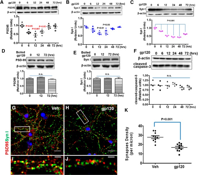
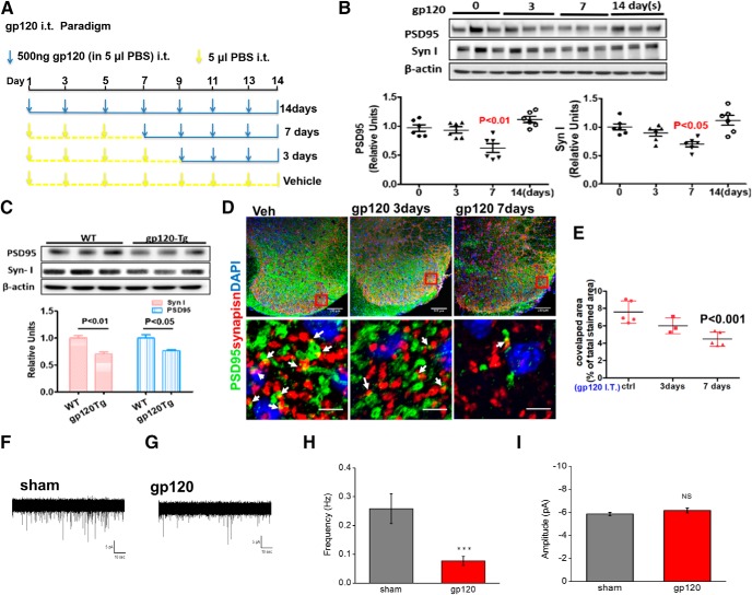
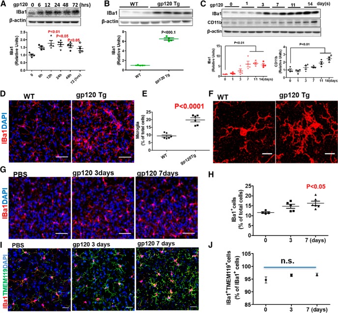
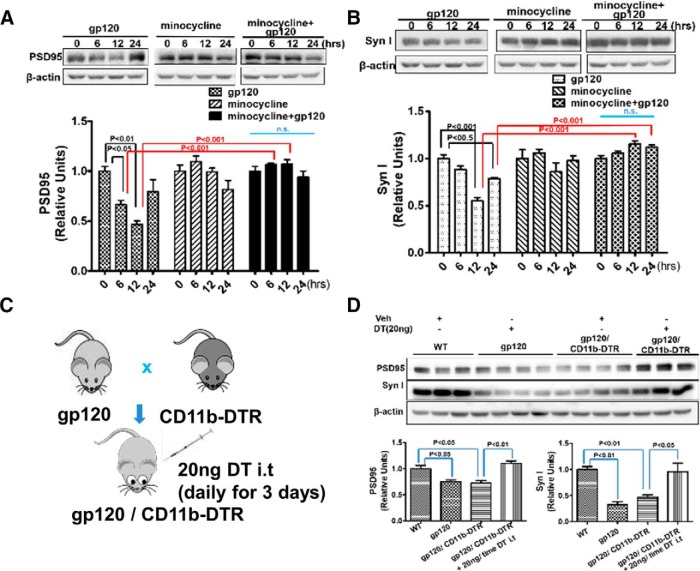
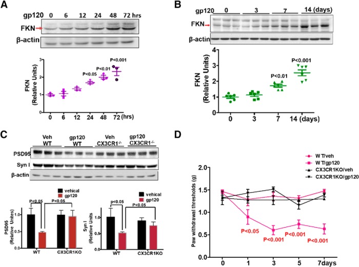
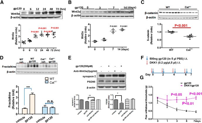
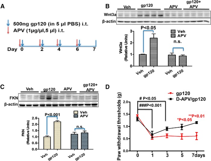
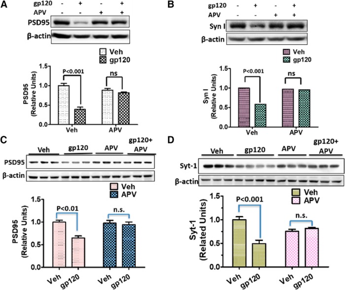
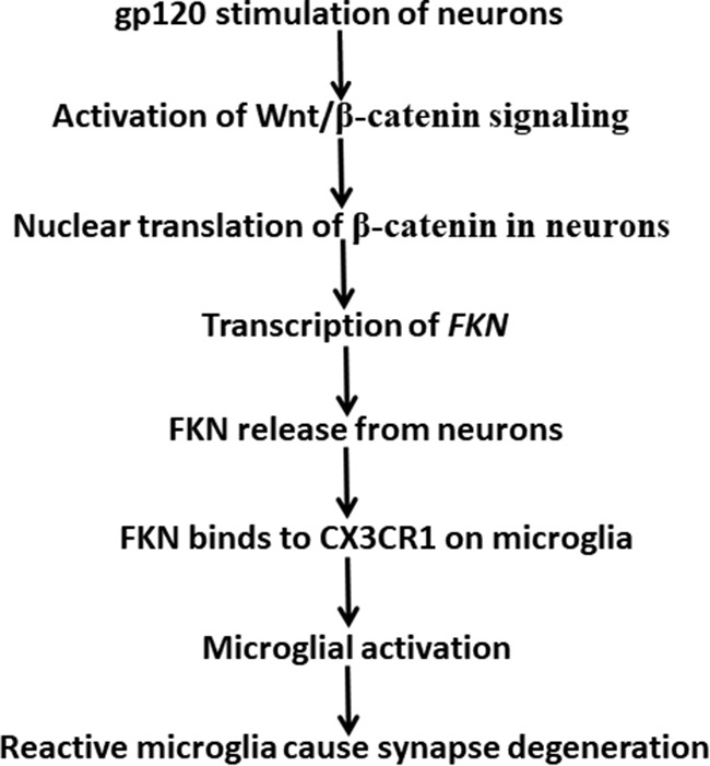
HIV-1 infection of the nervous system causes various neurological diseases, and synaptic degeneration is likely a critical step in the neuropathogenesis. Our prior studies revealed a significant decrease of synaptic protein, specifically in the spinal dorsal horn of patients with HIV-1 in whom pain developed, suggesting a potential contribution of synaptic degeneration to the pathogenesis of HIV-associated pain. However, the mechanism by which HIV-1 causes the spinal synaptic degeneration is unclear. Here, we identified a critical role of microglia in the synaptic degeneration. In primary cortical cultures (day in vitro 14) and spinal cords of 3- to 5-month-old mice (both sexes), microglial ablation inhibited gp120-induced synapse decrease. Fractalkine (FKN), a microglia activation chemokine specifically expressed in neurons, was upregulated by gp120, and knockout of the FKN receptor CX3CR1, which is predominantly expressed in microglia, protected synapses from gp120-induced toxicity. These results indicate that the neuron-to-microglia intercellular FKN/CX3CR1 signaling plays a role in gp120-induced synaptic degeneration. To elucidate the mechanism controlling this intercellular signaling, we tested the role of the Wnt/β-catenin pathway in regulating FKN expression. Inhibition of Wnt/β-catenin signaling blocked both gp120-induced FKN upregulation and synaptic degeneration, and gp120 stimulated Wnt/β-catenin-regulated FKN expression via NMDA receptors (NMDARs). Furthermore, NMDAR antagonist APV, Wnt/β-catenin signaling suppressor DKK1, or knockout of CX3CR1 alleviated gp120-induced mechanical allodynia in mice, suggesting a critical contribution of the Wnt/β-catenin/FKN/CX3R1 pathway to gp120-induced pain. These findings collectively suggest that HIV-1 gp120 induces synaptic degeneration in the spinal pain neural circuit by activating microglia via Wnt3a/β-catenin-regulated FKN expression in neurons.SIGNIFICANCE STATEMENT Synaptic degeneration develops in the spinal cord dorsal horn of HIV patients with chronic pain, but the patients without the pain disorder do not show this neuropathology, indicating a pathogenic contribution of the synaptic degeneration to the development of HIV-associated pain. However, the mechanism underlying the synaptic degeneration is unclear. We report here that HIV-1 gp120, a neurotoxic protein that is specifically associated with the manifestation of pain in HIV patients, induces synapse loss via microglia. Further studies elucidate that gp120 activates microglia by stimulating Wnt/β-catenin-regulated fractalkine in neuron. The results demonstrate a critical role of microglia in the pathogenesis of HIV-associated synaptic degeneration in the spinal pain neural circuit.

Keywords: HIV-1; Wnt; microglia; pain; spinal cord; synaptic degeneration.


Figures

Figure 1.
Figure 1.
HIV-1 gp120 induces synapse loss in a time-dependent manner in primary cortical cultures. A–C, Time course of gp120-induced decreases of synaptic proteins. Cortical neuron–glia cocultures (14 DIV) were incubated with 200 pm gp120 for the indicated times. Cell lysates were analyzed by immunoblotting using antibodies against PSD-95 (A), Syn I (B), or Syt-1 (C). For A and B, N = 4; for C, N = 3 cultures/condition. D, E, Heat-inactivated gp120 did not affect PSD-95 (D) and Syn I (E) in cultures; N = 3. F, Western blot of cleaved caspase-3 from gp120-treated cell lysates; N = 4. G–J, Representative immunofluorescence images showing PSD-95 and Syn I after 12 h vehicle (Veh) or gp120 treatment (PSD-95 is red, Syn I is green, and colocalized region is yellow). I, J, Higher magnifications of boxed region. Scale bars: G, H, 20 μm; I, J, 5 μm. K, Bar graph summarizes the effects of gp120 on synapse density. N = 12 neurons from three cultured coverslips/condition (mean ± SEM). n.s., no significant difference.
Figure 2.
Figure 2.
gp120 causes a synapse decrease in the spinal cord dorsal horn. A, The paradigm of intrathecal gp120 treatment. B, PSD-95 and Syn I protein levels in the L4–L5 spinal cord after intrathecal gp120 injection (N = 6 mice/time point). C, PSD-95 and Syn I protein levels in the L4–L5 spinal cord of the gp120 Tg and WT mice (N = 3 mice/condition). D, Confocal images of SDH sections showing anti-Syn I and anti-PSD-95 antibodies staining colocalized areas of Syn I and PSD-95 were calculated (white arrows). Scale bars: top, 100 μm; bottom, 5 μm. E, Quantification: images of layers 1–2 of the SDH from three to five mice were included for each group. For each mouse, three to four spinal slices were included, and two to three images were obtained from each section. F, G, Representative whole-cell recording of mEPSC from the SDH GABAergic interneurons (GFP labeled) from mice treated via intrathecal injection with vehicle (Sham; F) or gp120 (G). Five microliters of gp120 (500 ng) or PBS was intrathecally injected into GAD67-GFP transgenic mice once every other day for 7 d. Cells were held at Vm (membrane potential) = −70 mV. H, I, Summary graphs showing the differences of frequency (H) and amplitude (I) of mEPSCs between the sham- and gp120-treated mice. N = 15 (sham) and N = 31 (gp120) neurons were recorded from three sham and five intrathecally injected gp120 mice for mEPSCs. Error bars represent SEM. ns, no significant difference, ***P < 0.001.
Figure 3.
Figure 3.
Gp120 causes microglial activation. A, Levels of IBa1 in 200 pm gp120-treated primary cortical cultures (N = 4 cultures in each condition). The cell composition of the cocultures is shown in Figure 3-1. B, IBa1 in L4–L5 spinal cords from gp120 transgenic and WT mice. WT, N = 3 mice; gp120 Tg, N = 4 mice. C, IBa1 and CD11b in L4–L5 spinal cords from mice after intrathecal gp120 injection (500 ng/injection). N = 4 mice/time point. D, Confocal images of IBa1+ cells in the SDH from WT and gp120 Tg mice. Scale bars, 50 μm. E, Quantitative summary of D (mean ± SEM); N = 6 mice/group. F, High-magnification images showing morphological characteristics of reactive microglia. Scale bars, 10 μm. G, Confocal images of IBa1+ cells in the SDH of mice intrathecally injected with PBS or gp120. Scale bars, 50 μm. H, Quantitative summary of G (mean ± SEM); PBS, N = 4 mice; gp120/3 d, N = 5 mice; gp120/7 d, N = 6 mice. I, Confocal images of IBa1 and TMEM119 double-positive cells in the SDH of mice intrathecally injected with PBS or gp120. Scale bars, 30 μm. J, Quantitative summary of I (mean ± SEM); PBS, N = 4 mice; gp120/3 d; N = 5 mice; gp120/7 d, N = 6 mice. n.s., no significant difference.
Figure 4.
Figure 4.
Microglial activation is crucial for gp120-induced synapse loss. A, B, Cotreatment of primary cortical cultures with minocycline prevented gp120-induced decreases in PSD-95 (A) and Syn I (B). Cortical cultures (14 DIV) were treated with gp120 (200 pm), minocycline (30 μm), or gp120+minocycline for various time periods. Time 0 is the baseline without any drug treatment. N = 4 cultures/condition. C, Diagram showing the generation of gp120/CD11b-DTR mice for microglial ablation by DT. D, Effect of DT administration on PSD-95 and Syn I levels in the spinal cord of the gp120/CD11b-DTR mice. WT, gp120Tg, and gp120Tg/CD11b-DTR mice were administrated 20 ng DT (in 5 μl of PBS) or 5 μl of PBS intrathecally only once/day for 3 d. L4–L5 spinal cords were used for immunoblotting analysis of PSD-95 and Syn I. N, WT/ PBS, 3; gp120Tg/DT, 4; gp120gp/CD11b-DTR/PBS, 4; gp120gp/CD11b-DTR/DT, 3. Quantitative graphs are shown as mean ± SEM. DT-induced microglial atrophy in CD11b-DTR mice is shown in Figure 4-1. n.s., no significant difference.
Figure 5.
Figure 5.
FKN/CX3CR1 signaling is required for gp120-induced synaptic degeneration. A, gp120-stimulated FKN expression in cortical cultures (N = 3 cultures/treatment). B, gp120-stimulated (by intrathecal administration) FKN expression in the mouse spinal cords (N = 6 mice/time point). C, Protein levels of PSD 95 and Syn I in the spinal cord of CX3CR1−/− or WT mice at day 7 after intrathecal injection with vehicle (Veh) or gp120 (N = 5 mice/group). D, Behavioral tests of gp120-induced mechanical allodynia in WT and CX3CR1−/− mice. Threshold of mechanical sensitivity in the hindpaw was measured by von Frey tests (N = 6 mice/group). Error bars indicate the mean ± SEM.
Figure 6.
Figure 6.
Wnt3a/β-catenin pathway mediates gp120-induced FKN upregulation and synaptic degeneration. A, Temporal profiles of Wnt3a expression in gp120-treated primary cortical cultures (N = 6 cultures/group). B, Temporal profiles of Wnt3a expression in the spinal cords (L4–L5) from mice after intrathecal injection with gp120 (N = 3 mice/group). C, β-catenin protein level in spinal cords (L4–L5) from WT and Cat+/− mice (N = 6 mice for WT; and N = 7 mice for Cat+/−). D, Comparison of the effect of gp120 on FKN expression in WT and Cat+/− mice, with intrathecal injection with gp120 every other day for 7 d. Spinal (L4–L5) FKN protein was analyzed by Western blotting. N = 3 mice/group for Veh/WT, Veh/Cat+/−, and gp120/Cat+/−; N = 4 mice for gp120/WT. E, Effect of Wnt3a neutralizing antibody on gp120-induced synaptic degeneration in primary cortical cultures. Anti-Wnt3a antibody (2 μg/ml) was added to the cultures for 30 min, and then gp120 (200pΜ) was added for an additional 12 h (N = 3 independent cultures/group). F, Drug administration paradigm. DKK1 (1 μg/5 μl) was injected 30 min before gp120 (500 ng/5 μl, i.t., in PBS) injection. G, Behavioral tests of the effect of DKK1 on gp120-induced mechanical allodynia. The threshold of mechanical sensitivity in the hindpaw was measured by von Frey tests (N = 6 mice/group). Error bars indicated the mean ± SEM. n.s., no significant difference, *P < 0.05, **P < 0.01, ***P < 0.001.
Figure 7.
Figure 7.
The NMDAR is crucial for gp120 to induce Wnt3a and FKN expression. A, Mice were intrathecally injected with either APV (5 μg/5 μl in PBS) or vehicle (Veh; 5 μl of PBS) 30 min before gp120 injection (500 ng/5 μl/2 d). B, C, Spinal cords (L4–L5) were collected at day 7 for immunoblotting analysis of Wnt3a (B) and FKN (C). N = 6 mice for the groups of Veh and APV; N = 7 for the groups of gp120 and gp120+APV. D, Von Frey tests of the effect of APV on gp120-induced mechanical pain (N = 5 mice/group). *p < 0.05; **p < 0.01 (gp120 vs gp120/APV at the same time point); #p < 0.05; ###p < 0.001 (between different time point within the gp120/APV group). The involvement of gp120 coreceptor is suggested by Figure 7-1. n.s., no significant difference.
Figure 8.
Figure 8.
gp120-induced synapse loss depends on NMDAR activation. A, B, Effects of APV on gp120-induced decreases of PSD-95 (A) and Syn I (B) in primary cortical cultures. APV (100 nm) was added to the cultures (14 DIV) 30 min before gp120 (200 pΜ) application. The cultures were exposed to gp120 for 12 h before harvesting for immunoblotting (N = 4 independent cultures/group). C, D, Effects of APV on gp120-induced decreases of PSD-95 (C) and Syn I (D) in the spinal cords (L4–L5). Mice were intrathecally injected with APV (5 μg/5 μl PBS) or vehicle (Veh; 5 μl of PBS) 30 min before gp120 injection (500 ng/5 μl/2 d). L4–L5 spinal cords were collected for immunoblotting analysis of PSD-95 (C) and Syt-1 (D). N = 3 mice/group (mean ± SEM). n.s., no significant difference.
Figure 9.
Figure 9.
A model of gp120-induced synaptic degeneration in the spinal pain neural circuit. gp120 stimulates neurons and activates Wnt/β-catenin signaling that leads to β-catenin-mediated FKN transcription. FKN protein released from neurons binds to CX3CR1 to activate microglia to induce synaptic degeneration.

Similar articles

Cited by

References

    1. Avraham HK, Jiang S, Fu Y, Rockenstein E, Makriyannis A, Zvonok A, Masliah E, Avraham S (2014) The cannabinoid CB2 receptor agonist AM1241 enhances neurogenesis in GFAP/Gp120 transgenic mice displaying deficits in neurogenesis. Br J Pharmacol 171:468–479. 10.1111/bph.12478 - DOI - PMC - PubMed
    1. Bliss TV, Collingridge GL, Kaang BK, Zhuo M (2016) Synaptic plasticity in the anterior cingulate cortex in acute and chronic pain. Nat Rev Neurosci 17:485–496. 10.1038/nrn.2016.68 - DOI - PubMed
    1. Brown GC, Neher JJ (2014) Microglial phagocytosis of live neurons. Nat Rev Neurosci 15:209–216. 10.1038/nrn3710 - DOI - PubMed
    1. Cadwell CR, Palasantza A, Jiang X, Berens P, Deng Q, Yilmaz M, Reimer J, Shen S, Bethge M, Tolias KF, Sandberg R, Tolias AS (2016) Electrophysiological, transcriptomic and morphologic profiling of single neurons using patch-seq. Nat Biotechnol 34:199–203. 10.1038/nbt.3445 - DOI - PMC - PubMed
    1. Cailhier JF, Partolina M, Vuthoori S, Wu S, Ko K, Watson S, Savill J, Hughes J, Lang RA (2005) Conditional macrophage ablation demonstrates that resident macrophages initiate acute peritoneal inflammation. J Immunol 174:2336–2342. 10.4049/jimmunol.174.4.2336 - DOI - PubMed

Publication types