

Review
doi: 10.1007/s00232-019-00090-5.
Epub 2019 Aug 30.
Life During Wartime: A Personal Recollection of the Circa 1990 Prestegard Lab and Its Contributions to Membrane Biophysics
Affiliations
- PMID: 31471644
- PMCID: PMC6814499
- DOI: 10.1007/s00232-019-00090-5
Item in Clipboard
Review
Life During Wartime: A Personal Recollection of the Circa 1990 Prestegard Lab and Its Contributions to Membrane Biophysics
J Membr Biol.
2019 Dec.
Abstract
A subjective account is presented of challenges and excitement of being a postdoctoral trainee in the lab of James H. Prestegard at Yale University in New Haven, Connecticut from 1989 to 1991. This includes accounts of the early development of bicelles and of oriented sample NMR results that contributed to our modern understanding of the properties of the water-lipid interface of disordered phase biological membranes.
Keywords: Bicelles; Interface; Membranes; NMR; Spectroscopy.
Conflict of interest statement
Conflict of interest
The author declares that he has no conflict of interest.
Figures
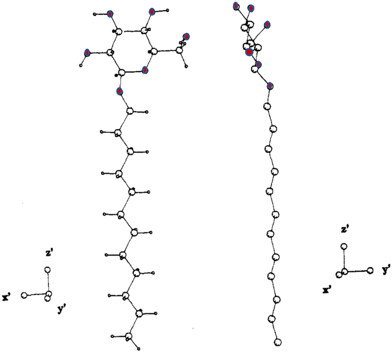
Depiction of the β-glucoside head group orientation of BDOG with respect to the membrane bilayer normal, as determined in magnetically aligned CHAPSO-DMPC bicelles (Sanders & Prestegard, 1991). The red atoms are oxygens. The dodecyl chain is depicted in fully extended form, aligned with the bilayer normal. The axes define the orientation of the order tensor with respect to the glucopyranoside head group. The z’ axis is coincident with the bilayer normal (which is also aligned with the alkyl chain). This figure is reprinted with permission from Journal of the American Chemical Society (Sanders & Prestegard, 1991), copyright 1991 American Chemical Society.
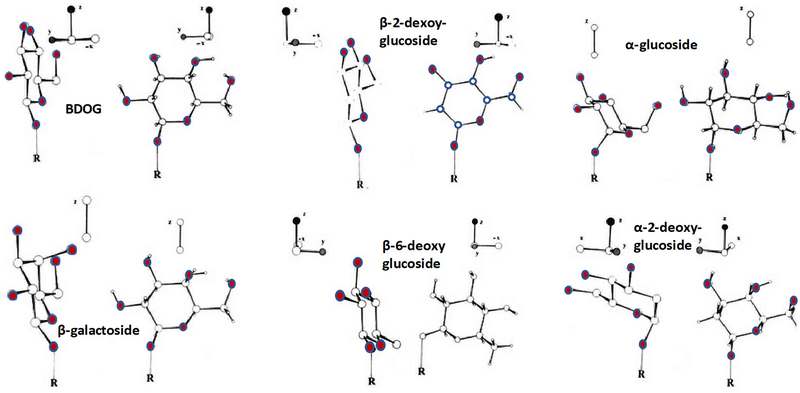
Glycoside head group orientations for a series of alkyl glycosides, as determined in magnetically-aligned CHAPSO-DMPC bicelles (Sanders & Prestegard, 1992). The red atoms are oxygens and “R” is the alkyl (dodecyl or tetradecyl) chain. Shown for each glycoside is its determined order tensor, with the z (vertical) axis in each case aligned with the bilayer normal. In some cases (where all three coordinate axes are shown) it was possible to determine the full 2nd-ranked order tensor. However, for the cases where only the z axis is depicted we collected only enough data to determine the orientation of the head group with respect to the bilayer normal, not also with respect to the other two eigenvectors of the order matrix. This figure is reprinted with permission from Journal of the American Chemical Society (Sanders & Prestegard, 1992), copyright 1992 American Chemical Society.
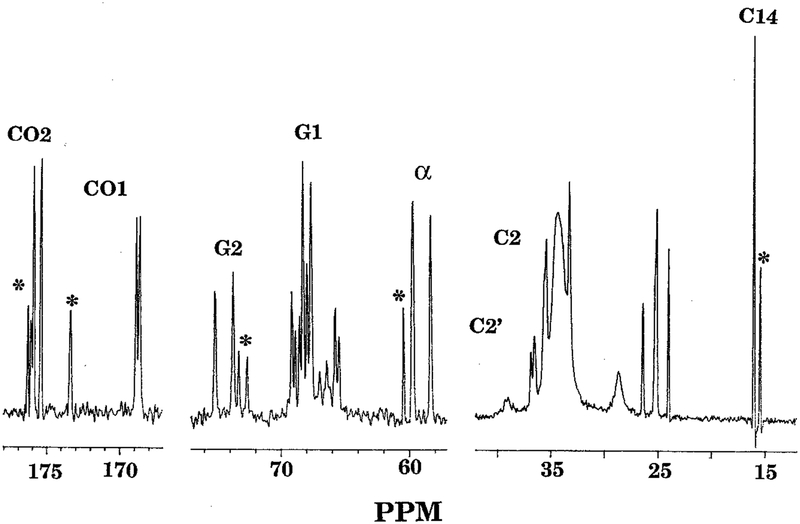
Previously-unpublished 13C (67.9 MHz) NMR spectrum from circa 1993 of magnetically-aligned bicelles containing a racemic mixture of racemic DMPC and l-DHPC at 40° C. The DHPC:DMPC molar ratio was 1:2.87 and the total amphiphile (DMPC+DHPC) concentration (w/v) was 25% in 0.1 M KCl/D2O. The free induction decay (FID) was collected with high power WALTZ 1H decoupling and 3 sec. delays. The spectrum was produced following gaussian apodization of the free induction decay and is externally referenced to ethylene glycol (63.95 PPM). Peaks that can be assigned with certainty (see (Sanders, 1993)) are labeled, while known DHPC resonances are labeled with asterix. CO1 and CO2 are the sn-1 and sn-2 carbonyls from the side chain ester linkages. G1 and G2 are peaks from the sn-1 and sn-2 glycerol backbone carbons. C2, and C2’ are from the side alpha methylene carbons, while C14 is the myristoyl tail terminal methyl group, which for the two tails is degenerate. This spectrum can be compared to the corresponding spectrum collected from an l-DMPC-only bicelle sample, as published in (Sanders, 1993), with spectral parameters compared in Table 1 of this paper. The doublet splitting of the peaks is due to distance and orientation-dependent through-space 31P-13C dipolar coupling.
References
-
- Blumenthal R, Farber MA (November 1, 1991) Policing New Haven: Patrols and Politics - A special report.; Chief With High Profile Uses Streets to Test New Theories. New York Times.
-
- Bolla JR, Agasid MT, Mehmood S & Robinson CV (2019) Membrane Protein-Lipid Interactions Probed Using Mass Spectrometry. Annu Rev Biochem, 88, 85–111. - PubMed
-
- Chang SB, Alben JO, Wisner DA & Tsai MD (1986) Phospholipids Chiral at Phosphorus .11. Phospholipids Chiral at Phosphorus - Fourier-Transform Infrared Study on the Gel-Liquid Crystalline Transition of Chiral Thiophosphatidylcholine. Biochemistry-US, 25, 3435–3440. - PubMed
-
- De Loof H, Harvey SC, Segrest JP, Pastor RW Mean field stochastic boundary molecular dynamics simulation of a phospholipid in a membrane. (1991) Biochemistry-US, 30, 2099–2113. - PubMed
Publication types
MeSH terms
Substances
Grants and funding
LinkOut - more resources
Full Text Sources
Miscellaneous
