

A comprehensive knowledge base of synaptic electrophysiology in the rodent hippocampal formation
- PMID: 31472001
- PMCID: PMC7875289
- DOI: 10.1002/hipo.23148
A comprehensive knowledge base of synaptic electrophysiology in the rodent hippocampal formation
Abstract
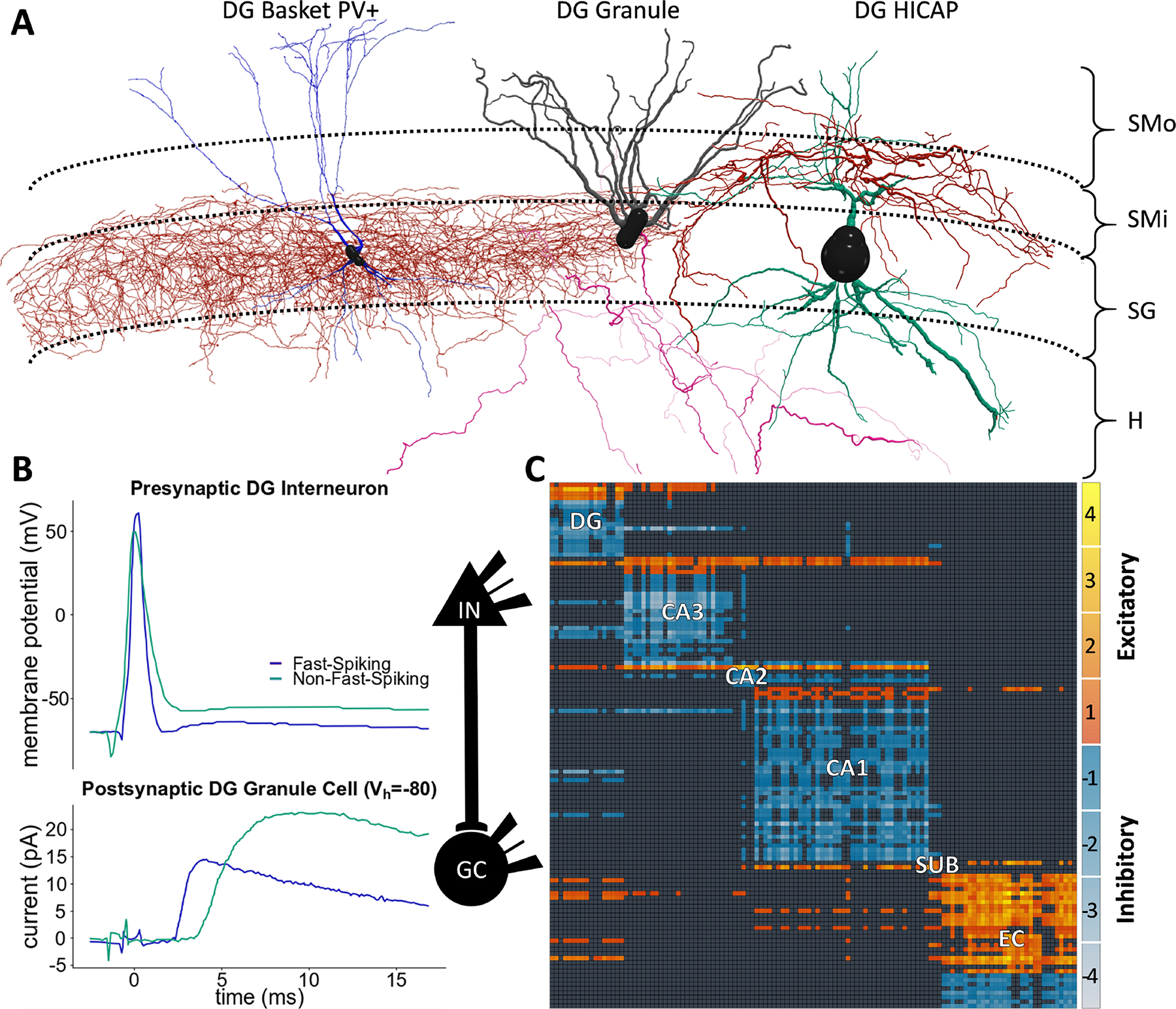
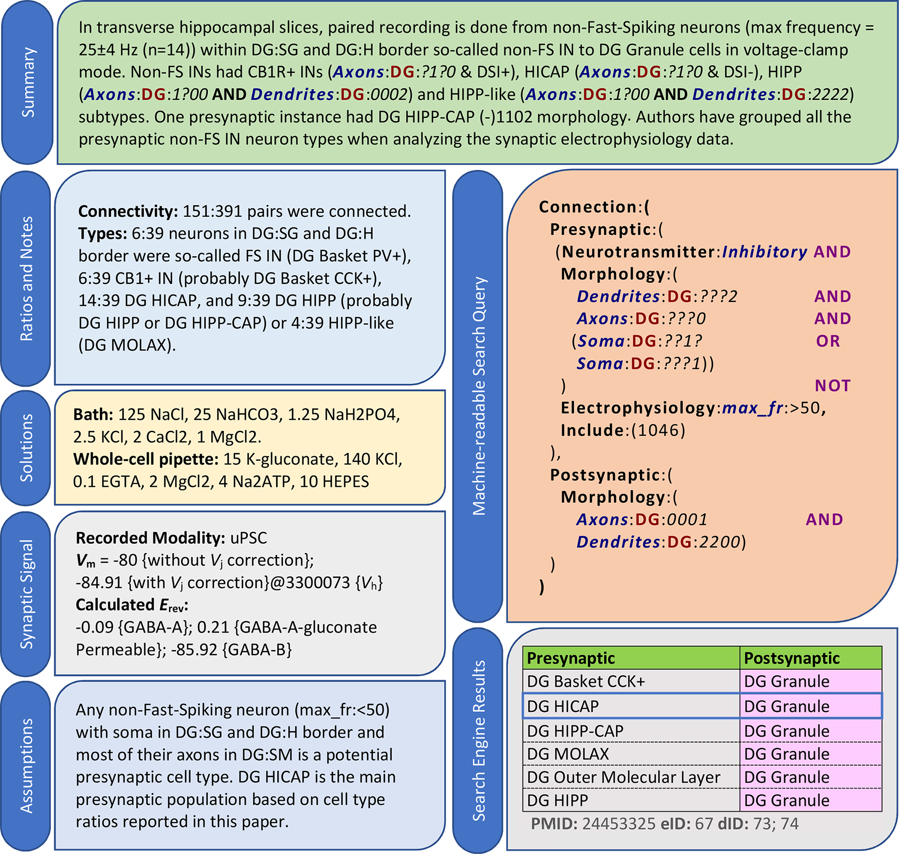
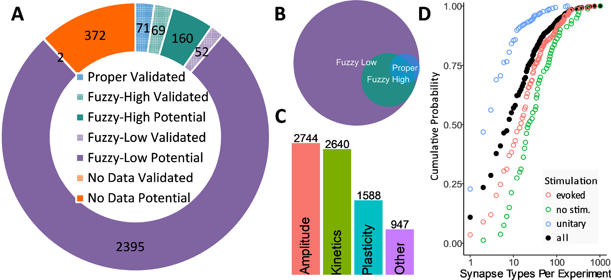
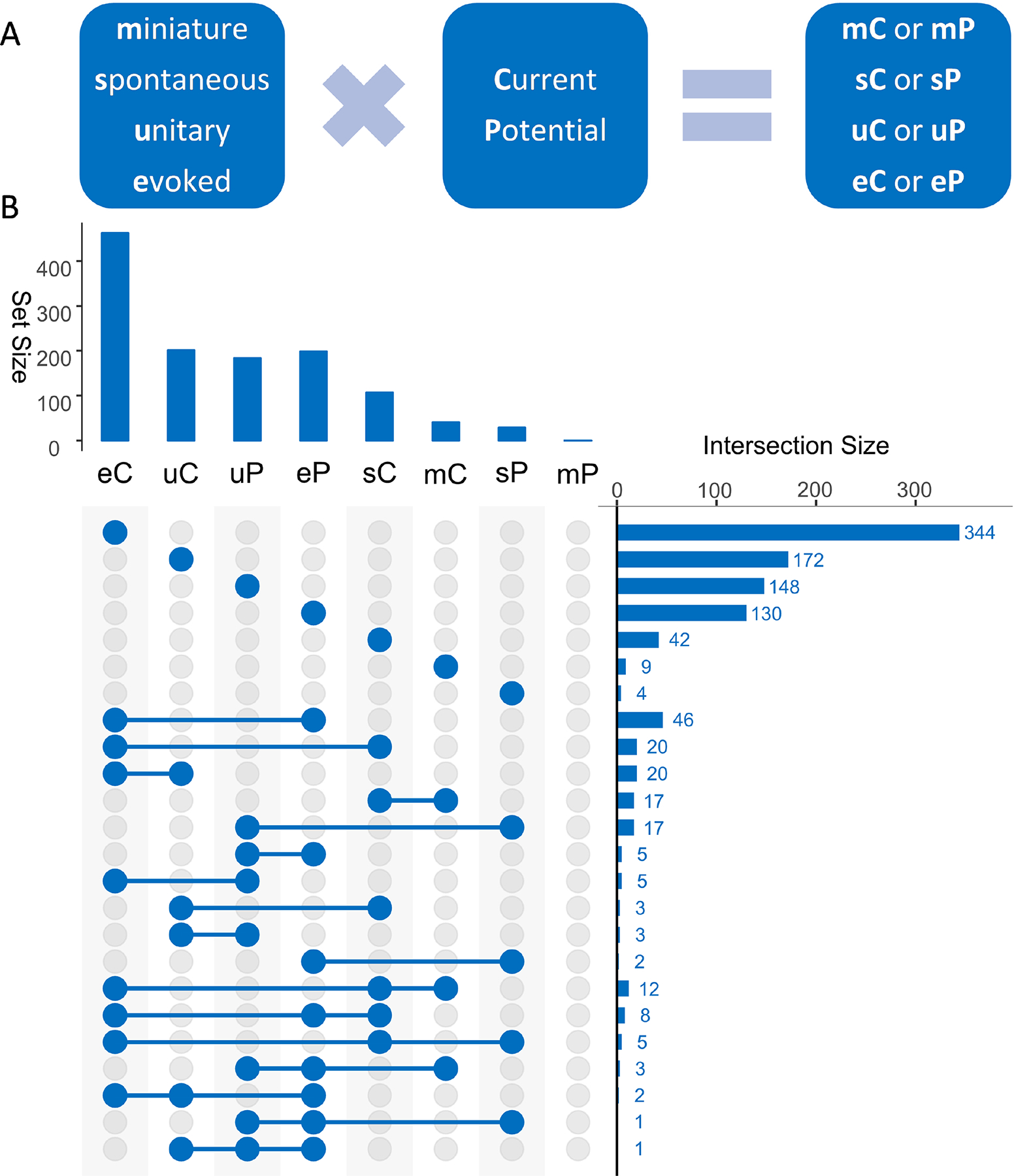
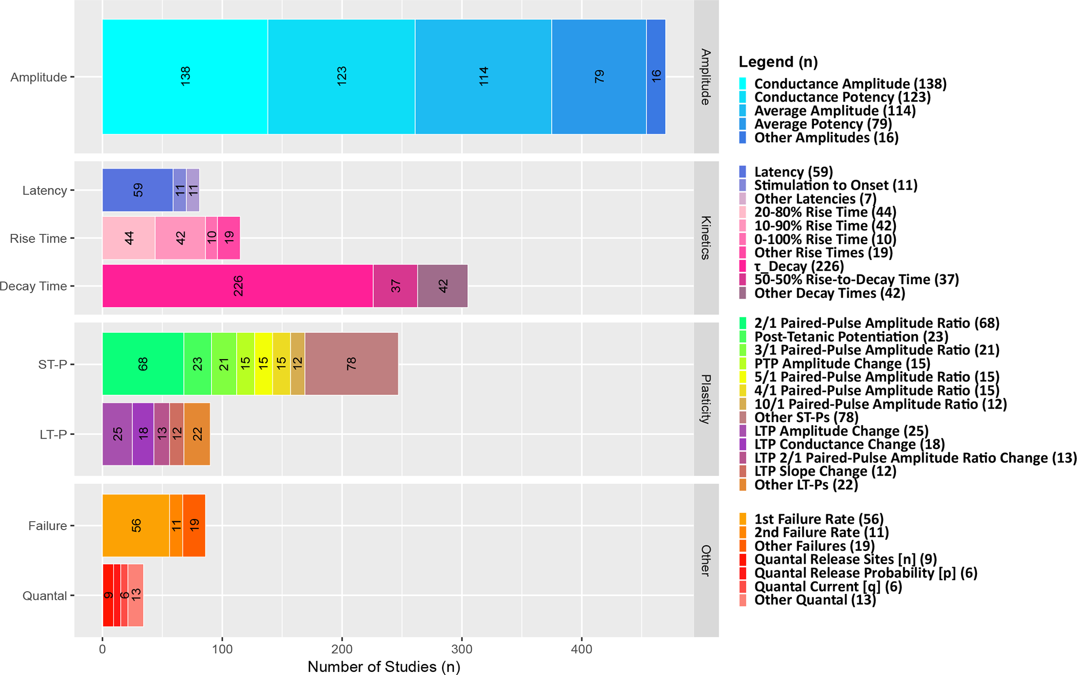
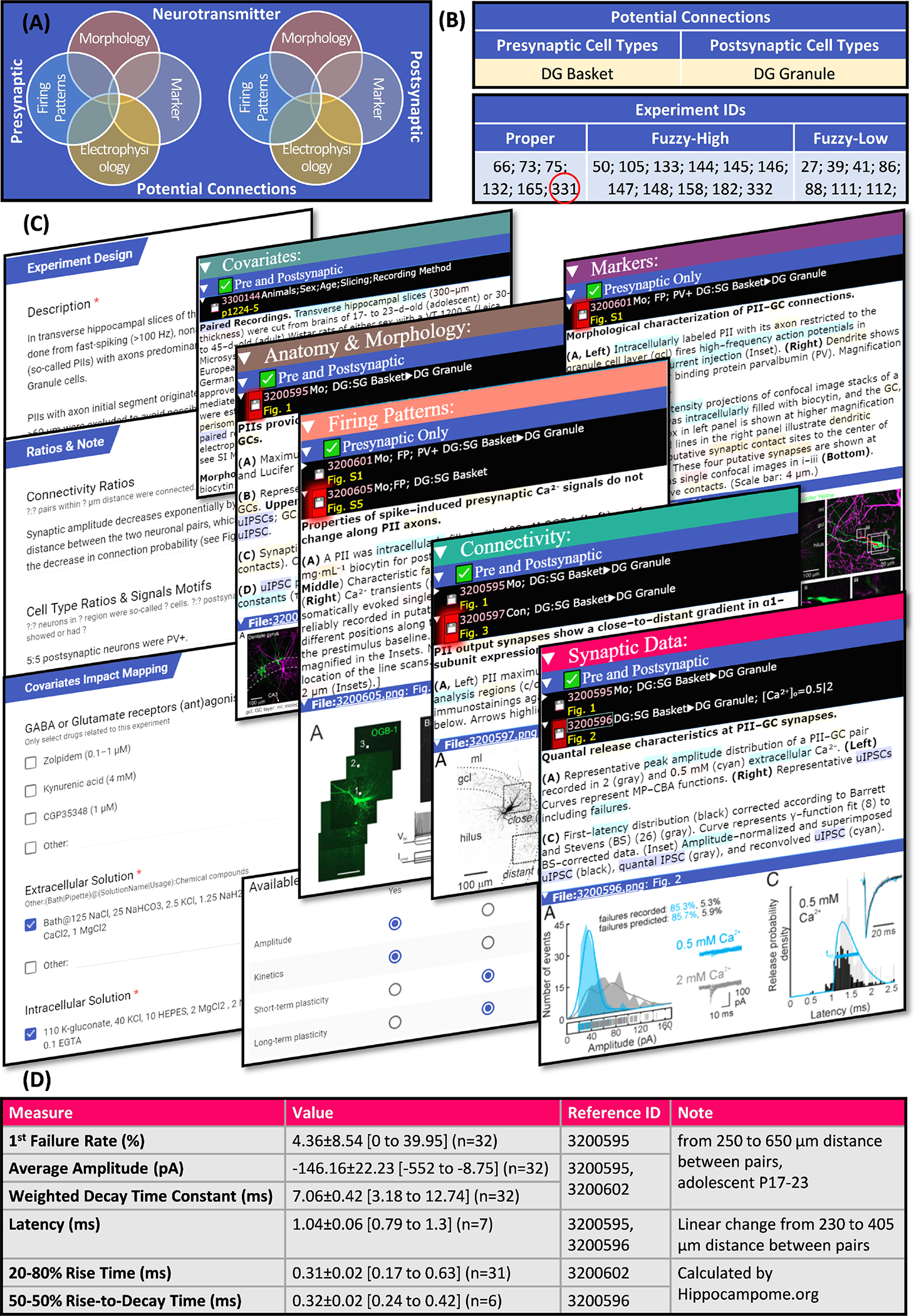
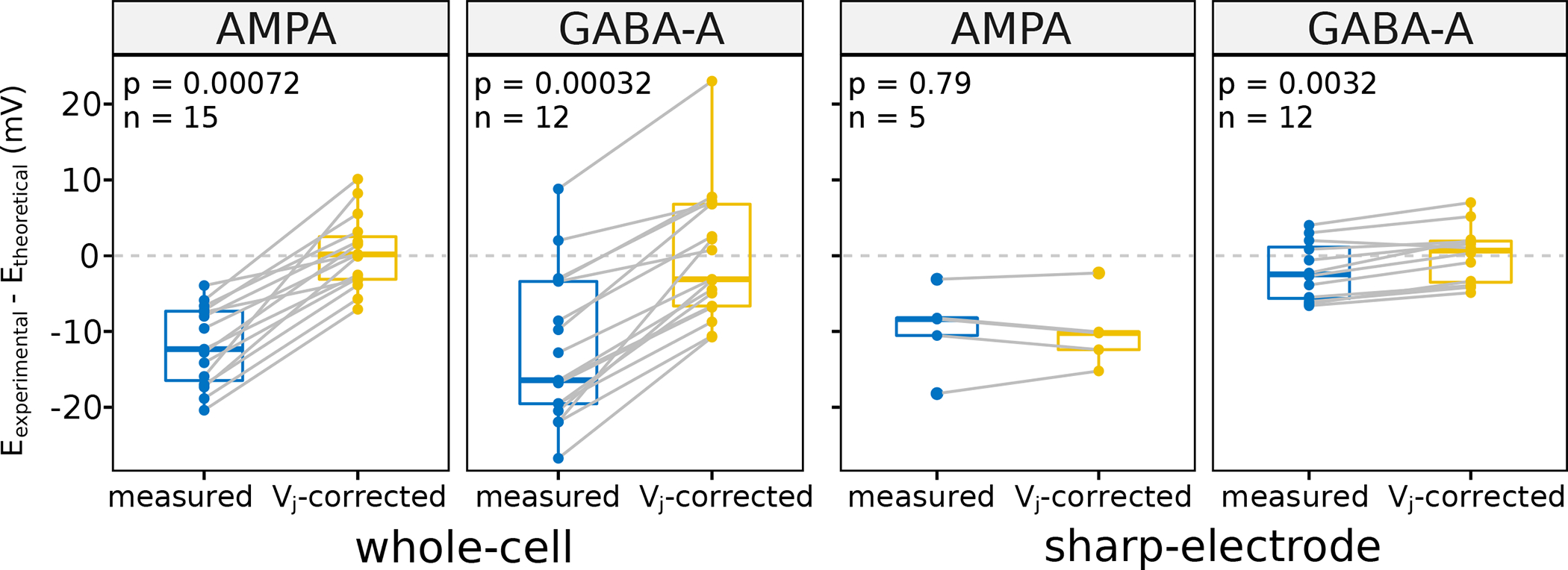
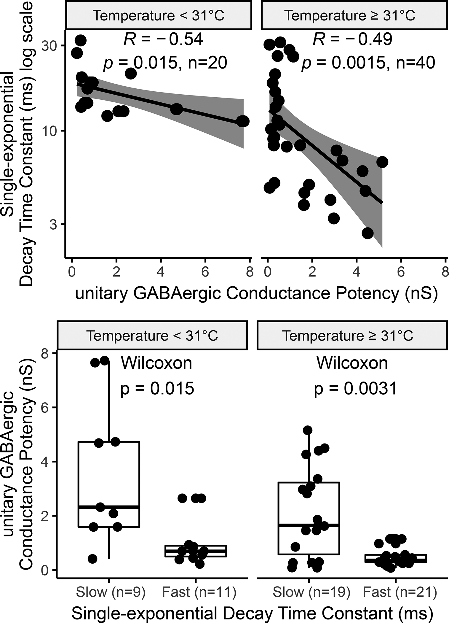
The cellular and synaptic architecture of the rodent hippocampus has been described in thousands of peer-reviewed publications. However, no human- or machine-readable public catalog of synaptic electrophysiology data exists for this or any other neural system. Harnessing state-of-the-art information technology, we have developed a cloud-based toolset for identifying empirical evidence from the scientific literature pertaining to synaptic electrophysiology, for extracting the experimental data of interest, and for linking each entry to relevant text or figure excerpts. Mining more than 1,200 published journal articles, we have identified eight different signal modalities quantified by 90 different methods to measure synaptic amplitude, kinetics, and plasticity in hippocampal neurons. We have designed a data structure that both reflects the differences and maintains the existing relations among experimental modalities. Moreover, we mapped every annotated experiment to identified potential connections, that is, specific pairs of presynaptic and postsynaptic neuron types. To this aim, we leveraged Hippocampome.org, an open-access knowledge base of morphologically, electrophysiologically, and molecularly characterized neuron types in the rodent hippocampal formation. Specifically, we have implemented a computational pipeline to systematically translate neuron type properties into formal queries in order to find all compatible potential connections. With this system, we have collected nearly 40,000 synaptic data entities covering 88% of the 3,120 potential connections in Hippocampome.org. Correcting membrane potentials with respect to liquid junction potentials significantly reduced the difference between theoretical and experimental reversal potentials, thereby enabling the accurate conversion of all synaptic amplitudes to conductance. This data set allows for large-scale hypothesis testing of the general rules governing synaptic signals. To illustrate these applications, we confirmed several expected correlations between synaptic measurements and their covariates while suggesting previously unreported ones. We release all data open-source at Hippocampome.org in order to further research across disciplines.
Keywords: circuit biophysics; computational biology; information storage and retrieval; knowledge bases; models; neuron types; synapses/physiology.
© 2019 Wiley Periodicals, Inc.
Figures
Similar articles
-
Hippocampome.org 2.0 is a knowledge base enabling data-driven spiking neural network simulations of rodent hippocampal circuits.Elife. 2024 Feb 12;12:RP90597. doi: 10.7554/eLife.90597. Elife. 2024. PMID: 38345923 Free PMC article.
-
Hippocampome.org: a knowledge base of neuron types in the rodent hippocampus.Elife. 2015 Sep 24;4:e09960. doi: 10.7554/eLife.09960. Elife. 2015. PMID: 26402459 Free PMC article.
-
Comprehensive Estimates of Potential Synaptic Connections in Local Circuits of the Rodent Hippocampal Formation by Axonal-Dendritic Overlap.J Neurosci. 2021 Feb 24;41(8):1665-1683. doi: 10.1523/JNEUROSCI.1193-20.2020. Epub 2020 Dec 23. J Neurosci. 2021. PMID: 33361464 Free PMC article.
-
Analysis of neural cell functions in gene knockout mice: electrophysiological investigation of synaptic plasticity in acute hippocampal slices.Methods Enzymol. 2006;417:52-66. doi: 10.1016/S0076-6879(06)17005-6. Methods Enzymol. 2006. PMID: 17132497 Review.
-
LTD, LTP, and the sliding threshold for long-term synaptic plasticity.Hippocampus. 1996;6(1):35-42. doi: 10.1002/(SICI)1098-1063(1996)6:1<35::AID-HIPO7>3.0.CO;2-6. Hippocampus. 1996. PMID: 8878740 Review.
Cited by
-
Robust Resting-State Dynamics in a Large-Scale Spiking Neural Network Model of Area CA3 in the Mouse Hippocampus.Cognit Comput. 2023 Jul;15(4):1190-1210. doi: 10.1007/s12559-021-09954-2. Epub 2022 Jan 28. Cognit Comput. 2023. PMID: 37663748 Free PMC article.
-
Formation and Retrieval of Cell Assemblies in a Biologically Realistic Spiking Neural Network Model of Area CA3 in the Mouse Hippocampus.bioRxiv [Preprint]. 2024 Mar 29:2024.03.27.586909. doi: 10.1101/2024.03.27.586909. bioRxiv. 2024. Update in: J Comput Neurosci. 2024 Nov;52(4):303-321. doi: 10.1007/s10827-024-00881-3. PMID: 38585941 Free PMC article. Updated. Preprint.
-
An update to Hippocampome.org by integrating single-cell phenotypes with circuit function in vivo.PLoS Biol. 2021 May 6;19(5):e3001213. doi: 10.1371/journal.pbio.3001213. eCollection 2021 May. PLoS Biol. 2021. PMID: 33956790 Free PMC article.
-
Hippocampome.org 2.0 is a knowledge base enabling data-driven spiking neural network simulations of rodent hippocampal circuits.Elife. 2024 Feb 12;12:RP90597. doi: 10.7554/eLife.90597. Elife. 2024. PMID: 38345923 Free PMC article.
-
A Continuous Attractor Model with Realistic Neural and Synaptic Properties Quantitatively Reproduces Grid Cell Physiology.bioRxiv [Preprint]. 2024 May 1:2024.04.29.591748. doi: 10.1101/2024.04.29.591748. bioRxiv. 2024. Update in: Int J Mol Sci. 2024 May 31;25(11):6059. doi: 10.3390/ijms25116059. PMID: 38746202 Free PMC article. Updated. Preprint.
References
-
- Ascoli GA, Donohue DE, & Halavi M (2007). NeuroMorpho.Org: a central resource for neuronal morphologies. J Neurosci, 27(35), 9247–9251. doi:10.1523/JNEUROSCI.2055-07.2007 - DOI - PMC - PubMed
-
- Barry PH (1994). JPCalc, a software package for calculating liquid junction potential corrections in patch-clamp, intracellular, epithelial and bilayer measurements and for correcting junction potential measurements. J Neurosci Methods, 51(1), 107–116. - PubMed
-
- Barry PH, & Lynch JW (1991). Liquid junction potentials and small cell effects in patch-clamp analysis. J Membr Biol, 121(2), 101–117. - PubMed
Publication types
MeSH terms
Grants and funding
LinkOut - more resources
Full Text Sources
