

Association between ultrasound morphologic features and histopathological findings of lobular carcinoma
- PMID: 31472006
- PMCID: PMC6745349
- DOI: 10.1002/jmrs.336
Association between ultrasound morphologic features and histopathological findings of lobular carcinoma
Erratum in
-
Erratum.J Med Radiat Sci. 2019 Dec;66(4):301. doi: 10.1002/jmrs.370. J Med Radiat Sci. 2019. PMID: 31854136 Free PMC article. No abstract available.
Abstract
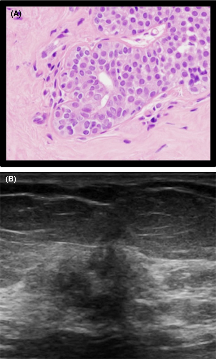
Introduction: Despite the incidence and recurrence rates of breast cancer, there are currently no biomarkers to predict which cases will develop into lobular carcinoma (LC). The purpose of this study was to determine the association between ultrasound morphologic characteristics of LC and histopathological classifications.
Methods: A retrospective, cross-sectional study was conducted on the ultrasound images and histopathological reports of 100 patients with a confirmed LC diagnosis between January 2013 and December 2016.
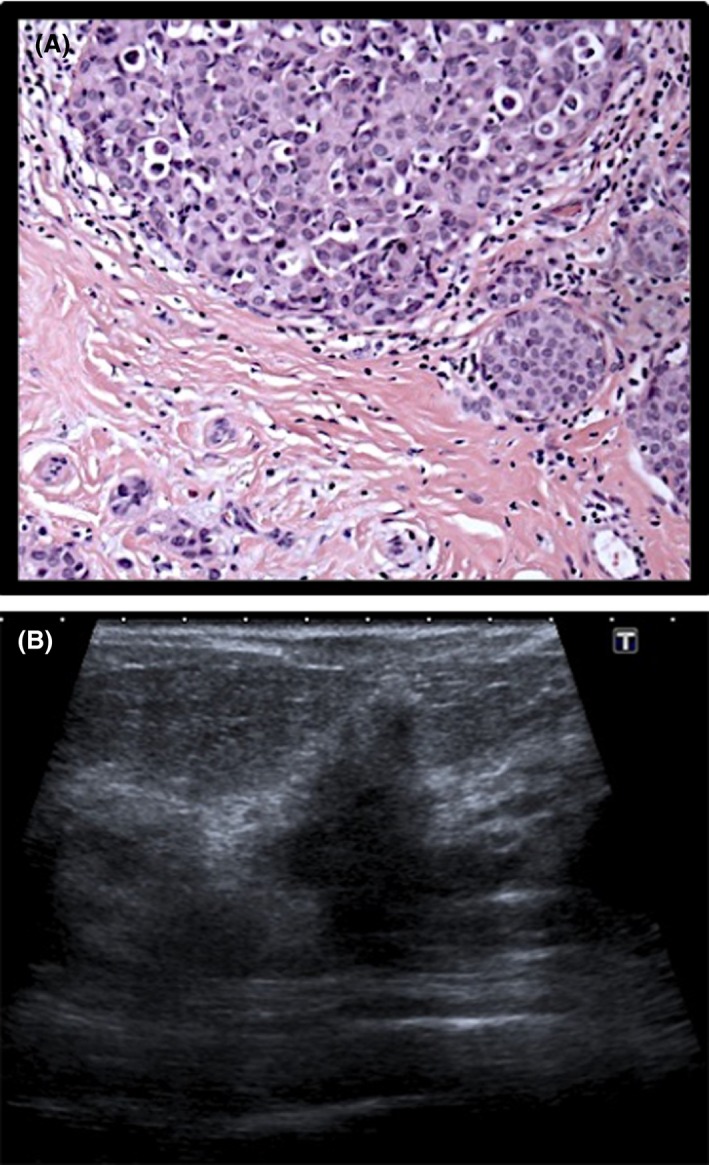
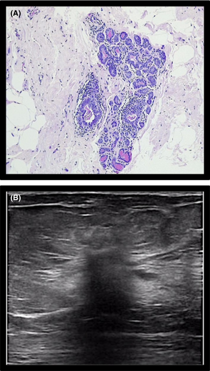
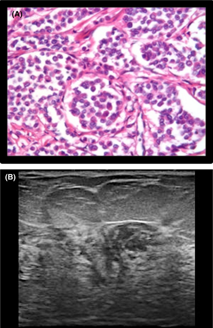
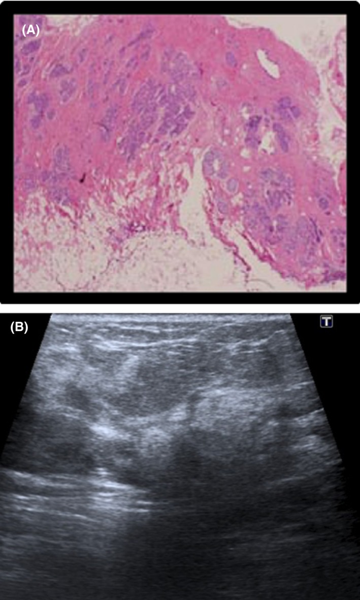
Results: Morphologic ultrasound characteristics most frequently reported in the dataset of positively diagnosed LC patients were; irregular ultrasound shape (86%), hypoechoic echogenicity (88%), poorly circumscribed margin (95%), posterior acoustic enhancement (93%) and absent calcifications (81%). Using Fisher's extract test, it was found that stromal fibrosis, single file type pattern, atypical lobular hyperplasia and LC Grade II were significantly correlated with irregular shape and hypoechoic echogenicity.
Conclusion: A prognostic predictor tool can be designed from this study's findings which can then be used in practice to raise awareness of the unique morphometric markers related to LC of the breast.
Keywords: Atypical lobular hyperplasia; histology; lobular carcinoma; morphology; predictors; ultrasound.
© 2019 University of Pretoria. Journal of Medical Radiation Sciences published by John Wiley & Sons Australia, Ltd on behalf of Australian Society of Medical Imaging and Radiation Therapy and New Zealand Institute of Medical Radiation Technology.
Conflict of interest statement
There is no financial support or relationships that may pose conflict of interest.
Figures
References
-
- Abdel‐Gawad EA, Khalil OA, Ragaee SM. Assessment of breast lesions using BI‐RADS US lexicon in mammographically dense breasts (ACR categories 3 and 4) with histopathological correlation. Egypt. J. Radiol. Nucl. Med. 2014; 45: 1301–7.
-
- Berg WA. Diagnostic accuracy of mammography, clinical examination, US, and MR imaging in preoperative assessment of breast cancer. Radiology 2004; 233: 830–49. - PubMed
-
- Yates LR, Jones A, Mackay A, et al. Genetic analysis of lobular carcinoma in situ and associated invasive lobular carcinoma. Breast Cancer Res 2010; 12.
-
- Krecke KN, Gisvold JJ. Invasive lobular carcinoma of the breast: Mammographic findings and extent of disease at diagnosis in 184 patients. Am J Roentgenol 1993; 161: 957–60. - PubMed
MeSH terms
LinkOut - more resources
Full Text Sources
Medical
