

Effect of channel separation and interaural mismatch on fusion and lateralization in normal-hearing and cochlear-implant listeners
- PMID: 31472555
- PMCID: PMC6713556
- DOI: 10.1121/1.5123464
Effect of channel separation and interaural mismatch on fusion and lateralization in normal-hearing and cochlear-implant listeners
Abstract
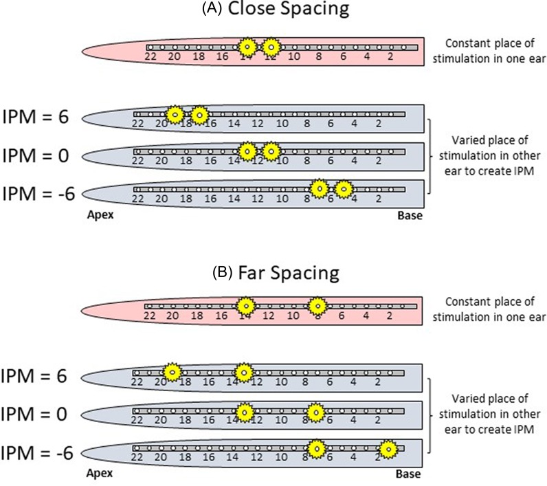
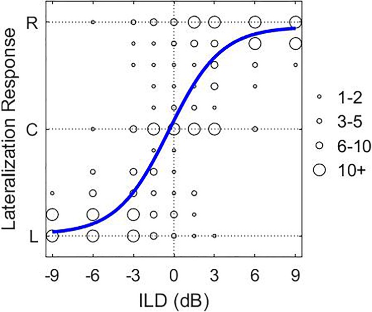
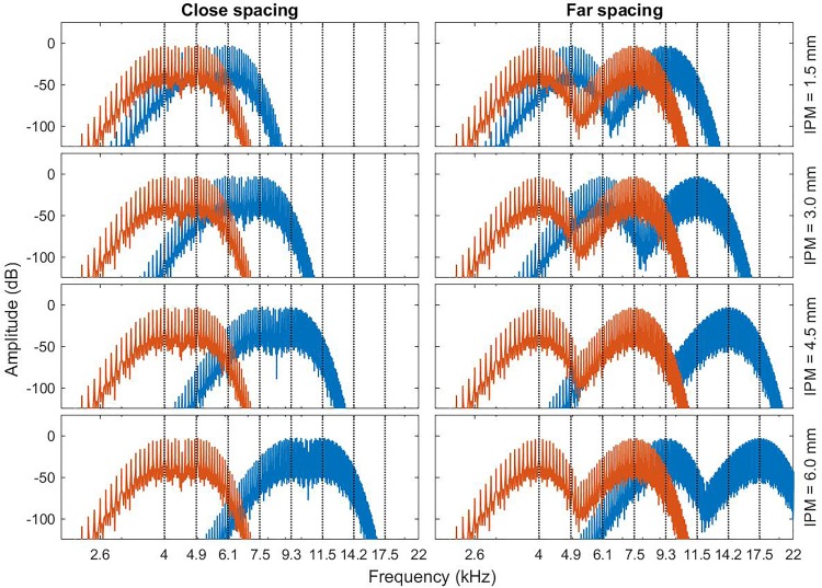
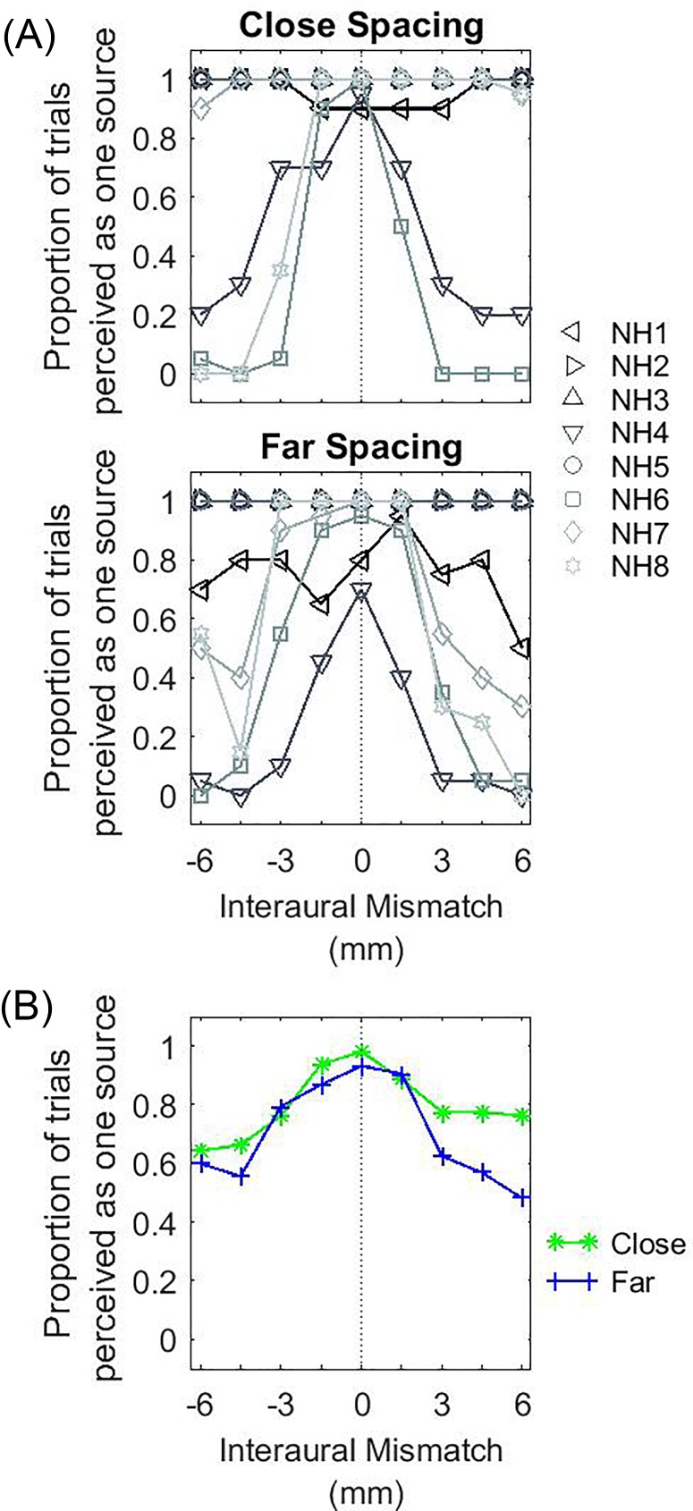
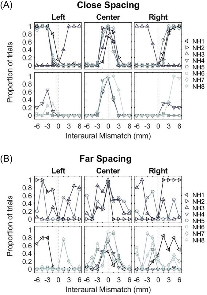
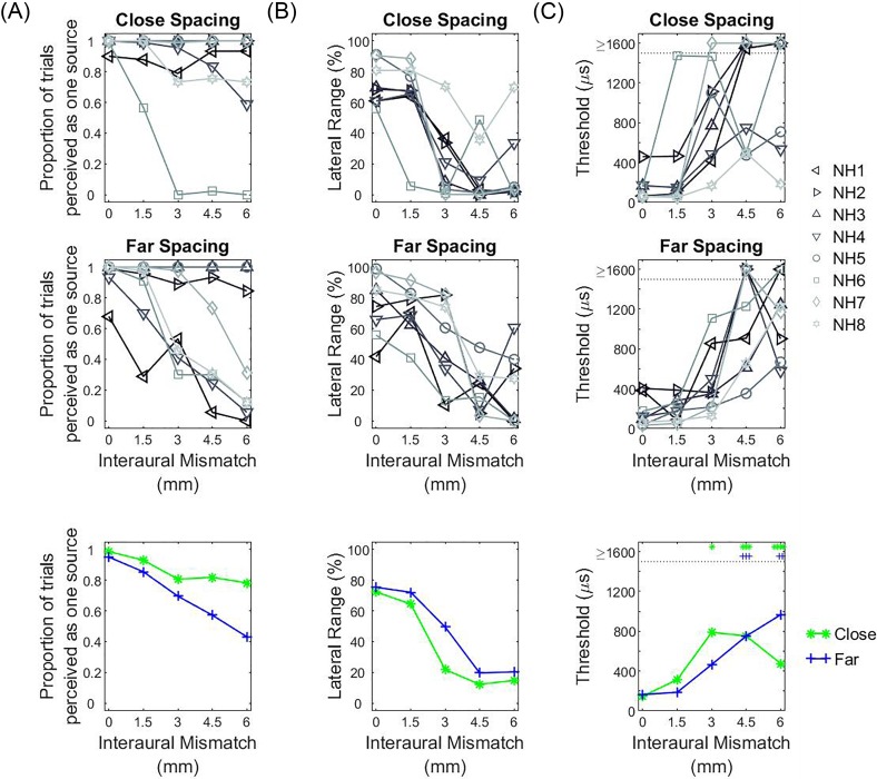
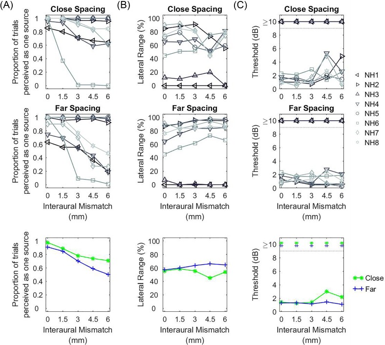
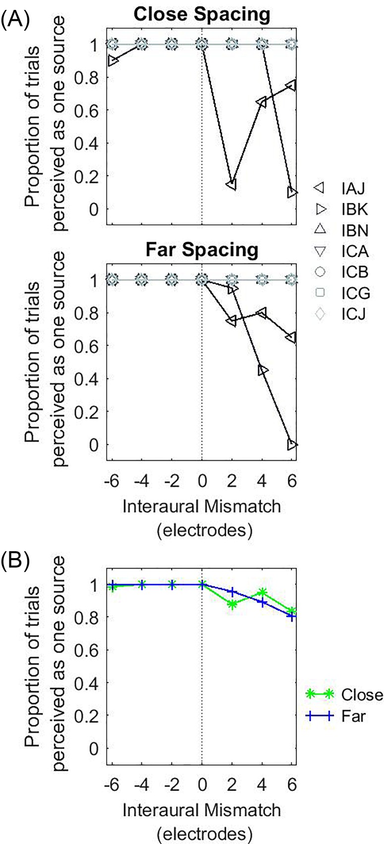
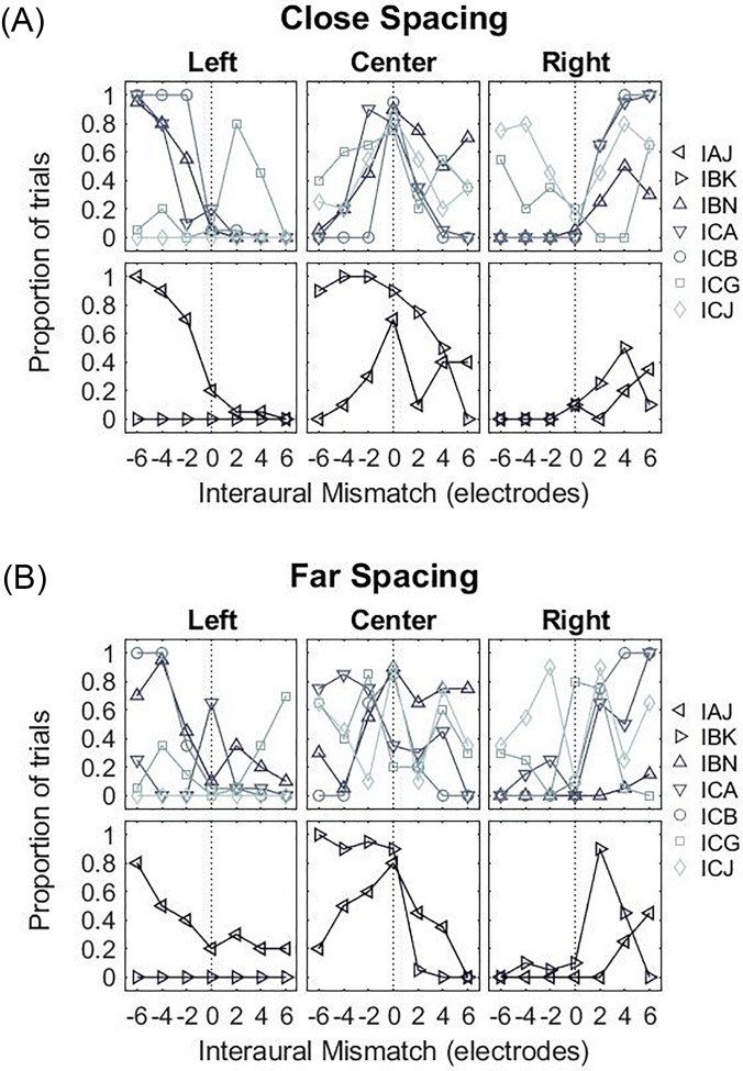
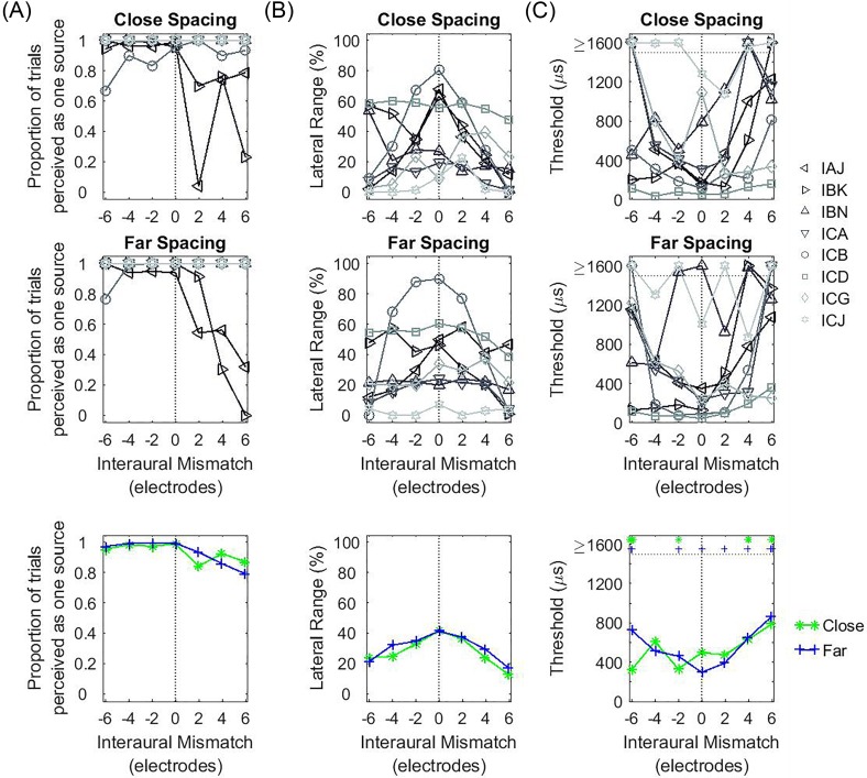
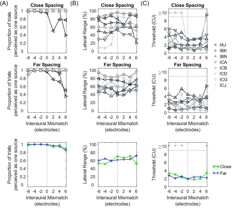
Bilateral cochlear implantation has provided access to some of the benefits of binaural hearing enjoyed by normal-hearing (NH) listeners. However, a gap in performance still exists between the two populations. Single-channel stimulation studies have shown that interaural place-of-stimulation mismatch (IPM) due to differences in implantation depth leads to decreased binaural fusion and lateralization of interaural time and level differences (ITDs and ILDs, respectively). While single-channel studies are informative, multi-channel stimulation is needed for good speech understanding with cochlear implants (CIs). Some multi-channel studies have shown that channel interaction due to current spread can affect ITD sensitivity. In this work, we studied the effect of IPM and channel spacing, along with their potential interaction, on binaural fusion and ITD/ILD lateralization. Experiments were conducted in adult NH listeners and CI listeners with a history of acoustic hearing. Results showed that IPM reduced the range of lateralization for ITDs but not ILDs. CI listeners were more likely to report a fused percept in the presence of IPM with multi-channel stimulation than NH listeners. However, no effect of channel spacing was found. These results suggest that IPM should be accounted for in clinical mapping practices in order to maximize bilateral CI benefits.
Figures
Similar articles
-
Sensitivity to interaural level and envelope time differences of two bilateral cochlear implant listeners using clinical sound processors.Ear Hear. 2004 Oct;25(5):488-500. doi: 10.1097/01.aud.0000145124.85517.e8. Ear Hear. 2004. PMID: 15599195
-
Effects of rate and age in processing interaural time and level differences in normal-hearing and bilateral cochlear-implant listeners.J Acoust Soc Am. 2019 Nov;146(5):3232. doi: 10.1121/1.5130384. J Acoust Soc Am. 2019. PMID: 31795662 Free PMC article.
-
Effects of interaural pitch matching and auditory image centering on binaural sensitivity in cochlear implant users.Ear Hear. 2015 May-Jun;36(3):e62-8. doi: 10.1097/AUD.0000000000000135. Ear Hear. 2015. PMID: 25565660 Free PMC article.
-
Perception and coding of interaural time differences with bilateral cochlear implants.Hear Res. 2015 Apr;322:138-50. doi: 10.1016/j.heares.2014.10.004. Epub 2014 Oct 19. Hear Res. 2015. PMID: 25456088 Review.
-
[Interaural stimulation timing mismatch in listeners provided with a cochlear implant and a hearing aid : A review focusing on quantification and compensation].HNO. 2023 Aug;71(8):513-520. doi: 10.1007/s00106-023-01308-8. Epub 2023 May 23. HNO. 2023. PMID: 37219567 Review. German.
Cited by
-
Binaural fusion: Complexities in definition and measurement.J Acoust Soc Am. 2024 Oct 1;156(4):2395-2408. doi: 10.1121/10.0030476. J Acoust Soc Am. 2024. PMID: 39392352 Free PMC article. Review.
-
Interaural asymmetry of dynamic range: Abnormal fusion, bilateral interference, and shifts in attention.Front Neurosci. 2023 Jan 9;16:1018190. doi: 10.3389/fnins.2022.1018190. eCollection 2022. Front Neurosci. 2023. PMID: 36699517 Free PMC article.
-
The Temporal Limits Encoder as a Sound Coding Strategy for Bilateral Cochlear Implants.IEEE/ACM Trans Audio Speech Lang Process. 2021;29:265-273. doi: 10.1109/taslp.2020.3039601. Epub 2020 Nov 20. IEEE/ACM Trans Audio Speech Lang Process. 2021. PMID: 33409339 Free PMC article.
-
Electro-Haptic Stimulation: A New Approach for Improving Cochlear-Implant Listening.Front Neurosci. 2021 Jun 9;15:581414. doi: 10.3389/fnins.2021.581414. eCollection 2021. Front Neurosci. 2021. PMID: 34177440 Free PMC article. Review.
-
The Relationship Between Interaural Insertion-Depth Differences, Scalar Location, and Interaural Time-Difference Processing in Adult Bilateral Cochlear-Implant Listeners.Trends Hear. 2022 Jan-Dec;26:23312165221129165. doi: 10.1177/23312165221129165. Trends Hear. 2022. PMID: 36379607 Free PMC article.
References
-
- Blanks, D. A. , Roberts, J. M. , Buss, E. , Hall, J. W. , and Fitzpatrick, D. C. (2007). “ Neural and behavioral sensitivity to interaural time differences using amplitude modulated tones with mismatched carrier frequencies,” J. Assoc. Res. Otolaryngol. 8, 393–408.10.1007/s10162-007-0088-5 - DOI - PMC - PubMed
-
- Carlyon, R. P. , Macherey, O. , Frijns, J. H. M. , Axon, P. R. , Kalkman, R. K. , Boyle, P. , Baguley, D. M. , Briggs, J. , Deeks, J. M. , Briaire, J. J. , Barreau, X. , and Dauman, R. (2010). “ Pitch comparisons between electrical stimulation of a cochlear implant and acoustic stimuli presented to a normal-hearing contralateral ear,” J. Assoc. Res. Otolaryngol. 11, 625–640.10.1007/s10162-010-0222-7 - DOI - PMC - PubMed
Publication types
MeSH terms
Grants and funding
LinkOut - more resources
Full Text Sources
Medical
Miscellaneous