. 2019 Oct;96(4):883-889.
doi: 10.1016/j.kint.2019.06.016. Epub 2019 Jul 10.

Mutations in KIRREL1, a slit diaphragm component, cause steroid-resistant nephrotic syndrome

Affiliations

Mutations in KIRREL1, a slit diaphragm component, cause steroid-resistant nephrotic syndrome

Ashish K Solanki et al. Kidney Int. 2019 Oct.

Abstract

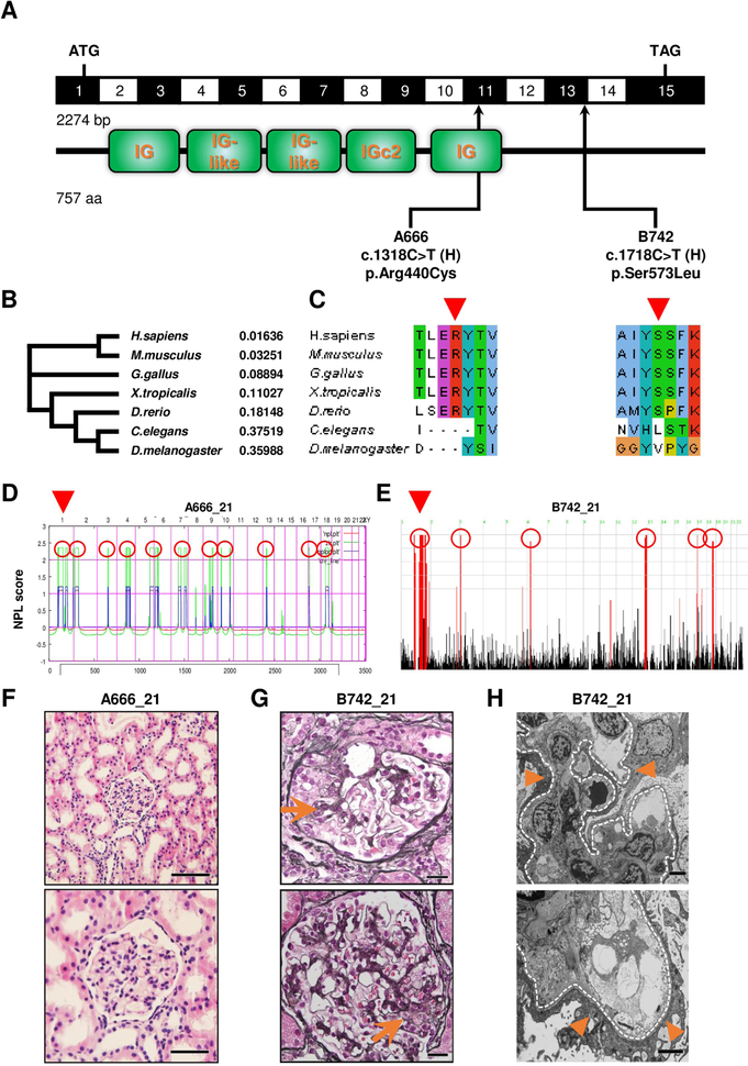
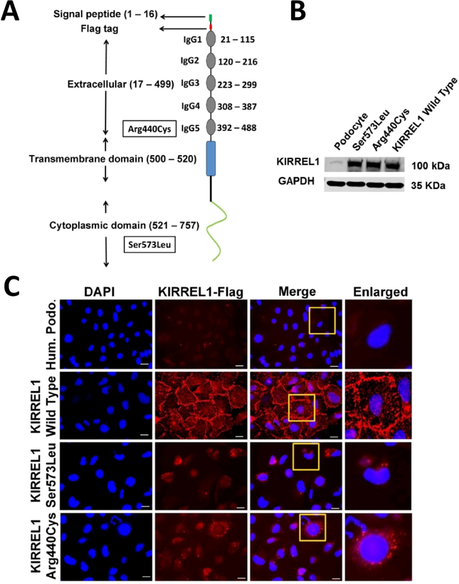
Steroid-resistant nephrotic syndrome is a frequent cause of chronic kidney disease almost inevitably progressing to end-stage renal disease. More than 58 monogenic causes of SRNS have been discovered and majority of known steroid-resistant nephrotic syndrome causing genes are predominantly expressed in glomerular podocytes, placing them at the center of disease pathogenesis. Herein, we describe two unrelated families with steroid-resistant nephrotic syndrome with homozygous mutations in the KIRREL1 gene. One mutation showed high frequency in the European population (minor allele frequency 0.0011) and this patient achieved complete remission following treatment, but later progressed to chronic kidney disease. We found that mutant KIRREL1 proteins failed to localize to the podocyte cell membrane, indicating defective trafficking and impaired podocytes function. Thus, the KIRREL1 gene product has an important role in modulating the integrity of the slit diaphragm and maintaining glomerular filtration function.

Keywords: KIRREL1; focal segmental glomerulosclerosis; minimal change disease; steroid-resistant nephrotic syndrome.


Figures

Figure 1.
Figure 1.. Homozygosity mapping and whole exome sequencing identify recessive mutations of KIRREL1 in 2 families with steroid-resistant nephrotic syndrome
(A) KIRREL1 (NM_018240.6/NP_060710.3). Exon structure of human cDNA with the corresponding protein including its functional domains obtained from SMART. Positions of start codons and of stop codon are indicated. Arrows indicate positions of pathogenic mutations detected in families with SRNS. H, homozygous. (B) Phylogram of KIRREL1 protein was generated using multiple sequence alignment in Clustal omega. Numbers are representing the percent identity graph data comparing evolutionary relationship among different organisms to human reference for KIRREL1 protein (0: 100% identical; 1: 0% identical). (C) Evolutionary conservation of amino acid residues that are altered in patients with SRNS. Altered amino acid residues of KIRREL1 (p.Arg440Cys, p.Ser573Leu). (D) Homozygosity mapping identifies regions of homozygosity with recessive candidate loci (red circles) in patient A666_21. Nonparametric LOD (NPL) scores and SNP positions (Affymetrix 250K StyI array) are plotted on human chromosomes concatenated from p-ter (left) to q-ter (right). Genetic distance is given in centimorgans (cM). Whole exome sequencing identifies a homozygous mutation of KIRREL1 (p.Arg440Cys) that is positioned within the maximum NPL peak on chromosome 1 (arrowhead). (E) Homozygosity mapping identifies regions of homozygosity with recessive candidate loci (red circles) in patient B742_21. Nonparametric LOD (log of the odds ratio) (NPL) score profile is plotted across the human genome. Within the maximum NPL peak on chromosome 1 (arrowhead), we identified a homozygous mutation in KIRREL1 (p.Ser573Leu). (F) Renal histology of individual A666_21 with KIRREL1 mutation. Presence of only minor findings of mesangial hypercellularity is consistent with minimal change disease on light microscopy (H&E staining). (Scale bars: upper panel 200 μm and lower panel 100 μm). (G) Renal histology of individual B742_21 with KIRREL1 mutation shows focal segmental glomerular sclerosis (arrows) on light microscopy (MPAS staining). (Scale bars: upper and lower panels 20 μm). (H) Renal histology of B742_21 with KIRREL1 mutation showing podocyte foot process effacement (arrowheads) on transmission electron microscopy (TEM). Glomerular basement membrane is highlighted by a dotted line. (Scale bars: upper and lower panels 2 μm).
Figure 2.
Figure 2.. KIRREL1 mutants mislocalize in human podocytes.
(A) Schematic domain distribution, position of Flag tag and mutations in the KIRREL1 protein. (B) Western blot analysis showing the expression of the wild type control and KIRREL1 mutants plasmid constructs as indicated. (C) Unpermeabilized wild type human podocytes, human podocytes stably expressing flag tagged wild type KIRREL1 and flag tagged KIRREL1 mutants p.Ser573Leu and p.Arg440Cys were stained with Flag antibody, that labeled Flag-KIRREL1 extracellularly. Their membrane localization was evaluated by immunofluorescence microscopy using a confocal microscope. KIRREL1 mutants p.Ser573Leu and p.Arg440Cys show mislocalization compared to wild type control in cultured human podocytes cell lines. (Scale bars: 25 μm).

References

    1. Smith JM, Stablein DM, Munoz R, Hebert D & McDonald RA Contributions of the Transplant Registry: The 2006 Annual Report of the North American Pediatric Renal Trials and Collaborative Studies (NAPRTCS). Pediatr Transplant 11, 366–73 (2007). - PubMed
    1. Greenbaum LA, Benndorf R & Smoyer WE Childhood nephrotic syndrome--current and future therapies. Nat Rev Nephrol 8, 445–58 (2012). - PubMed
    1. Lovric S, Ashraf S, Tan W & Hildebrandt F Genetic testing in steroid-resistant nephrotic syndrome: when and how? Nephrol Dial Transplant 31, 1802–1813 (2016). - PMC - PubMed
    1. Machuca E, Benoit G & Antignac C Genetics of nephrotic syndrome: connecting molecular genetics to podocyte physiology. Hum Mol Genet 18, R185–94 (2009). - PubMed
    1. Tryggvason K, Patrakka J & Wartiovaara J Hereditary proteinuria syndromes and mechanisms of proteinuria. N Engl J Med 354, 1387–401 (2006). - PubMed

Publication types

MeSH terms