Review
. 2019 Oct 15;51(4):609-624.
doi: 10.1016/j.immuni.2019.08.005. Epub 2019 Aug 28.

Inflammasomes: Threat-Assessment Organelles of the Innate Immune System

Affiliations
Review

Inflammasomes: Threat-Assessment Organelles of the Innate Immune System

Charles L Evavold et al. Immunity. .

Abstract

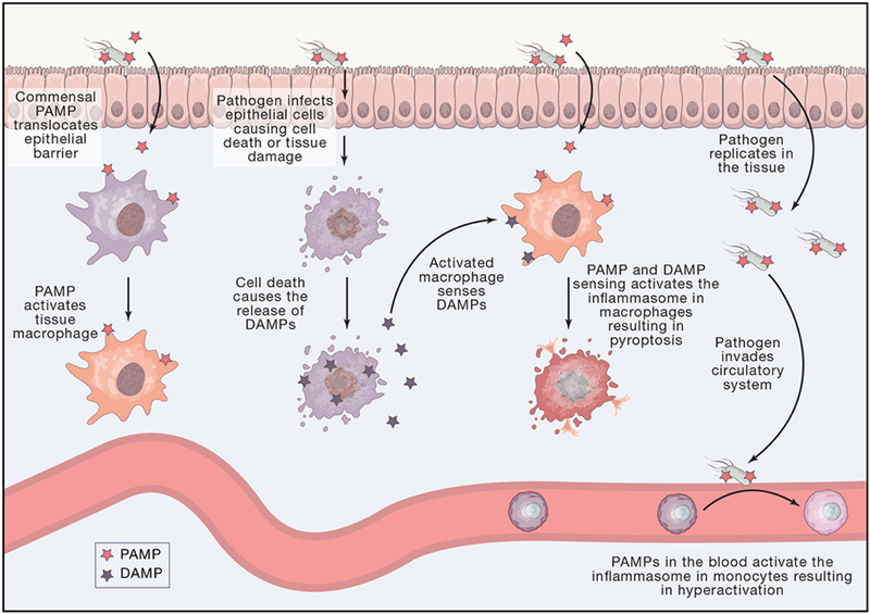
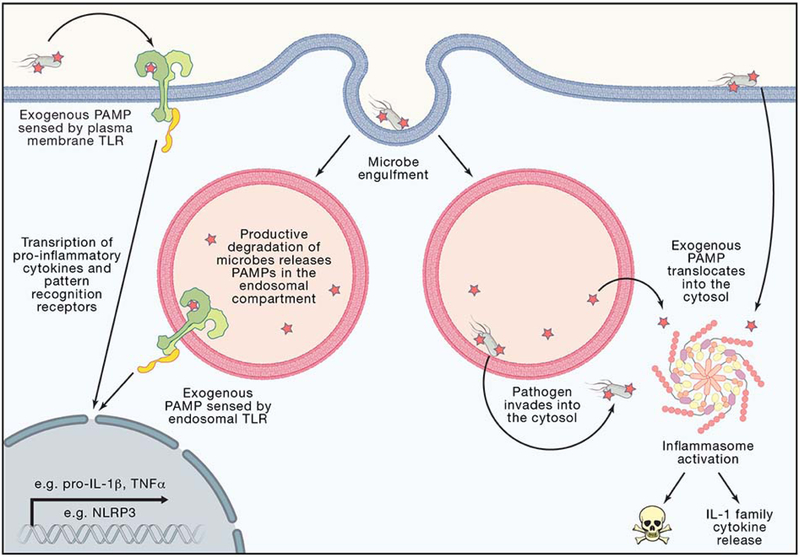
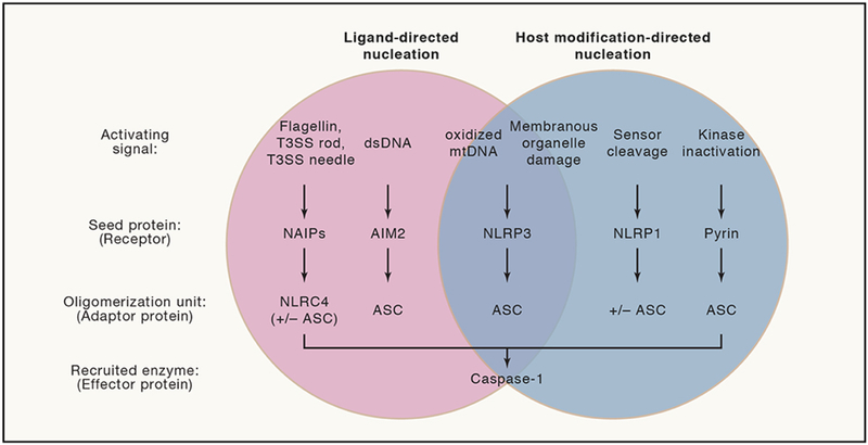
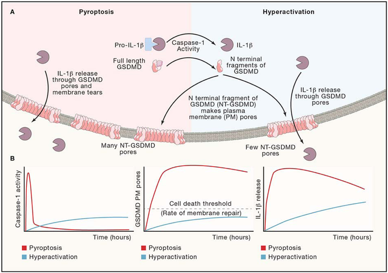
Inflammasomes are supramolecular organizing centers that operate to drive interleukin-1 (IL-1)-dependent inflammation. Depending on context, inflammatory caspases act upstream or downstream of inflammasome assembly, serving as the principal enzymes that control activities of these organelles. In this review, we discuss mechanisms of inflammasome assembly and signaling. We posit that upstream regulatory proteins, classically known as pattern-recognition receptors, operate to assess infectious and non-infectious threats to the host. Threat assessment is achieved through two general strategies: (1) direct binding of receptors to microbial or host-derived ligands or (2) indirect detection of changes in cellular homeostasis. Upon activation, these upstream regulatory factors seed the assembly of inflammasomes, leading to IL-1 family cytokine release from living (hyperactive) or dead (pyroptotic) cells. The molecular and physiological consequences of these distinct cell fate decisions are discussed.


Conflict of interest statement

Declaration of Interests

J.C.K. holds is a member of the scientific advisory board at IFM therapeutics and holds a patent on the therapeutic potential of hyperactivating stimuli (PCT/US2016/012994).

Figures

Figure 1:
Figure 1:. Tissue and Cellular Level Topology Affect Threat Level Classification.
Gut epithelial barrier and blood vessels are sites of microbial recognition with different inputs and outcomes. Tissue resident macrophages can become activated to secrete conventional pro-inflammatory cytokines by sensing PAMPs such as those from pathogens or commensal microbes. Pathogens can infect many cell types in the gut such as epithelial cells, resulting is tissue damage and cell death and the release of DAMPs. An activated tissue resident macrophage that now senses DAMPs can form an inflammasome and undergo pyroptosis. A pathogen can replicate in the tissue. The infected tissue can allow whole pathogens or PAMPs to translocate into blood vessels. PAMPs in the blood vessels represent a threat of systemic infection through the circulatory system. A monocyte that senses a PAMP in the blood can form an inflammasome to become hyperactive.
Figure 2:
Figure 2:. Cellular Level Topology Affect Threat Level Classification.
The extracellular space, endosomal lumen, and cytosol are sites of microbial recognition with different sensors and outcomes. Plasma membrane TLRs survey the extracellular space for PAMPs or DAMPs representing a lower threat level as the molecular patterns could derive from commensal bacteria or sterile injury. Endosomal TLRs survey the endosomal lumen that topologically is considered continuous with the extracellular space, representing a lower threat level as the molecular patterns could derive from productive degradation of commensals or host cell debris. TLR activation leads to transcription of pro-inflammatory cytokines, such as pro-IL-1β and TNFα and up regulation of pattern recognition receptors, such as the inflammasome receptor NLRP3. Pathogens can inject effectors and translocate PAMPs into the cytosol through the plasma membrane or endosomal membrane. Pathogens can also directly translocate from the endosomal lumen into the sterile cytosol. Exogenous PAMPs or pathogens found in the cytosol cause inflammasome activation, representing a higher threat level as the host is being invaded or intoxicated by pathogenic microbes. Inflammasome activation leads to release of IL-1β, IL-18, and pyroptotic cell death.
Figure 3:
Figure 3:. Modes of Inflammasome Nucleation Reflect Diverse Mechanisms to Contextualize Intent of Pathogens.
Inflammasomes by definition serve as platforms to activate the inflammatory caspase, caspase-1. The NAIP inflammasome recognizes bacterial PAMPs in the cytosol through oligomerization of NLRC4 to directly recruit caspase-1 or in combination with ASC. The AIM2 inflammasome recognizes mislocalized dsDNA in the cytosol that may represent PAMPs or DAMPs to seed an inflammasome using ASC to recruit caspase-1. The literature suggests that NLRP3 may directly sense ligands or may sense broad dysfunction of host processes to seed an inflammasome using ASC to recruit caspase-1. The NLRP1 inflammasome serves as a “bait” protein to indirectly sense pathogens via sensing pathogen-derived protease activity. NLRP1 may directly recruit caspase-1, but ASC serves to amplify caspase-1 activation. The pyrin inflammasome is sequestered during homeostasis. Disruption of cytoskeleton-associated RhoA kinase activity indirectly leads to pyrin release to seed an inflammasome using ASC to recruit caspase-1.
Figure 4:
Figure 4:. Cell Fate Decisions after Inflammasome Activation.
(A) Inflammasome activation can lead to the cell fates of “hyperactivation” or “pyroptosis” depending on the cell-type and stimulation. Magnitude and kinetics of caspase-1 activation may affect the rates and quantity of GSDMD cleaved, leading to different magnitude and kinetics of GSDMD pore residency on the plasma membrane. Membrane repair processes serve to remove GSDMD pores from the plasma membrane. (B) Theoretical magnitudes and kinetics of caspase-1 activity, GSDMD cleavage, and IL-1β release for pyroptotic and hyperactive cell fates. Caspase-1 curves are extrapolated based on a comparison of hyperactive neutrophils and pyroptotic macrophages treated with the same stimulus (Boucher et al., 2018). GSDMD pore curves are extrapolated based on a membrane permeability level of hyperactive macrophages treated with oxidized phospholipids or infected with ΔoatA S. aureus compared to pyroptotic macrophages treated with nigericin or FlaTox (a flagellin, anthrax lethal toxin fusion protein) (Evavold et al., 2018). IL-1β release curves are extrapolated based on comparison of hyperactive macrophages treated with oxidized phospholipids and pyroptotic macrophages treated with ATP (Zanoni et al., 2017).

References

    1. Arbore G, West EE, Spolski R, Robertson AAB, Klos A, Rheinheimer C, Dutow P, Woodruff TM, Yu ZX, O’Neill LA, et al. (2016). T helper 1 immunity requires complement-driven NLRP3 inflammasome activity in CD4⁺ T cells. Science 352, aad1210. - PMC - PubMed
    1. Ayala JM, Yamin TT, Egger LA, Chin J, Kostura MJ, and Miller DK (1994). IL-1 beta-converting enzyme is present in monocytic cells as an inactive 45-kDa precursor. The Journal of Immunology 153, 2592–2599. - PubMed
    1. Bauernfeind FG, Horvath G, Stutz A, Alnemri ES, MacDonald K, Speert D, Fernandes-Alnemri T, Wu J, Monks BG, Fitzgerald KA, et al. (2009). Cutting edge: NF-kappaB activating pattern recognition and cytokine receptors license NLRP3 inflammasome activation by regulating NLRP3 expression. J. Immunol 183, 787–791. - PMC - PubMed
    1. Baum R, Sharma S, Carpenter S, Li Q-Z, Busto P, Fitzgerald KA, Marshak-Rothstein A, and Gravallese EM (2015). Cutting edge: AIM2 and endosomal TLRs differentially regulate arthritis and autoantibody production in DNase II-deficient mice. J. Immunol 194, 873–877. - PMC - PubMed
    1. Blander JM, and Sander LE (2012). Beyond pattern recognition: five immune checkpoints for scaling the microbial threat. Nature Publishing Group 12, 215–225. - PubMed

Publication types