. 2020 Jun;11(3):418-432.
doi: 10.1007/s12975-019-00729-4. Epub 2019 Aug 31.

Ischemic Neuroprotectant PKCε Restores Mitochondrial Glutamate Oxaloacetate Transaminase in the Neuronal NADH Shuttle after Ischemic Injury

Affiliations

Ischemic Neuroprotectant PKCε Restores Mitochondrial Glutamate Oxaloacetate Transaminase in the Neuronal NADH Shuttle after Ischemic Injury

Jing Xu et al. Transl Stroke Res. 2020 Jun.

Abstract

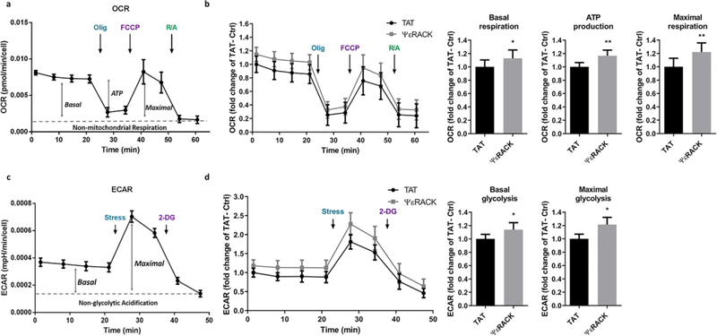
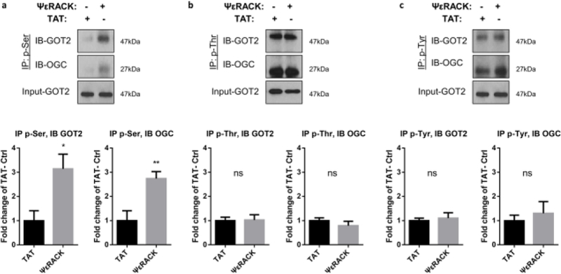
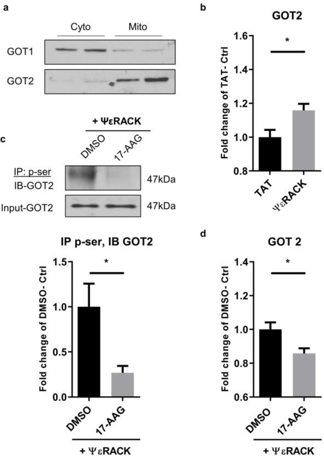
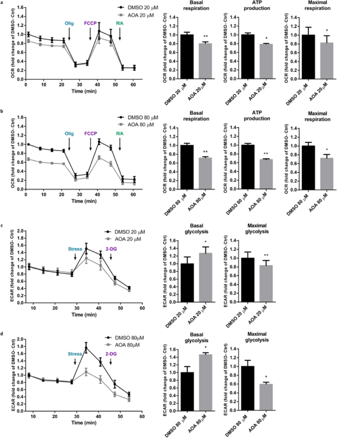
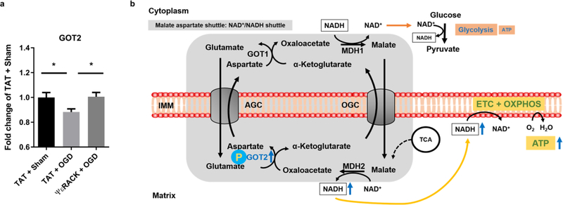
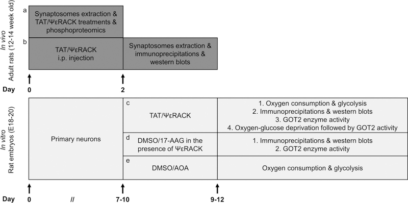
The preservation of mitochondrial function is a major protective strategy for cerebral ischemic injuries. Previously, our laboratory demonstrated that protein kinase C epsilon (PKCε) promotes the synthesis of mitochondrial nicotinamide adenine dinucleotide (NAD+). NAD+ along with its reducing equivalent, NADH, is an essential co-factor needed for energy production from glycolysis and oxidative phosphorylation. Yet, NAD+/NADH are impermeable to the inner mitochondrial membrane and their import into the mitochondria requires the activity of specific shuttles. The most important neuronal NAD+/NADH shuttle is the malate-aspartate shuttle (MAS). The MAS has been implicated in synaptic function and is potentially dysregulated during cerebral ischemia. The aim of this study was to determine if metabolic changes induced by PKCε preconditioning involved regulation of the MAS. Using primary neuronal cultures, we observed that the activation of PKCε enhanced mitochondrial respiration and glycolysis in vitro. Conversely, inhibition of the MAS resulted in decreased oxidative phosphorylation and glycolytic capacity. We further demonstrated that activation of PKCε increased the phosphorylation of key components of the MAS in rat brain synaptosomal fractions. Additionally, PKCε increased the enzyme activity of glutamic oxaloacetic transaminase 2 (GOT2), an effect that was dependent on the import of PKCε into the mitochondria and phosphorylation of GOT2. Furthermore, PKCε activation was able to rescue decreased GOT2 activity induced by ischemia. These findings reveal novel protective targets and mechanisms against ischemic injury, which involves PKCε-mediated phosphorylation and activation of GOT2 in the MAS.

Keywords: Cerebral ischemia; Conditioning; Ischemia tolerance; Ischemic preconditioning; Malate aspartate shuttle; Mitochondria.


Conflict of interest statement

Conflict of Interest

The authors declare that they have no conflict of interest.

The authors declare no potential conflicts of interest.

Figures

Fig. 1
Fig. 1. Activation of PKCε increases the respiratory and glycolytic rate in primary neurons.
(a) Representative profile of the oxygen consumption rate (OCR) showing the sequential injection of metabolic inhibitors: complex V inhibition by oligomycin (Olig), mitochondrial uncoupling by FCCP, complex I and complex III inhibition by rotenone and antimycin (R/A); arrow indicates application. Basal: Basal respiration rate; ATP: oxygen consumption rate linked to ATP production; Maximal: maximal uncoupled respiration rate. (b) Primary neurons were exposed to control -TAT or specific PKCε activator-ΨεRACK (500 nM) for 1 hour. OCR was measured 48 hours later using the Seahorse Analyzer. Values were normalized to cell counts and represented as fold change of TAT control (n=4, mean ± SEM, *P<0.05, Two-tailed paired Student’s t-test). (c) Representative profile of the extracellular acidification rate (ECAR) showing the sequential injection of metabolic inhibitors. Energetic stress cocktail: complex V inhibition by oligomycin and mitochondrial uncoupling by FCCP; glycolysis inhibition by 2-Deoxyglucose (2-DG); arrow indicates application. Basal: Basal glycolysis rate; Maximal: maximal glycolytic rate. (d) Primary neurons were treated with TAT or ΨεRACK (500 nM) for 1 hour. ECAR was measured 48 hours later using the Seahorse Analyzer. Values were normalized to cell counts and represented as fold change of TAT control (n=4, mean ± SEM, * P <0.05, Two-tailed paired Student’s t-test).
Fig. 2
Fig. 2. Activation of PKCε increases serine phosphorylation of the MAS components GOT2 and OGC in synaptosomes.
Synaptosomes from rat cortices were isolated 48 hours after TAT or ΨεRACK (0.2 mg/kg i.p.) treatment. Proteins were immunoprecipitated with (a) phospho-serine, (b) phospho-threonine, (c) phospho-tyrosine, and immunoblotted for GOT2 and OGC. Input-GOT2 acts as a loading control. Quantification fold change of TAT control and representative blot as shown above (n=4–5, mean ± SEM, * P <0.05, ** P <0.01, ns = non-significant, Two-tailed unpaired Student’s t-test). GOT2: Glutamic oxaloacetic transaminase 2; OGC: Oxoglutarate carrier.
Fig. 3
Fig. 3. Effects of PKCε activation on the phosphorylation levels of GOT2 in primary neurons.
(a-c) Neurons were exposed to TAT or ΨεRACK (500 nM) for 1 hour. Forty-eight hours after treatment, protein from cells was immunoprecipitated with (a) phospho-serine, (b) phospho-threonine, (c) phospho-tyrosine, and immunoblotted for GOT2. Input-GOT2 represents as a loading control. Quantification fold change of TAT control and representative blot as shown above. (d-f) PKCε activation does not alter the expression of MAS components in neurons. Real-time qPCR was performed (d) 24 hours and (e) 48 hours following the 1-hour treatment of TAT or ΨεRACK (500 nM) in primary neurons. mRNA levels were normalized to GAPDH and shown as fold change of TAT control. (f) Western blots of GOT2 48 hours following the 1-hour treatment with TAT or ΨεRACK (500 nM) in neurons. Actin acts as a loading control. Quantification fold change of TAT control and representative blot as shown above (n=5–7, mean ± SEM, * P <0.05, ns = non-significant, Two-tailed unpaired Student’s t-test). MDH1: Malate dehydrogenase 1; MDH2: Malate dehydrogenase 2; AGC: Aspartate glutamate carrier; GOT1: Glutamic oxaloacetic transaminase 1; GOT2: Glutamic oxaloacetic transaminase 2; OGC: Oxoglutarate carrier.
Fig. 4
Fig. 4. PKCε activation increases the enzyme activity of GOT2 via phosphorylation.
(a) Western blot reveals enriched GOT1 in the cytosolic fraction and GOT2 in the mitochondrial fraction. (b) Primary neurons were exposed to TAT or ΨεRACK (500 nM) for 1 hour. Forty-eight hours later, activity of GOT2 (GOT in the mitochondrial fraction) was measured using Biovision AST colorimetric assay kit and normalized to protein concentration. Values are shown as fold-change of TAT control. (c, d) Neurons were treated with 500 nM vehicle (DMSO) or Hsp90 inhibitor (17-AAG) in the presence of ΨεRACK (500 nM). (c) Representative Western blot for serine-phosphorylation of GOT2. Input-GOT2 acts as a loading control. Quantification fold change of TAT control as shown above. (d) Activity of GOT2 is represented as fold change of control (n=5–7, mean ± SEM, * P <0.05, ns = non-significant, Two-tailed unpaired Student’s t-test).
Fig. 5
Fig. 5. Respiratory and glycolytic rates are impaired with the inhibition of GOT in neurons.
Primary neurons were treated with vehicle (DMSO) or GOT enzyme inhibitor, Aminooxyacetic acid (AOA), for 48 hours. OCR and ECAR were assessed using the Seahorse Biosciences Technology. Values were normalized to cell counts and represented as fold change of DMSO control. (a, b) Measurements and quantifications of OCR with (a) 20 µM and (b) 80 µM DMSO or AOA. (c, d) Measurements and quantifications of ECAR with (c) 20 µM, and (d) 80 µM DMSO or AOA (n=4–5, mean ± SEM, * P <0.05, ** P <0.01, Two-tailed paired Student’s t-test).
Fig. 6
Fig. 6. PKCε rescues decreased GOT2 activity after ischemia reperfusion injury.
Primary neurons exposed to TAT or ΨεRACK (500 nM) were subjected to oxygen and glucose deprivation (OGD) for 2 hours. Activity of GOT2 was assessed 12 hours following OGD using Biovision AST colorimetric assay and normalized to protein concentration. Values are represented as fold change of control (TAT + sham). (a) In TAT-treated neurons, GOT2 activity was decreased after 2 hours of OGD compared to Sham OGD. In neurons subjected to OGD, PKCε preconditioning rescued the decreased activity of GOT2 compared to TAT treated group (n=10, mean ± SEM, * P <0.05, One-way ANOVA, post-hoc Bonferroni). (b) Schematic diagram of the proposed effects of PKCε on GOT2. Our results suggest that PKCε activation increases the phosphorylation and enzyme activity of GOT2. This leads to increased activity of the NAD+/NADH shuttle, and enhanced respiratory and glycolytic rates. Ischemic injury reduces the enzyme activity of GOT2 that gives rise to the impaired respiratory and glycolytic rates. GOT1: Glutamic oxaloacetic transaminase 1; GOT2: Glutamic oxaloacetic transaminase 2; AGC: Aspartate glutamate carrier; OGC: Oxoglutarate carrier; MDH1: Malate dehydrogenase 1; MDH2: Malate dehydrogenase 2; NAD+/NADH: Nicotinamide adenine dinucleotide; ATP: Adenosine triphosphate; IMM: Inner mitochondrial membrane; ETC: Electron transport chain; OXPHOS: Oxidative phosphorylation; TCA: Tricarboxylic acid cycle.
Fig. 7
Fig. 7. Schematic diagram of the experimental design.
Briefly, (a, b) synaptosomes were extracted from adult rats. Phosphoproteomic analysis, immunoprecipitation and immunoblots were used to examine PKCε-activated phosphorylation. (c-e) Primary neurons were prepared from embryonic 18–20 day old pups. Oxygen consumption and glycolysis were evaluated by Seahorse Bioscience Technology. PKCε-mediated phosphorylation was determined by immunoprecipitation and western blots. A colorimetric assay was utilized to assess the GOT2 activity. We used oxygen-glucose deprivation as an in vitro model to evaluate the effects of ischemic injury on GOT2 activity. For pharmacological treatments and experimental details please refer to “Materials and Methods”.

References

    1. Benjamin EJ, Muntner P, Alonso A, Bittencourt MS, Callaway CW, Carson AP et al. Heart Disease and Stroke Statistics-2019 Update: A Report From the American Heart Association. Circulation. 2019;139(10):e56–e66. 10.1161/CIR.0000000000000659. - DOI - PubMed
    1. Jackson CW, Escobar I, Xu J, Perez-Pinzon MA. Effects of ischemic preconditioning on mitochondrial and metabolic neruoprotection: 5’ adenosine monophosphate-activated protein kinase and sirtuins. Brain Circ. 2018;4(2):54–61. 10.4103/bc.bc_7_18. - DOI - PMC - PubMed
    1. Kristin V. Ischemic conditioning in organ transplantation. Cond Med. 2018;1(4):212–9.
    1. Cuomo Ornella, Vinciguerra Antonio, Cepparulo Pasquale, Anzilotti Serenella, Brancaccio Paola, Formisano Luigi et al. Differences and similarities in neuroprotective molecular pathways activated by distinct preconditioning inducers. Cond Med. 2018;1(4):187–203.
    1. Raval AP, Dave KR, Mochly-Rosen D, Sick TJ, Perez-Pinzon MA. Epsilon PKC is required for the induction of tolerance by ischemic and NMDA-mediated preconditioning in the organotypic hippocampal slice. J Neurosci. 2003;23(2):384–91. - PMC - PubMed

Publication types

LinkOut - more resources