. 2019 Sep 5;178(6):1375-1386.e11.
doi: 10.1016/j.cell.2019.07.034. Epub 2019 Aug 29.

A Cold-Sensing Receptor Encoded by a Glutamate Receptor Gene

Affiliations

A Cold-Sensing Receptor Encoded by a Glutamate Receptor Gene

Jianke Gong et al. Cell. .

Abstract

In search of the molecular identities of cold-sensing receptors, we carried out an unbiased genetic screen for cold-sensing mutants in C. elegans and isolated a mutant allele of glr-3 gene that encodes a kainate-type glutamate receptor. While glutamate receptors are best known to transmit chemical synaptic signals in the CNS, we show that GLR-3 senses cold in the peripheral sensory neuron ASER to trigger cold-avoidance behavior. GLR-3 transmits cold signals via G protein signaling independently of its glutamate-gated channel function, suggesting GLR-3 as a metabotropic cold receptor. The vertebrate GLR-3 homolog GluK2 from zebrafish, mouse, and human can all function as a cold receptor in heterologous systems. Mouse DRG sensory neurons express GluK2, and GluK2 knockdown in these neurons suppresses their sensitivity to cold but not cool temperatures. Our study identifies an evolutionarily conserved cold receptor, revealing that a central chemical receptor unexpectedly functions as a thermal receptor in the periphery.


Conflict of interest statement

Declaration of Interests: The authors declare no competing interests.

Figures

Figure 1.
Figure 1.. An unbiased activity-based genetic screen identifies a key role for GLR-3 in cold sensation.
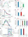
(A) Schematic describing the design of the screen. Worms carrying a transgene co-expressing GCaMP3 and DsRed in the intestine were mutagenized with EMS and their progeny was screened using a real-time PCR thermocycler for candidates defective in cooling-evoked calcium increase in the intestine. Sample traces were shown to the right, highlighting glr-3(xu261) mutant. The temperature was cooled from 23 to 10°C. (B–C) Calcium imaging shows that the glr-3(tm6403) deletion mutant exhibited a severe defect in cooling-evoked calcium response in the intestine. This phenotype was rescued by expressing wild-type glr-3 gene specifically in the intestine using the ges-1 promoter. (B) Sample traces. (C) Bar graph. Error bars: SEM. n≥13. ***p<0.0001 (ANOVA with Bonferroni test). (D–E) glr-3 is expressed in the intestine and the ASER sensory neuron, in addition to the central interneuron RIA. A genomic DNA fragment encompassing 3.2 kb of 5’UTR and the entire coding region of glr-3 was used to drive YFP expression. (D) Low magnification image. (E) High magnification image highlighting neuronal expression in the head. Arrow heads point to ASER and RIAR (RIAL not visible on this focal plane). Scale bars: 100 µm (D); 20 µm (E). (D) and (E) are two independent images taken with different objectives. See also Figure S1.
Figure 2.
Figure 2.. GLR-3 acts in ASER neuron to mediate cold sensation and cold-avoidance behavior.
(A–B) ASER neuron is cold-sensitive and requires GLR-3 for cold sensation. Cooling evoked robust calcium response in ASER neuron, which is defective in glr-3(tm6403) mutant worms. Transgenic expression of wild-type glr-3 cDNA in ASER using the ASER-specific gcy-5 promoter rescued the mutant phenotype. (A) Sample traces. (B) Bar graph. Error bars: SEM. n≥12. ***p<0.0001 (ANOVA with Bonferroni test). (C–D) Loss of GLR-3 does not affect the sensitivity of ASER neuron to salt. glr-3(tm6403) mutant worms showed normal response to NaCl gradient compared to WT, while che-2(e1033) mutant worms were defective in this response. (C) Sample traces. (B) Bar graph. Error bars: SEM. n≥11. ***p<0.0001 (ANOVA with Bonferroni test). (E–F) Cooling-evoked calcium response persists in unc-13, unc-31 and eat-4 mutant backgrounds. (C) Sample traces. (D) Bar graphs. Error bars: SEM. n≥11. ***p<0.0001 (ANOVA with Bonferroni test). (G) GLR-3 mediates cooling-evoked avoidance behavior in ASER neuron. Cooling triggered turns in worms during swimming, and glr-3 mutant worms showed a strong defect in this behavioral response, which was rescued by transgenic expression of glr-3 cDNA in ASER. Error bars: SEM. n≥15. (H) glr-3(tm6403) mutant worms show normal salt chemotaxis behavior, while che-2(e1033) mutant worms are defective in this behavior. The chemotaxis assay was run in triplicates, and the experiment was repeated four times. Error bars: SEM. ***p<0.0001 (ANOVA with Bonferroni test).\ See also Figure S2.
Figure 3.
Figure 3.. GLR-3 and mouse GluK2 function as a cold senor in vitro, and mouse GluK2 can functionally substitute for GLR-3 in cold sensation in vivo.
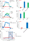
(A–B) Ectopic expression of GLR-3 and mouse GluK2 in worm muscle cells confers cold-sensitivity to these cells. GLR-3 and mouse GluK2 were expressed as a transgene in worm body-wall muscles using the myo-3 promoter. Cooling evoked calcium response in muscle cells of transgenic worms. All genotypes carried a transgene expressing GCaMP6. (A) Sample traces. (B) Bar graph. Error bars: SEM. n≥11. ***p<0.0001 (ANOVA with Bonferroni test). (C–D) Heterologous expression of GLR-3 and mouse GluK2 in CHO cells confers cold sensitivity to these cells. GLR-3 and mouse GluK2 were transfected into CHO cells, and cooling evoked calcium response in these cells as shown by Fura-2 imaging. (C) Sample traces. (D) Bar graph. Error bars: SEM. n≥20. ***p<0.0001 (ANOVA with Bonferroni test). (E–F) Mouse GluK2 can functionally substitute for GLR-3 in cold sensation in ASER neuron. Transgenic expression of mouse GluK2 in ASER neuron of glr-3 mutant worms rescued the glr-3 mutant phenotype in cooling-evoked calcium response in ASER neuron. (E) Sample traces. (F) Bar graph. Error bars: SEM. n≥12. ***p<0.0001 (ANOVA with Bonferroni test). (G) Mouse GluK2 can functionally substitute for GLR-3 in cold-avoidance behavior mediated by ASER neuron. Transgenic expression of mouse GluK2 in ASER neuron of glr-3 mutant worms rescued the glr-3 mutant phenotype in cooling-evoked avoidance behavior. Error bars: SEM. n≥15. See also Figure S2–3.
Figure 4.
Figure 4.. The sensitivity of GLR-3/GluK2 and TRPM8 to cold, glutamate and menthol.
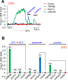
Cooling from 32°C to 20°C activated TRPM8 but not GLR-3 or mouse GluK2 when transfected in CHO cells. Mouse GluK2, but not GLR-3 or TRPM8, can be activated by glutamate (10 mM), while TRPM8 was also sensitive to menthol (100 µM) but not glutamate (10 mM). (A) Sample traces. (B) Bar graph. Error bars: SEM. n≥20. ***p<0.0001 (ANOVA with Bonferroni test). See also Figure S4–5.
Figure 5.
Figure 5.. The cold sensitivity of GLR-3/GluK2 is independent of its channel function.
(A–C) Channel-dead GLR-3/GluK2 variants show normal cold sensitivity in CHO cells. The two channel-dead variants for mouse GluK2 (A), as well as GLR-3 (B), showed similar cold sensitivity compared to wild-type GluK2/GLR-3 when transfected in CHO cells. (A-B) Sample trances. (C) Bar graph. Error bars: SEM. n≥20. ***p<0.0001 (ANOVA with Bonferroni test). (D–F) Channel-dead GLR-3/GluK2 variants show normal cold sensitivity in ASER neuron in vivo. The two channel-dead variants for GLR-3 (D), as well as mouse GluK2 (E), showed similar cold sensitivity compared to wild-type GluK2/GLR-3 when expressed as a transgene in ASER neuron of glr-3 mutant worms. (D-E) Sample traces. (F) Bar graph. Error bars: SEM. n≥12. ***p<0.0001 (ANOVA with Bonferroni test). (G–I) Channel-dead GLR-3/GluK2 variants show normal cold sensitivity in worm muscle cells. The two channel-dead variants for GLR-3 (G), as well as mouse GluK2 (H), showed similar cold sensitivity compared to wild-type GluK2/GLR-3 when expressed as a transgene in worm muscle cells. (G-H) Sample traces. (I) Bar graph. Error bars: SEM. n≥12. ***p<0.0001 (ANOVA with Bonferroni test). (J–L) Cold-insensitive GLR-3/GluK2 mutants display glutamate-gated channel activity. (J) GLR-3(P121L) and GLR-3(P130L) were no longer sensitive to cold. (K) GluK2(P151L) was insensitive to cold but sensitive to glutamate. (L) Bar graph. Error bars: SEM. n≥22. ***p<0.0001 (ANOVA with Bonferroni test). See also Figure S5.
Figure 6.
Figure 6.. GLR-3 relies on G protein signaling to transmit cold signals.
(A–B) G protein inhibitors blocked the cold sensitivity of GLR-3 in CHO cells. The pan-G protein inhibitor mSIRK (50 µM), the Gi/o inhibitor PTX (100 ng/ml), but not the Gq/11 inhibitor YM-254890 (10 µM), blocked cooling-evoked calcium response mediated by GLR-3 in CHO cells. (A) Sample traces. (B) Bar graph. Error bars: SEM. n≥20. ***p<0.0001 (ANOVA with Bonferroni test). (C–D) G protein inhibitors blocked the cold sensitivity of mouse GluK2 in CHO cells. The pan-G protein inhibitor mSIRK (50 μM), the Gi/o inhibitor PTX (100 ng/ml), but not the Gq/11 inhibitor YM-254890 (10 μM), blocked cooling-evoked calcium response mediated by mouse GluK2 in CHO cells. (C) Sample traces. (D) Bar graph. Error bars: SEM. n≥20. ***p<0.0001 (ANOVA with Bonferroni test). (E–F) The cold sensitivity of ASER neuron requires the Gi/o protein gene goa-1. goa-1(n1134) mutant worms showed a strong defect in cooling-evoked calcium response in ASER neuron, a phenotype that was rescued by transgenic expression of wild-type goa-1 gene specifically in ASER neuron. (E) Sample traces. (F) Bar graph. Error bars: SEM. n≥11. ***p<0.0001 (ANOVA with Bonferroni test). See also Figure S6.
Figure 7.
Figure 7.. Mouse DRG neurons express GluK2 and knockdown of GluK2 in DRG neurons suppresses their sensitivity to cold but not cool temperatures.
(A–B) Mouse DRG neurons express GluK2 mRNA transcripts. (A) In situ hybridization using RNAscope assay detected GluK2 mRNA signals (puncta) in DRG tissue. The dotted ovals denote the shape of the soma of the neurons expressing GluK2 transcripts. Only those neurons with at least 4 puncta inside the soma were scored positive, as the background signals detected by the control probe varied between 0–2 puncta/cell. The inset image in (A) highlights one GluK2-positive neuron. Green: GluK2 transcripts. Blue: DAPI. (B) Negative control probe staining (provided by Advanced Cell Diagnostics). Scale bar: 50 µm. (C) Real-time quantitative RT-PCR (qPCR) analysis shows that siRNA knockdown of GluK2 in mouse DRG neurons greatly reduced the mRNA level of GluK2. qPCR reactions were run in triplicates. The experiment was repeated five times. Error bars: SEM. ***p<0.0001 (t test). (D–F) siRNA knockdown of GluK2 in mouse DRG neurons greatly reduces the sensitivity of these neurons to cold but not cool temperatures. (D) Sample trace showing calcium response to cool temperature (cooling to 22°C). (E) Sample trace showing calcium response to cold temperature (cooling to 10°C). (F) Bar graph. Sample sizes were shown on top of each bar. ***p<0.0001 (χ2 test). (G–H) Fish and human GluK2 can also function as a cold receptor in vitro. Heterologous expression of fish and human GluK2 in CHO cells confers cold sensitivity to these cells. (E) Sample traces. (F) Bar graph. Error bars: SEM. n≥20. ***p<0.0001 (ANOVA with Bonferroni test).

References

    1. Bandell M, Macpherson LJ, and Patapoutian A (2007). From chills to chilis: mechanisms for thermosensation and chemesthesis via thermoTRPs. Curr Opin Neurobiol 17, 490–497. - PMC - PubMed
    1. Bargmann CI (2006). Chemosensation in C. elegans. WormBook, 1–29. - PMC - PubMed
    1. Bautista DM, Siemens J, Glazer JM, Tsuruda PR, Basbaum AI, Stucky CL, Jordt SE, and Julius D (2007). The menthol receptor TRPM8 is the principal detector of environmental cold. Nature 448, 204–208. - PubMed
    1. Bowie D (2008). Ionotropic glutamate receptors & CNS disorders. CNS & neurological disorders drug targets 7, 129–143. - PMC - PubMed
    1. Brockie PJ, Madsen DM, Zheng Y, Mellem J, and Maricq AV (2001). Differential expression of glutamate receptor subunits in the nervous system of Caenorhabditis elegans and their regulation by the homeodomain protein UNC-42. The Journal of neuroscience : the official journal of the Society for Neuroscience 21, 1510–1522. - PMC - PubMed

Publication types

MeSH terms

Substances