Comparative Study
. 2019 Oct 3;76(1):96-109.e9.
doi: 10.1016/j.molcel.2019.07.016. Epub 2019 Aug 29.

N6-Methyladenosine Modification Controls Circular RNA Immunity

Affiliations
Comparative Study

N6-Methyladenosine Modification Controls Circular RNA Immunity

Y Grace Chen et al. Mol Cell. .

Abstract

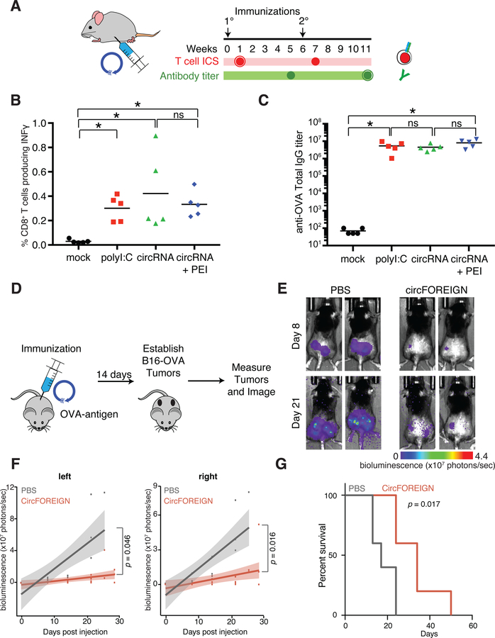
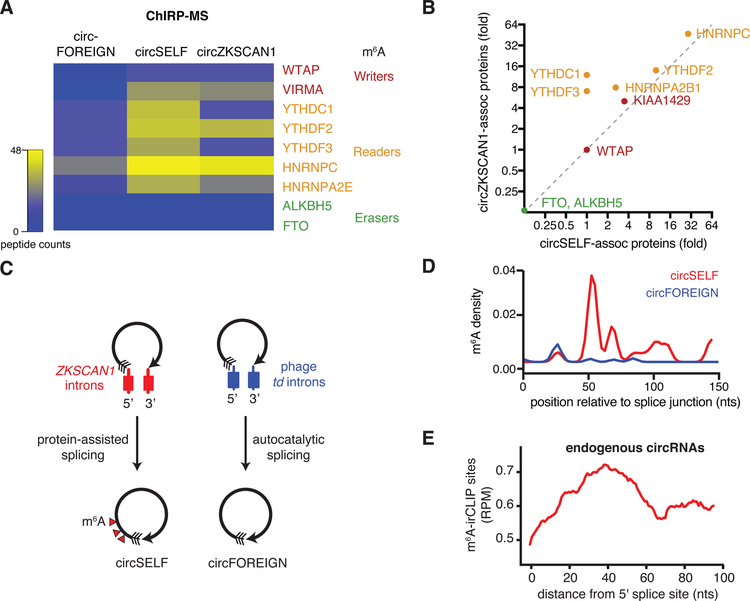
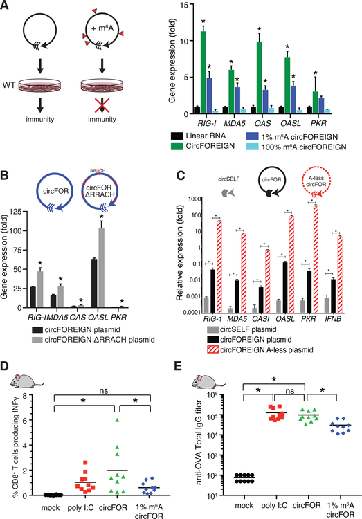
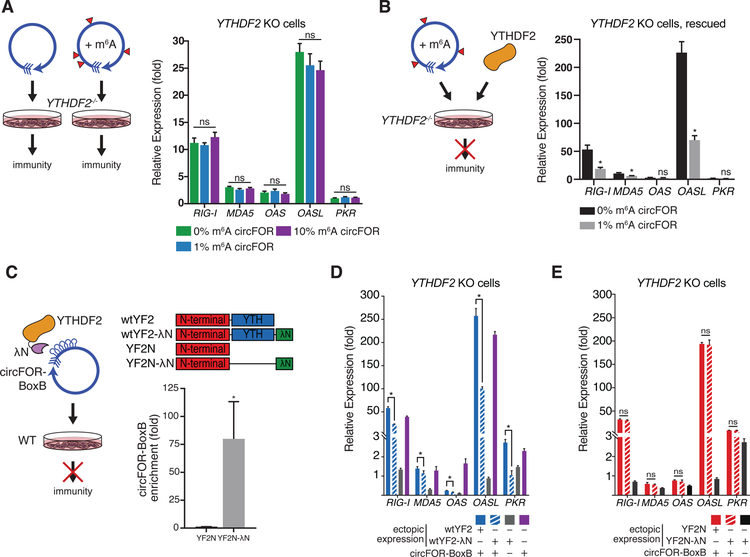
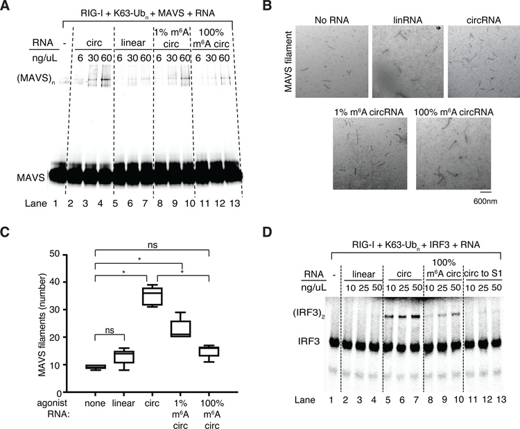
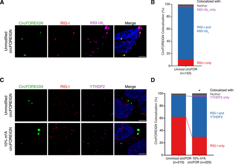
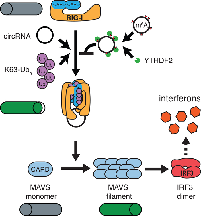
Circular RNAs (circRNAs) are prevalent in eukaryotic cells and viral genomes. Mammalian cells possess innate immunity to detect foreign circRNAs, but the molecular basis of self versus foreign identity in circRNA immunity is unknown. Here, we show that N6-methyladenosine (m6A) RNA modification on human circRNAs inhibits innate immunity. Foreign circRNAs are potent adjuvants to induce antigen-specific T cell activation, antibody production, and anti-tumor immunity in vivo, and m6A modification abrogates immune gene activation and adjuvant activity. m6A reader YTHDF2 sequesters m6A-circRNA and is essential for suppression of innate immunity. Unmodified circRNA, but not m6A-modified circRNA, directly activates RNA pattern recognition receptor RIG-I in the presence of lysine-63-linked polyubiquitin chain to cause filamentation of the adaptor protein MAVS and activation of the downstream transcription factor IRF3. CircRNA immunity has considerable parallel to prokaryotic DNA restriction modification system that transforms nucleic acid chemical modification into organismal innate immunity.

Keywords: N6-methyladenosine; RIG-I; YTHDF; cancer immunotherapy; circular RNA; self/non-self; vaccine.


Conflict of interest statement

DECLARATION OF INTERESTS

Stanford University and Emory University have filed patent applications based on this work, on which H.Y.C., Y.G.C., R.C., L.A., B.P. and S.K. are named as co-inventors. H.Y.C. is a co-founder and advisor of Accent Therapeutics and Pretzel Therapeutics. H.Y.C. is an advisor of 10X Genomics, Arsenal Biosciences, and Spring Discovery.

Figures

Figure 1.
Figure 1.. CircFOREIGN induces immune response but m6A-modified circFOREIGN attenuates stimulation in vivo
A. Agonist RNA in conjunction with OVA is delivered by subcutaneous injection. T cell ICS and antibody titers are measured at the indicated times following primary and secondary immunizations. B. CircRNA stimulates anti-OVA T cell responses independent of transfection agent following primary vaccination. Means are shown (n = 5), *p < 0.05, Kruskal-Wallis test. C. CircRNA stimulates anti-OVA antibody titers independent of transfection agent following secondary vaccination. Means are shown (n = 5), *p < 0.05, Anova-Tukey’s test. D. CircFOREIGN vaccination in conjunction with OVA is delivered by subcutaneous injection. 14 days later, OVA-expressing B16-melanoma cells are established in right and left flanks. Tumors are measured and imaged. E. Quantification of bioluminescence measurements in left and right tumors for mice vaccinated with PBS or circFOREIGN prior to tumor establishment. p value calculated by Wilcoxon signed-rank test. n=5 mice in each group. F. Quantification of bioluminescence measurements in left and right tumors for mice vaccinated with PBS or circFOREIGN prior to tumor establishment. p value calculated by Wilcoxon signed-rank test. n=5 mice in each group. G. Mice vaccinated with circFOREIGN survive twice as long as negative control mice. Survival curves for mice vaccinated with PBS or circFOREIGN prior to tumor establishment. p value calculated by log-rank test. n=5 mice in each group.
Figure 2.
Figure 2.. CircSELF but not circFOREIGN associate with m6A machinery and are modified by m6A
A. Heatmap of peptide counts from ChIRP-MS of circZKSCAN1, circSELF, and circFOREIGN. Enzymes are classified as m6A writers, readers, and erasers. B. m6A machinery associates with circZKSCAN1 and circSELF but not circFOREIGN, as indicated by ChIRP-MS. Fold enrichment over RNase-treated control is shown. C. Model showing ZKSCAN1 introns directing protein-assisted splicing to yield m6A-modified circSELF and phage td introns directing autocatalytic splicing to form unmodified circFOREIGN. D. m6A-irCLIP identifies high-confidence m6A positions proximal to circRNA splice junction. ZKSCAN1 introns suffice to direct m6A modification on circSELF (red) compared with td intron-directed circFOREIGN (blue). Density of m6A-irCLIP reads normalized to reads per million. E. m6A-irCLIP read density near circRNA splice junction of endogenous human circRNAs in HeLa cells. Density of m6A-irCLIP reads normalized to reads per million for reads proximal to circRNA splice junctions.
Figure 3.
Figure 3.. m6A-modified circFOREIGN attenuates immune response in vitro and in vivo
A. (Left) Model of unmodified and m6A-modified circFOREIGN effects of immunogenicity. (Right) Transfection of unmodified circFOREIGN into wild-type HeLa cells stimulates immune response, but m6A-modified circFOREIGN does not. Graph showing gene expression of innate immune genes 24 hours following RNA transfection. Relative expression of the indicated mRNA and transfected RNA are measured by qRT-PCR, results normalized to expression following mock transfection. Means ± SEM are shown (n = 3), *p < 0.05, Student’s t-test, comparing gene stimulation of linear RNA to indicated RNA. B. Transfection of circFOREIGN plasmid lacking RRACH m6A consensus motifs stimulates immune response at a greater level than circFOREIGN. RRACH motifs (n = 12 sites) were mutated to RRUCH throughout the exon sequence. Mutating every adenosine to uracil within the first 200 bases (n = 37 sites) after the splice junction further increased immunogenicity. Graph showing gene expression of innate immune genes following DNA plasmid transfection. Relative expression of the indicated mRNA and transfected RNA are measured by qRT-PCR, results normalized to expression following mock transfection. Means ± SEM are shown (n = 3), **p < 0.01, ***p < 0.001, Student’s t-test, comparing circFOREIGN to indicated RNA transfection. C. Transfection of circFOREIGN plasmid with all adenosines replaced by uracil results in elevated immunogenicity. Relative expression of the indicated mRNA and transfected RNA are measured by qRT-PCR, results normalized to expression following mock transfection. Means ± SEM are shown (n = 3), *p<0.01, Student’s t-test, comparing circFOREIGN to indicated RNA transfection. D. m6A-modified circFOREIGN attenuates anti-OVA T cell responses following primary vaccination. Means are shown (n = 10), *p < 0.05, Anova-Tukey’s test. E. m6A-modified circRNA attenuates anti-OVA antibody titers following secondary vaccination. Means are shown (n = 10), *p < 0.05, ANOVA-Tukey’s test.
Figure 4.
Figure 4.. m6A-modified circFOREIGN evades non-self detection through the binding of YTHDF2
A. YTHDF2 is necessary for differential response to unmodified vs. m6A-modified circFOREIGN. Transfection of unmodified or m6A-modified circFOREIGN into YTHDF2−/− HeLa cells stimulate immune response. Left: Model showing the responses to unmodified or m6A-modified circFOREIGN. Right: Graph showing gene expression of innate immune genes 24 hours following RNA transfection. Relative expression of the indicated mRNA and transfected RNA are measured by qRT-PCR, results normalized to expression following mock transfection. Means ± SEM are shown (n = 3). Student’s t-test, comparing circFOREIGN with 0% m6A to indicated RNA transfection. B. Ectopic expression of YTHDF2 rescues response to unmodified vs. m6A-modified circFOREIGN in YTHDF2 KO HeLa cells. Left: Model showing response to m6A-modified circFOREIGN following rescue. Right: Graph showing gene expression of innate immune genes 24 hours following RNA transfection. Relative expression of the indicated mRNA and transfected RNA are measured by qRT-PCR, results normalized to expression following mock transfection. Means ± SEM are shown (n = 3). *p<0.05, Student’s t-test, comparing 0% m6A circFOREIGN to 1% m6A circFOREIGN. C. Tethering of YTHDF2 to unmodified circFOREIGN mask circRNA immunity. Left: Model showing in vivo tethering of protein to RNA via lambdaN and BoxB leading to attenuation of immunogencity. Right top: Protein domain architecture of full-length wild-type YTHDF2 with and without lambdaN tethering tag, and YTHDF2 N-terminal domain with and without lambdaN tethering tag. Right bottom: RIP-qPCR enrichment of indicated YTH protein followed by qRT-PCR of circRNA-BoxB or control actin RNA. Means ± SEM are shown (n = 3). *p<0.05, Student’s t-test, comparing YTHDF2 N-terminus with lambdaN tethering to YTHDF2 N-terminus without tethering. D. Transfection of unmodified circBoxB tethered to full length wild-type YTHDF2 into wild-type HeLa cells attenuates immune response. Graph showing gene expression of innate immune genes 24 hours following RNA transfection. Relative expression of the indicated mRNA and transfected RNA are measured by qRT-PCR, results normalized mock transfection. Wild-type YTHDF2-lambdaN (grey) was ectopically expressed as immunogenicity negative control. Transfection with solely circBoxB (purple) serves as immunogenicity positive control. Means ± SEM are shown (n = 3). *p<0.05, Student’s t-test, comparing circBoxB with wild-type YTHDF2 with lambdaN tethering to wild-type YTHDF2 without tethering. E. Transfection of unmodified circBoxB tethered to N-terminal domain of YTHDF2 into YTHDF2 KO cells is insufficient to attenuate immune response. Graph showing gene expression of innate immune genes 24 hours following RNA transfection. Relative expression of the indicated mRNA and transfected RNA are measured by qRT-PCR, results normalized mock transfection. N-terminal domain of YTHDF2-lambdaN (black) was ectopically expressed as immunogenicity negative control. Means ± SEM are shown (n = 3). Student’s t-test, comparing circBoxB with YTHDF2 N-terminus with lambdaN tethering to YTHDF2 N-terminus without tethering.
Figure 5.
Figure 5.. RIG-I directly distinguishes unmodified vs. m6A-circFOREIGN and initiates innate immune signaling cascade.
A. In vitro reconstitution with purified RIG-I, MAVS, K63-Ubn and the indicated RNA ligands. Native gel of fluorescently-labeled MAVS 2CARD domain is shown. B. Representative electron microscopy images of MAVS filaments after MAVS polymerization assay with indicated RNAs. Scale bar indicates 600 nm. C. Quantification of the total number of MAVS filaments observed in five electron microscopy images for each agonist RNA. *p<0.05, Student’s t-test. D. In vitro reconstitution of the circRNA-mediated induction of IRF3 dimerization. Native gel of radiolabeled-IRF3 with the indicated RNA ligands is shown. S1 is cellular extract.
Figure 6.
Figure 6.. Immunofluorescence reveals circFOREIGN co-localization with RIG-I and K63-Ubn and YTHDF2 recruitment to m6A-modified circFOREIGN
A. CircFOREIGN co-localizes with RIG-I and K63-polyubiquitin chain. Representative field of view is shown. B. Quantification of circFOREIGN colocalization with RIG-I and K63-Ubn (n = 152). Foci were collected across 10 fields of view across biological replicates and representative of replicate experiments. C. 10% m6A circFOREIGN has increased co-localization with YTHDF2. Representative field of view is shown. Foci were collected across >10 fields of view and representative of replicate experiments. D. Quantification of circFOREIGN and 10% m6A circFOREIGN colocalization with YTHDF2 and RIG-I. *p<0.05, Pearson’s χ² test.
Figure 7.
Figure 7.. Model of circRNA induction of innate immune response through RIG-I
Unmodified circRNA interacts with RIG-I in the presence of K63-Ubn to induce MAVS filamentation, which triggers IRF3 dimerization and interferon production. m6A-modified circRNAs together with YTHDF2 repel RIG-I and does not initiate signaling.

Comment in

References

    1. Ahmad S, Mu X, Yang F, Greenwald E, Park JW, Jacob E, Zhang C-Z, and Hur S (2018). Breaching Self-Tolerance to Alu Duplex RNA Underlies MDA5-Mediated Inflammation. Cell 172, 797–810.e713. - PMC - PubMed
    1. Baron-Benhamou J, Gehring NH, Kulozik AE, and Hentze MW (2004). Using the λN Peptide to Tether Proteins to RNAs. In mRNA Processing and Metabolism: Methods and Protocols, Schoenberg DR, ed. (Totowa, NJ: Humana Press; ), pp. 135–153. - PubMed
    1. Batista PJ, Molinie B, Wang J, Qu K, Zhang J, Li L, Bouley DM, Lujan E, Haddad B, Daneshvar K, et al. (2014). m(6)A RNA modification controls cell fate transition in mammalian embryonic stem cells. Cell stem cell 15, 707–719. - PMC - PubMed
    1. Budhu S, Loike JD, Pandolfi A, Han S, Catalano G, Constantinescu A, Clynes R, and Silverstein SC (2010). CD8+ T cell concentration determines their efficiency in killing cognate antigen–expressing syngeneic mammalian cells in vitro and in mouse tissues. The Journal of Experimental Medicine 207, 223–235. - PMC - PubMed
    1. Chen YG, Kim MV, Chen X, Batista PJ, Aoyama S, Wilusz JE, Iwasaki A, and Chang HY (2017). Sensing Self and Foreign Circular RNAs by Intron Identity. Molecular cell 67, 228–238.e225. - PMC - PubMed

Publication types

MeSH terms