. 2019 Oct 3;76(1):82-95.e7.
doi: 10.1016/j.molcel.2019.07.017. Epub 2019 Aug 29.

Disease-Causing Mutations in SF3B1 Alter Splicing by Disrupting Interaction with SUGP1

Affiliations

Disease-Causing Mutations in SF3B1 Alter Splicing by Disrupting Interaction with SUGP1

Jian Zhang et al. Mol Cell. .

Abstract

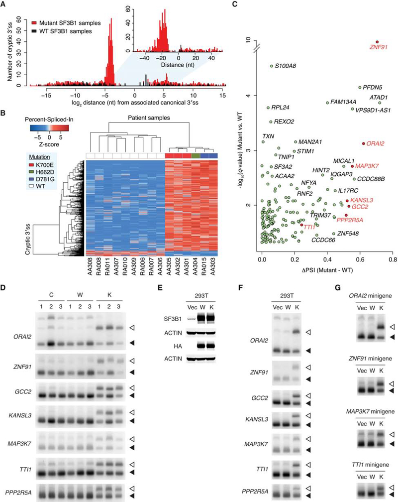
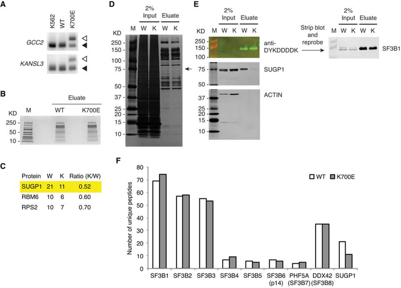
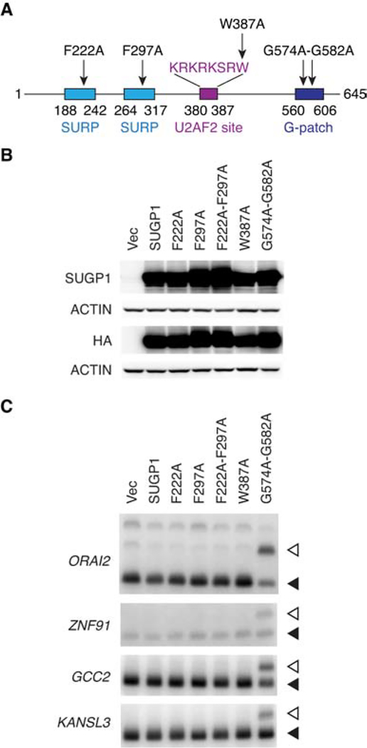
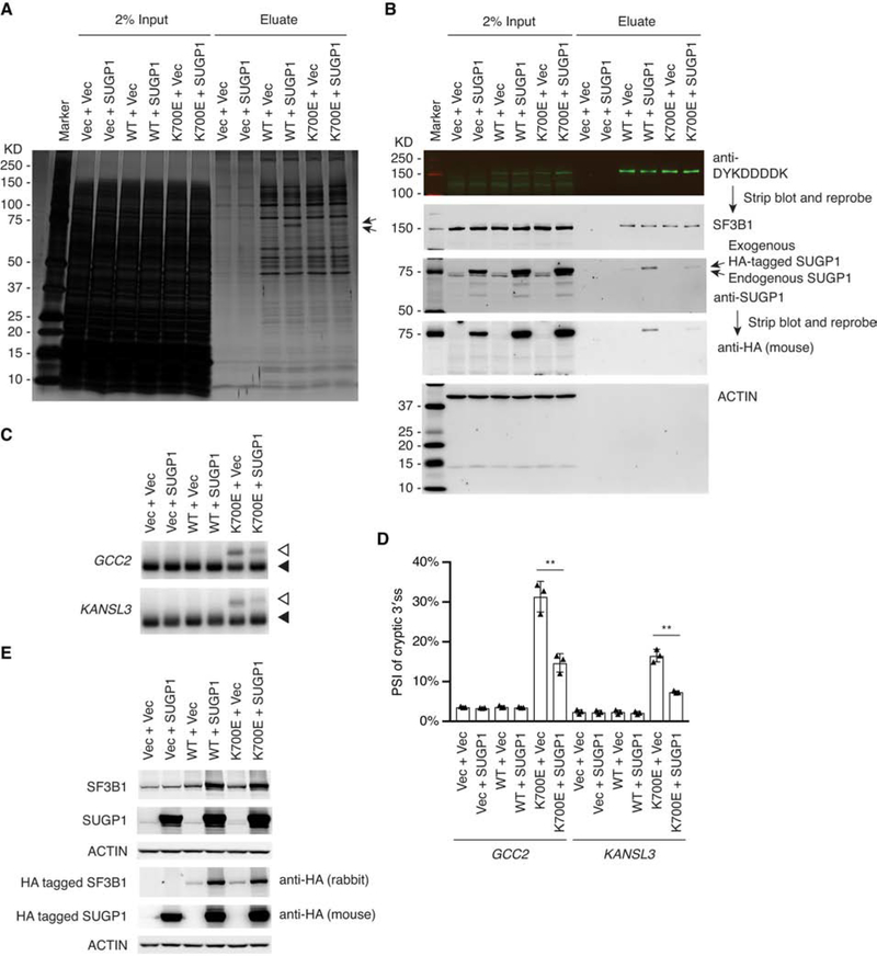
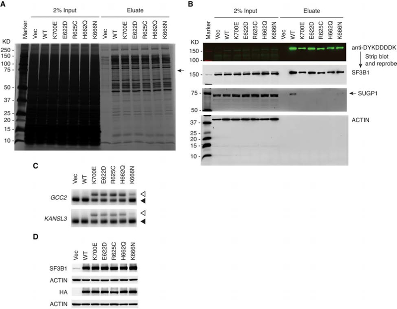
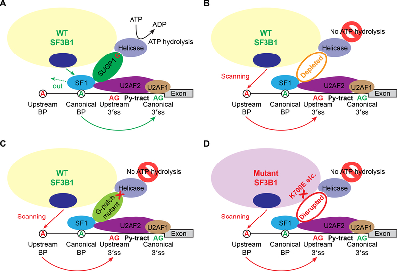
SF3B1, which encodes an essential spliceosomal protein, is frequently mutated in myelodysplastic syndromes (MDS) and many cancers. However, the defect of mutant SF3B1 is unknown. Here, we analyzed RNA sequencing data from MDS patients and confirmed that SF3B1 mutants use aberrant 3' splice sites. To elucidate the underlying mechanism, we purified complexes containing either wild-type or the hotspot K700E mutant SF3B1 and found that levels of a poorly studied spliceosomal protein, SUGP1, were reduced in mutant spliceosomes. Strikingly, SUGP1 knockdown completely recapitulated the splicing errors, whereas SUGP1 overexpression drove the protein, which our data suggest plays an important role in branchsite recognition, into the mutant spliceosome and partially rescued splicing. Other hotspot SF3B1 mutants showed similar altered splicing and diminished interaction with SUGP1. Our study demonstrates that SUGP1 loss is a common defect of spliceosomes with disease-causing SF3B1 mutations and, because this defect can be rescued, suggests possibilities for therapeutic intervention.

Keywords: SF1; SRSF2; U2 snRNP; U2AF1; U2AF2; branch point; leukemia; myelodysplastic syndromes; p14; spliceosome.


Conflict of interest statement

DECLARATION OF INTERESTS

The authors declare no competing interests.

Figures

Figure 1.
Figure 1.. SF3B1 Mutations Lead to Use of Upstream Cryptic 3′ Splice Sites
(A) Distribution of cryptic 3′ss around the associated canonical 3′ss. Red and black histograms indicate 1,145 cryptic 3′ss more frequently used (q value < 0.05) in six mutant SF3B1 MDS patient samples and 186 cryptic 3′ss in nine WT SF3B1 samples, respectively. (B) Hierarchical clustering and heatmap analysis of the 627 cryptic 3′ss differentially used in six mutant vs. nine WT SF3B1 samples (q value < 0.05, and cryptic 3′ss closer than 100 nt upstream of the associated canonical 3′ss). Each row represents one cryptic 3′ss, and each column one MDS patient sample. Z-scores in the matrix represent normalized Percent-Spliced-In values. The color bars above the heatmap indicate the SF3B1 mutations. (C) Volcano plot representation of genes associated with the 169 cryptic 3′ss differentially used in mutant vs. WT SF3B1 samples (q value < 0.05, closer than 50 nt upstream of the associated canonical 3′ss, and more than 15 supporting reads averaged over mutant SF3B1 samples). The horizontal axis shows the Percent-Spliced-In (PSI) difference between mutant and WT SF3B1 samples, and the vertical axis shows the significance. Genes selected for further experimental validation are highlighted in red. (D) RT-PCR products of the cryptic 3′ss (open arrowheads) and canonical 3′ss (solid arrowheads) of seven select genes (ORAI2, ZNF91, GCC2, KANSL3, MAP3K7, TTI1, and PPP2R5A) in normal control samples (C), WT SF3B1 MDS patient samples (W), and K700E SF3B1 MDS patient samples (K). (E) HEK293T cells (293T) were transfected with empty vector plasmid (Vec), or expression plasmid for HA-tagged WT (W) or K700E (K) SF3B1, followed by western blotting. (F) Total RNA was extracted from HEK293T cells as in (E), followed by RT-PCR of the cryptic 3′ss (open arrowheads) and canonical 3′ss (solid arrowheads) of the indicated genes. (G) Each minigene was cotransfected with empty vector plasmid (Vec), or expression plasmid for HA-tagged WT (W) or K700E (K) SF3B1 to HEK293T cells, followed by RT-PCR of the cryptic 3′ss (open arrowheads) and canonical 3′ss (solid arrowheads) from the minigenes. See also Figure S7 and Table S1.
Figure 2.
Figure 2.. The K700E Mutation Specifically Impairs Association with SUGP1
(A) Total RNA was extracted from K562 parental cells and CRISPR/Cas9 engineered K562 cells expressing mono-allelic His6-FLAG-tagged WT or K700E SF3B1, followed by RT-PCR of the cryptic 3′ss (open arrowheads) and canonical 3′ss (solid arrowheads) of two select genes (GCC2 and KANSL3). (B) SF3B1-associated proteins were purified from CRISPR/Cas9 engineered K562 cells expressing mono-allelic His6-FLAG-tagged WT or K700E SF3B1 using the large-scale affinity purification protocol, followed by SDS-PAGE and staining with QC Colloidal Coomassie Stain (Bio-Rad). M, Precision Plus Protein marker (Bio-Rad). (C) Mass spectrometry results of three candidate proteins differentially associated with WT (W) and K700E (K) SF3B1, with the second and third columns indicating the numbers of unique peptides and the fourth column the peptide ratio. (D and E) SF3B1-associated proteins were purified from CRISPR/Cas9 engineered K562 cells expressing mono-allelic His6-FLAG-tagged WT (W) or K700E (K) SF3B1 using the small-scale protocol, and then resolved by SDS-PAGE, followed by silver staining (D) or western blotting (E). M, Precision Plus Protein marker (Bio-Rad). (F) Mass spectrometry results of SF3b subunits (as well as SUGP1) associated with WT and K700E SF3B1. Note that in the K700E SF3B1 sample there was one unique peptide that belongs to a novel isoform (fragment) of SF3B2, but this single peptide was not included in the total number of unique peptides of SF3B2, which does not affect our conclusion. See also Figure S1 and Table S2.
Figure 3.
Figure 3.. Loss of SUGP1 Recapitulates the Splicing Defects of K700E SF3B1
(A) HEK293T cells were transfected with a negative control siRNA (siC), or one of two independent siRNAs targeting SUGP1, followed by western blotting. (B) RT-PCR products of the cryptic 3′ss (open arrowheads) and canonical 3′ss (solid arrowheads) of the indicated genes in HEK293T cells as in (A). (C) Each of the indicated WT (shown in green) and mutant (shown in red) minigenes was cotransfected either with expression plasmid for HA-tagged WT (W) or K700E (K) SF3B1 (left panels), or with one of two independent siRNAs targeting SUGP1 (right panels) to HEK293T cells, followed by RT-PCR of the cryptic 3′ss (open arrowheads) and canonical 3′ss (solid arrowheads) from the minigenes. As indicated, an empty vector plasmid (Vec) and a negative control siRNA (siC) were also used. See also Figures S2 and S3.
Figure 4.
Figure 4.. A Dominant Negative Mutant of SUGP1 Phenocopies Mutant SF3B1
(A) Schematic representation of SUGP1 functional domains and mutants. (B) HEK293T cells were transfected with empty vector plasmid (Vec), or expression plasmid for HA-tagged SUGP1 or one of the SUGP1 mutants (F222A, F297A, F222A-F297A, W387A, and G574A-G582A), followed by western blotting. (C) RT-PCR products of the cryptic 3′ss (open arrowheads) and canonical 3′ss (solid arrowheads) of the indicated genes in HEK293T cells as in (B). See also Figure S4.
Figure 5.
Figure 5.. Overexpression of SUGP1 Partially Rescues the Mutant Spliceosome
(A and B) The indicated plasmids were cotransfected to HEK293T cells in 10-cm plates, followed by affinity purification using the small-scale protocol. The SF3B1-associated proteins were resolved by SDS-PAGE, followed by silver staining (A) or western blotting (B). Marker, Precision Plus Protein marker (Bio-Rad); Vec, empty vector plasmid; SUGP1, expression plasmid for HA-tagged SUGP1; WT, expression plasmid for His6-FLAG-tagged WT SF3B1; K700E, expression plasmid for His6-FLAG-tagged K700E SF3B1. (C) The indicated plasmids were cotransfected to HEK293T cells in six-well plates. Total RNA was extracted from the cells, followed by RT-PCR of the cryptic 3′ss (open arrowheads) and canonical 3′ss (solid arrowheads) of two select genes (GCC2 and KANSL3). Vec, empty vector plasmid; SUGP1, expression plasmid for HA-tagged SUGP1; WT, expression plasmid for HA-tagged WT SF3B1; K700E, expression plasmid for HA-tagged K700E SF3B1. Note that the PCR products of the cryptic 3′ss of GCC2 and KANSL3 (open arrowheads) in the sixth lane appeared to migrate slightly more slowly than the PCR products in the fifth lane. However, DNA sequencing results indicated that the cryptic 3′ss and canonical 3′ss of GCC2 and KANSL3 in the last two lanes were exactly the same. (D) Quantification of RT-PCR products in (C). The bar graph was made using Prism (GraphPad). Error bars represent SDs of the means (n = 3, three independent experiments). **p < 0.01 (unpaired, two-tailed, and unequal variance t test, calculated using Microsoft Excel). (E) Protein extracts from HEK293T cells as in (C) were resolved by SDS-PAGE, followed by western blotting. See also Figures S5 and S6.
Figure 6.
Figure 6.. Other SF3B1 Mutations Also Weaken Interaction with SUGP1 in the Spliceosome
(A and B) HEK293T cells in 10-cm plates were transfected with empty vector plasmid (Vec), or expression plasmid for His6-FLAG-tagged WT, K700E, E622D, R625C, H662Q, or K666N SF3B1, followed by affinity purification using the small-scale protocol. The SF3B1-associated proteins were resolved by SDS-PAGE, followed by silver staining (A) or western blotting (B). Marker, Precision Plus Protein marker (Bio-Rad). (C and D) HEK293T cells in 6-well plates were transfected with empty vector plasmid (Vec), or expression plasmid for HA-tagged WT, K700E, E622D, R625C, H662Q, or K666N SF3B1, followed by RT-PCR of the cryptic 3′ss (open arrowheads) and canonical 3′ss (solid arrowheads) of GCC2 and KANSL3 (C) or western blotting (D).
Figure 7.
Figure 7.. The Mechanistic Model
(A) Under normal conditions, the WT SF3B1 spliceosome uses a canonical BP and 3′ss for splicing (see main text for a detailed explanation). The G-patch domain of SUGP1 is shown as “G” in red. Py-tract, polypyrimidine tract. (B and C) When SUGP1 is depleted (B), or mutated in the G-patch domain (C), the WT SF3B1 spliceosome uses an upstream cryptic BP and cryptic 3′ss for splicing. (D) Because the interaction between SF3B1 and SUGP1 is disrupted by SF3B1 mutations, the mutant SF3B1 spliceosome uses an upstream BP and cryptic 3′ss for splicing.

Similar articles

Cited by

References

    1. Aakre CD, Herrou J, Phung TN, Perchuk BS, Crosson S, and Laub MT (2015). Evolving new protein-protein interaction specificity through promiscuous intermediates. Cell 163, 594–606. - PMC - PubMed
    1. Alsafadi S, Houy A, Battistella A, Popova T, Wassef M, Henry E, Tirode F, Constantinou A, Piperno-Neumann S, Roman-Roman S, et al. (2016). Cancer-associated SF3B1 mutations affect alternative splicing by promoting alternative branchpoint usage. Nat. Commun 7, 10615. - PMC - PubMed
    1. Andrei SA, Sijbesma E, Hann M, Davis J, O’Mahony G, Perry MWD, Karawajczyk A, Eickhoff J, Brunsveld L, Doveston RG, et al. (2017). Stabilization of protein-protein interactions in drug discovery. Expert Opin. Drug Discov 12, 925–940. - PubMed
    1. Aravind L, and Koonin EV (1999). G-patch: a new conserved domain in eukaryotic RNA-processing proteins and type D retroviral polyproteins. Trends Biochem. Sci 24, 342–344. - PubMed
    1. Barreyro L, Chlon TM, and Starczynowski DT (2018). Chronic immune response dysregulation in MDS pathogenesis. Blood 132, 1553–1560. - PMC - PubMed

Publication types

MeSH terms