. 2019 Sep 2;10(9):638.
doi: 10.1038/s41419-019-1833-5.

The effect of cysteine oxidation on DJ-1 cytoprotective function in human alveolar type II cells

Affiliations

The effect of cysteine oxidation on DJ-1 cytoprotective function in human alveolar type II cells

Karim Bahmed et al. Cell Death Dis. .

Abstract

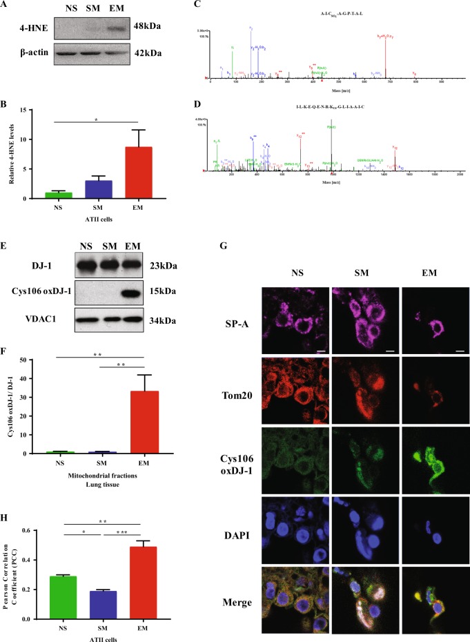
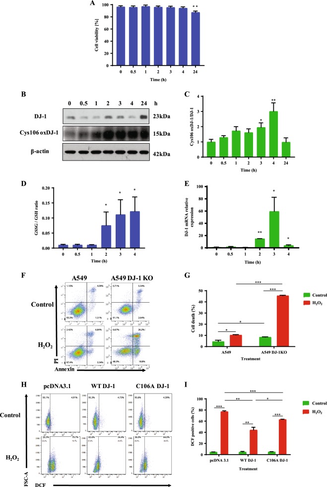
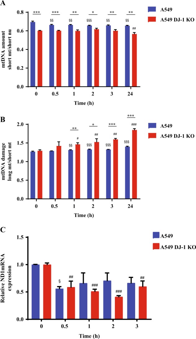
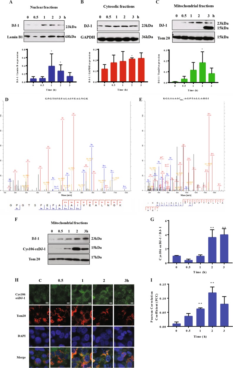
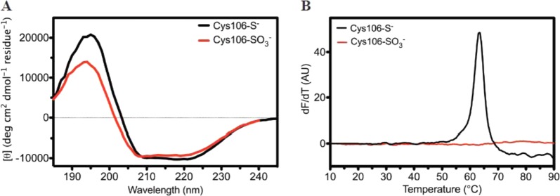
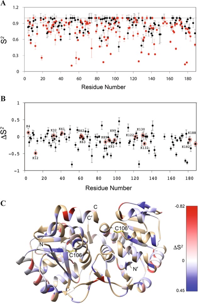
DJ-1 is a multifunctional protein with cytoprotective functions. It is localized in the cytoplasm, nucleus, and mitochondria. The conserved cysteine residue at position 106 (Cys106) within DJ-1 serves as a sensor of redox state and can be oxidized to both the sulfinate (-SO2-) and sulfonate (-SO3-) forms. DJ-1 with Cys106-SO2- has cytoprotective activity but high levels of reactive oxygen species can induce its overoxidation to Cys106-SO3-. We found increased oxidative stress in alveolar type II (ATII) cells isolated from emphysema patients as determined by 4-HNE expression. DJ-1 with Cys106-SO3- was detected in these cells by mass spectrometry analysis. Moreover, ubiquitination of Cys106-SO3- DJ-1 was identified, which suggests that this oxidized isoform is targeted for proteasomal destruction. Furthermore, we performed controlled oxidation using H2O2 in A549 cells with DJ-1 knockout generated using CRISPR-Cas9 strategy. Lack of DJ-1 sensitized cells to apoptosis induced by H2O2 as detected using Annexin V and propidium iodide by flow cytometry analysis. This treatment also decreased both mitochondrial DNA amount and mitochondrial ND1 (NADH dehydrogenase 1, subunit 1) gene expression, as well as increased mitochondrial DNA damage. Consistent with the decreased cytoprotective function of overoxidized DJ-1, recombinant Cys106-SO3- DJ-1 exhibited a loss of its thermal unfolding transition, mild diminution of secondary structure in CD spectroscopy, and an increase in picosecond-nanosecond timescale dynamics as determined using NMR. Altogether, our data indicate that very high oxidative stress in ATII cells in emphysema patients induces DJ-1 overoxidation to the Cys106-SO3- form, leading to increased protein flexibility and loss of its cytoprotective function, which may contribute to this disease pathogenesis.


Conflict of interest statement

The authors declare that they have no conflict of interest.

Figures

Fig. 1
Fig. 1. High oxidative stress induces Cys106-SO3 within DJ-1 in ATII cells in emphysema patients.
ATII cells were isolated from non-smokers, smokers, and emphysema patients. a 4-HNE expression was determined by western blotting. b Densitometric quantification is also shown. c Cys106-SO3 within DJ-1 peptide AICSO3-AGPTAL in freshly isolated ATII cells from emphysema patients as detected by mass spectrometry analysis. d Ubiquitination of lysine residue at position 99 within DJ-1 peptide ILKEQENRKUbGLIAA in ATII cells obtained from individuals with emphysema. e DJ-1 expression and Cys106-oxidation within DJ-1 in mitochondrial fractions obtained from lung tissue of emphysema patients by western blotting. f Relative expression is also shown. g DJ-1 with oxidized Cys106 (green) in mitochondria identified by Tom20 expression (red) in ATII cells detected using SP-A antibody (magenta). Nuclei were stained with DAPI (blue). The scale bar is 5 μm. h Co-localization of DJ-1 with oxidized Cys106 and Tom20 (N = 3–7 lungs per group; *P < 0.05; **P < 0.01; ***P < 0.001). Data are shown as means ± s.e.m.
Fig. 2
Fig. 2. The cytoprotective role of DJ-1 against oxidative stress.
A549 cells were treated with 1 mM H2O2 for indicated times. a Viability was estimated using Hoechst 33342 and PI double staining. b Expression of DJ-1 and Cys106-oxidized DJ-1 determined by western blotting. c Relative expression is also shown. d Oxidative stress analyzed by GSSG/GSH ratio. e DJ-1 mRNA expression was determined by RT-PCR. f Representative flow cytometry images using Annexin V and PI double staining in A549 cells and A549 cells with DJ-1 KO. g Quantification is also shown. h A549 cells with DJ-1 KO were transfected with pcDNA3.1, WT DJ-1, or C106A DJ-1 plasmids followed by treatment with 1 mM H2O2 for 2 h and analyzed using DCF staining. i Quantification is also shown. N = 3 replicates; *P < 0.05, **P < 0.01; ***P < 0.001. Data are shown as means ± s.e.m.
Fig. 3
Fig. 3. Mitochondrial dysfunction in A549 cells with DJ-1 knockout.
A549 cells with DJ-1 KO were generated using CRISPR-Cas9 strategy. A549 cells and DJ-1 KO A549 cells were treated with 1 mM H2O2 for indicated times to determine mtDNA amount by qPCR (a), mtDNA damage using qPCR (b), and ND1 mRNA expression by RT-PCR (c). *P < 0.05, **P < 0.01, and ***P < 0.001. $P < 0.05, $$P < 0.01, and $$$P < 0.001 compared to control A549 cell line. #P < 0.05, ##P < 0.01, and ###P < 0.001 compared to control A549 cells with DJ-1 KO. N = 3 replicates. Data are shown as means ± s.e.m.
Fig. 4
Fig. 4. H2O2 induces Cys106-SO3 DJ-1 formation in mitochondria.
A549 cells were treated with 1 mM H2O2 for indicated times and DJ-1 expression was determined by western blotting in nuclear (a), cytosolic (b), and mitochondrial (c) fractions. Densitometric quantifications are also shown. Bands detected at 23 kDa and 15 kDa in mitochondrial fractions by western blotting were analyzed by mass spectrometry analysis. The presence of DJ-1 in the band at 23 kDa (d) and Cys106-SO3 DJ-1 at 15 kDa (e) is shown. f Cys106-oxidized DJ-1 in mitochondrial fractions in A549 cells by western blotting. g Densitometric quantification is also shown. h Cys106-oxidized DJ-1 (green) in mitochondria identified by Tom20 (red) using immunocytofluorescence. Nuclei were stained with DAPI (blue). The scale bar is 5 μm. i Cys106-oxidized DJ-1 and Tom20 co-localization is shown. N = 3 replicates; *P < 0.05; **P < 0.01. Data are shown as means ± s.e.m.
Fig. 5
Fig. 5. Cys106-SO3 DJ-1 formation preserves secondary structure but destabilizes the protein.
a Circular dichroism (CD) spectra are shown for reduced (black) and Cys106-SO3 DJ-1 (red) DJ-1. Formation of Cys106-SO3 results in a minor decrease in secondary structural content. The y-axis shows per-residue molar ellipticity ([θ]). b The Thermofluor scanning fluorimetry assay was used to measure the melting temperature (Tm) of reduced (black) and Cys106-SO3 (red) DJ-1. The first derivative of the fluorescence as a function of temperature (dF/dT) is shown. Reduced DJ-1 has a Tm of 63 °C, while there is no measured transition for Cys106-SO3 DJ-1, indicating substantial destabilization
Fig. 6
Fig. 6. NMR relaxation analysis of picosecond–nanosecond (ps–ns) timescale dynamics in Cys106-SO3 DJ-1.
a The backbone 1H-15N amide bond vector order parameters (S2) in reduced (black) and Cys106-SO3 DJ-1 (red). S2 range from 1 for immobile residues on the ps–ns timescale to 0 for highly mobile bond vectors with no preferred orientation. Cys106-SO3 formation results in a marked lowering of S2 values in several regions of DJ-1. b The ΔS2 values calculated as: ΔS2 = S2(Cys106-SO3)- S2(reduced) for each residue. Negative values indicate residues that become more mobile in Cys106-SO3 DJ-1 and positive values indicate residues that become less mobile. Lysine residues are circled and labeled. c The ΔS2 values onto the ribbon diagram of the DJ-1 dimer. Red values indicate increased mobility and blue values indicated decreased mobility in Cys106-SO3 DJ-1. The core of the dimer experiences a slight decrease in mobility upon formation of Cys106-SO3, while the solvent-exposed regions have strongly increased mobility

Similar articles

Cited by

References

    1. Barnes PJ. Chronic obstructive pulmonary disease. N. Engl. J. Med. 2000;343:269–280. doi: 10.1056/NEJM200007273430407. - DOI - PubMed
    1. Tuder RM, Petrache I. Pathogenesis of chronic obstructive pulmonary disease. J. Clin. Invest. 2012;122:2749–2755. doi: 10.1172/JCI60324. - DOI - PMC - PubMed
    1. Hogg JC, Senior RM. Chronic obstructive pulmonary disease-part 2: pathology and biochemistry of emphysema. Thorax. 2002;57:830–834. doi: 10.1136/thorax.57.9.830. - DOI - PMC - PubMed
    1. Mason RJ. Biology of alveolar type II cells. Respirology. 2006;11:S12–S15. doi: 10.1111/j.1440-1843.2006.00800.x. - DOI - PubMed
    1. MacNee W. Oxidants/antioxidants and COPD. Chest. 2000;117:303S–317S. doi: 10.1378/chest.117.5_suppl_1.303S-a. - DOI - PubMed

Publication types