

Detailed and applied anatomy for improved rectal cancer treatment
- PMID: 31474788
- PMCID: PMC6686088
- DOI: 10.20524/aog.2019.0407
Detailed and applied anatomy for improved rectal cancer treatment
Abstract
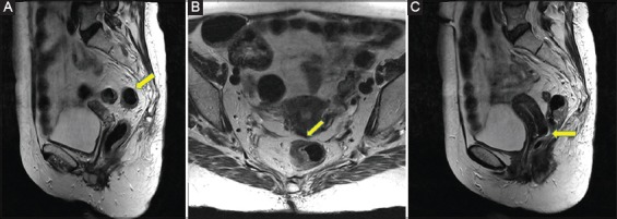
Rectal anatomy is one of the most challenging concepts of visceral anatomy, even though currently there are more than 23,000 papers indexed in PubMed regarding this topic. Nonetheless, even though there is a plethora of information meant to assist clinicians to achieve a better practice, there is no universal understanding of its complexity. This in turn increases the morbidity rates due to iatrogenic causes, as mistakes that could be avoided are repeated. For this reason, this review attempts to gather current knowledge regarding the detailed anatomy of the rectum and to organize and present it in a manner that focuses on its clinical implications, not only for the colorectal surgeon, but most importantly for all colorectal cancer-related specialties.
Keywords: Anatomy; cancer; rectum; surgery.
Conflict of interest statement
Conflict of Interest: None
Figures


Similar articles
-
Aspects of survival from colorectal cancer in Denmark.Dan Med J. 2012 Apr;59(4):B4428. Dan Med J. 2012. PMID: 22459726
-
Applied vascular anatomy of the colon and rectum: clinical implications for the surgical oncologist.Surg Oncol. 2006 Dec;15(4):243-55. doi: 10.1016/j.suronc.2007.03.002. Surg Oncol. 2006. PMID: 17531744 Review.
-
Surgical management of deep infiltrating endometriosis of the rectum: pleading for a symptom-guided approach.Hum Reprod. 2011 Feb;26(2):274-81. doi: 10.1093/humrep/deq332. Epub 2010 Dec 2. Hum Reprod. 2011. PMID: 21131296
-
Anatomic basis of sharp pelvic dissection for curative resection of rectal cancer.Yonsei Med J. 2005 Dec 31;46(6):737-49. doi: 10.3349/ymj.2005.46.6.737. Yonsei Med J. 2005. PMID: 16385648 Free PMC article. Review.
-
[Anatomical relationship between fascia propria of the rectum and visceral pelvic fascia in the view of continuity of fasciae].Zhonghua Wei Chang Wai Ke Za Zhi. 2019 Oct 25;22(10):949-954. doi: 10.3760/cma.j.issn.1671-0274.2019.10.009. Zhonghua Wei Chang Wai Ke Za Zhi. 2019. PMID: 31630492 Chinese.
Cited by
-
Rectal Artery Embolization for the Treatment of Hemorrhoidal Disease.Semin Intervent Radiol. 2025 Jan 24;42(1):93-100. doi: 10.1055/s-0044-1801360. eCollection 2025 Feb. Semin Intervent Radiol. 2025. PMID: 40342391 Review.
-
Genomic and transcriptomic determinants of response to neoadjuvant therapy in rectal cancer.Nat Med. 2022 Aug;28(8):1646-1655. doi: 10.1038/s41591-022-01930-z. Epub 2022 Aug 15. Nat Med. 2022. PMID: 35970919 Free PMC article.
-
Review: Pelvic nerves - from anatomy and physiology to clinical applications.Transl Neurosci. 2021 Oct 8;12(1):362-378. doi: 10.1515/tnsci-2020-0184. eCollection 2021 Jan 1. Transl Neurosci. 2021. PMID: 34707906 Free PMC article. Review.
-
MRI of rectal cancer-relevant anatomy and staging key points.Insights Imaging. 2020 Sep 3;11(1):100. doi: 10.1186/s13244-020-00890-7. Insights Imaging. 2020. PMID: 32880782 Free PMC article. Review.
-
MRI-detected extramural venous invasion of rectal cancer: Multimodality performance and implications at baseline imaging and after neoadjuvant therapy.Insights Imaging. 2021 Aug 9;12(1):110. doi: 10.1186/s13244-021-01023-4. Insights Imaging. 2021. PMID: 34370093 Free PMC article. Review.
References
-
- Kin C, Snyder K, Kiran RP, Remzi FH, Vogel JD. Accidental puncture or laceration in colorectal surgery:a quality indicator or a complexity measure? Dis Colon Rectum. 2013;56:219–225. - PubMed
-
- Octavian Neagoe C, Mazilu O. Pelvic intraoperative iatrogenic oncosurgical injuries:single-center experience. J BUON. 2016;21:498–504. - PubMed
-
- Jorge JM, Wexner SD. Anatomy and physiology of the rectum and anus. Eur J Surg. 1997;163:723–731. - PubMed
-
- Salerno G, Sinnatamby C, Branagan G, Daniels IR, Heald RJ, Moran BJ. Defining the rectum:surgically, radiologically and anatomically. Colorectal Dis. 2006;8(Suppl 3):5–9. - PubMed
Publication types
LinkOut - more resources
Full Text Sources