Review
. 2019 Sep-Oct;32(5):441-450.
doi: 10.20524/aog.2019.0404. Epub 2019 Jul 22.

Complications of endoscopic ultrasound-guided transmural drainage of pancreatic fluid collections and their management

Affiliations
Review

Complications of endoscopic ultrasound-guided transmural drainage of pancreatic fluid collections and their management

Surinder S Rana et al. Ann Gastroenterol. 2019 Sep-Oct.

Abstract

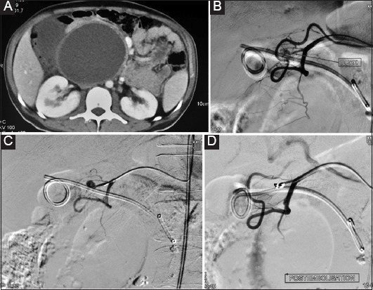
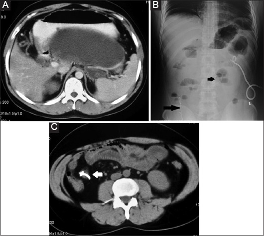
The development of endoscopic ultrasound (EUS)-guided drainage techniques and lumen-apposing metal stents (LAMS) has markedly reduced the complication rate of endoscopic transmural drainage of pancreatic collections and made these procedures safer and more effective. Despite its improved safety profile, various types of complications, some even life-threatening, can occur after EUS-guided drainage of pancreatic fluid collections. Stent maldeployment/migration, bleeding, gastrointestinal perforation, and air embolism are important complications of EUS-guided drainage of pancreatic collections. Delayed complications weeks after the procedure, such as bleeding and buried LAMS due to the presence of prolonged indwelling transmural stents, have also been described. Careful patient selection, with proper assessment of the size, solid necrotic content and location of the collection, as well as an in-depth understanding of various risk factors that predict complications, are important for a safer and more effective endoscopic transmural drainage. For a better clinical outcome, it is important for the endoscopist to know about various complications of EUS-guided drainage of pancreatic collections, as well as their appropriate management strategies.

Keywords: Endosonography; bleeding; embolism; perforation; pseudoaneurysm; stents.


Conflict of interest statement

Conflict of Interest: None

Figures

Figure 1
Figure 1
(A) Endoscopic ultrasound. Patient with chronic pseudocyst having the collection located more than 1 cm from the gastric wall. (B) Endoscopic ultrasound. Patient with chronic pseudocyst and splenic vein thrombosis having extensive collaterals leading to failure to find a safe non-vascular window for needle puncture
Figure 2
Figure 2
(A) Internal maldeployment of double pigtail plastic stent into collection. The transmural puncture site id cannulated with a guidewire using a gastroscope. (B) The transmural tract dilated with a 15 mm balloon. (C) The gastroscope inserted into the cavity. An inward migrated plastic stent is observed in the cavity, with surrounding residual fluid. (D) The stent grasped with grasping forceps and removed
Figure 3
Figure 3
Endoscopic ultrasound. (A) Abnormal vessel in the wall of the pancreatic fluid collection. (B) Abnormal blood vessel running through the pseudocyst cavity
Figure 4
Figure 4
(A) Computed tomography (CT) of the abdomen: large walled-off necrosis. (B) Patient had gastrointestinal bleeding 48 h after endoscopic ultrasound-guided transmural drainage. CT shows residual cavity with hyperdense contents suggestive of blood. Transmural stents are seen in situ. (C) CT angiography: no pseudoaneurysm. (D) Endoscopy: minimal ooze of blood is seen around the stents. Bleeding stopped spontaneously after 24 h
Figure 5
Figure 5
(A) Computed tomography: large walled-off necrosis. (B) Patient develops massive gastrointestinal bleed immediately after the procedure. Digital subtraction angiography (DSA) shows bleeding from the gastroduodenal artery around the plastic transmural stent (arrow). (C) DSA: selective cannulation of the bleeding vessel. (D) SA: successful coil embolization
Figure 6
Figure 6
(A) Computed tomography (CT) of the abdomen: large walled-off necrosis. (B) Patient had increased abdominal pain 48 h after endoscopic transmural drainage with multiple plastic stents. It was accompanied by abdominal distension. Abdominal X-ray reveals air under right dome of diaphragm along with multiple air fluid levels. Both the transmural stents have migrated externally into the small bowel (arrows). Patient was managed with percutaneous drainage of walled-off necrosis and conservative management of small bowel obstruction. (C) CT: one of the migrated plastic stent is causing small-bowel obstruction (arrows)

Similar articles

Cited by

References

    1. Banks PA, Bollen TL, Dervenis C, et al. Classification of acute pancreatitis—2012:revision of the Atlanta classification and definitions by international consensus. Gut. 2013;62:102–111. - PubMed
    1. Cui ML, Kim KH, Kim HG, et al. Incidence, risk factors and clinical course of pancreatic fluid collections in acute pancreatitis. Dig Dis Sci. 2014;59:1055–1062. - PubMed
    1. Rana SS, Bhasin DK, Reddy YR, et al. Morphological features of fluid collections on endoscopic ultrasound in acute necrotizing pancreatitis:do they change over time? Ann Gastroenterol. 2014;27:258–261. - PMC - PubMed
    1. Rana SS, Sharma RK, Gupta P, Gupta R. Natural course of asymptomatic walled off pancreatic necrosis. Dig Liver Dis. 2019;51:730734. - PubMed
    1. Rana SS. An overview of walled-off pancreatic necrosis for clinicians. Expert Rev Gastroenterol Hepatol. 2019;13:331–343. - PubMed

LinkOut - more resources