. 2019 Aug 16:12:192.
doi: 10.3389/fnmol.2019.00192. eCollection 2019.

ALWPs Improve Cognitive Function and Regulate Aβ Plaque and Tau Hyperphosphorylation in a Mouse Model of Alzheimer's Disease

Affiliations

ALWPs Improve Cognitive Function and Regulate Aβ Plaque and Tau Hyperphosphorylation in a Mouse Model of Alzheimer's Disease

Youngpyo Nam et al. Front Mol Neurosci. .

Abstract

Recently, we reported that ALWPs, which we developed by combining Liuwei Dihuang pills (LWPs) with antler, regulate the LPS-induced neuroinflammatory response and rescue LPS-induced short- and long-term memory impairment in wild-type (WT) mice. In the present study, we examined the effects of ALWPs on Alzheimer's disease (AD) pathology and cognitive function in WT mice as well as 5x FAD mice (a mouse model of AD). We found that administration of ALWPs significantly reduced amyloid plaque levels in 5x FAD mice and significantly decreased amyloid β (Aβ) levels in amyloid precursor protein (APP)-overexpressing H4 cells. In addition, ALWPs administration significantly suppressed tau hyperphosphorylation in 5x FAD mice. Oral administration of ALWPs significantly improved long-term memory in scopolamine (SCO)-injected WT mice and 5x FAD mice by altering dendritic spine density. Importantly, ALWPs promoted spinogenesis in primary hippocampal neurons and WT mice and modulated the dendritic spine number in an extracellular signal-regulated kinase (ERK)-dependent manner. Taken together, our results suggest that ALWPs are a candidate therapeutic drug for AD that can modulate amyloid plaque load, tau phosphorylation, and synaptic/cognitive function.

Keywords: Alzheimer’s disease; Aβ; amyloid plaque; dendritic spines; long-term memory; tau.


Figures

Figure 1
Figure 1
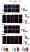
ALWPs significantly decrease amyloid plaque levels in 5x FAD mice. (A) 5x FAD mice were orally administered ALWPs (200 mg/kg, p.o.) or PBS daily for 2 weeks, followed by immunostaining of brain slices with an anti-4G8 antibody. Representative images of the cortex are shown. (B) Quantification of data from (A; con, n = 6 mice; ALWPs, n = 6 mice). (C) Representative images of the hippocampus of 5x FAD mice administered ALWPs (200 mg/kg, p. o.) or PBS daily for 2 weeks. (D,E) Quantification of data from (C; CA1 and DG; con, n = 6 mice; ALWPs, n = 6 mice). (F) Amyloid precursor protein (APP)-H4 cells (H4 cells overexpressing human APP and producing high levels of Aβ were treated with ALWPs (500 μg/ml) or PBS for 24 h, and human Aβ ELISA was performed (con, n = 22; ALWPs, n = 22). **p < 0.01, ***p < 0.0001.
Figure 2
Figure 2
ALWPs significantly increase cell-surface levels of APP. (A) APP-H4 cells were treated with ALWPs (500 μg/ml) or PBS for 24 h, and cell-surface biotinylation assays were conducted with an anti-6E10 antibody. (B,C) Quantification of data from (A; surface APP and total APP; con, n = 8; ALWPs, n = 8). (D,E) Primary hippocampal neurons were transfected with GFP plasmid DNA for 24 h, followed by treatment with ALWPs (500 μg/ml) or PBS for 24 h and live cell-surface staining (con, n = 134 dendrites; ALWPs, n = 120 dendrites). (F) Primary hippocampal neurons were transfected with GFP plasmid DNA for 24 h, treated with ALWPs (500 μg/ml) or PBS for 24 h, and immunostained with an anti-ADAM17 antibody. (G) Quantification of data from (F; con, n = 67 dendrites; ALWPs, n = 71 dendrites). (H,J,L,N) Primary hippocampal neurons were treated with ALWPs (500 μg/ml) or PBS for 24 h and immunostained with anti-ADAM8, ADAM9, ADAM10, or ADAM12 antibodies. (I,K,M,O) Quantification of data from (H,J,L,N; con, n = 60 dendrites; ALWPs, n = 60 dendrites). *p < 0.05, ***p < 0.0001.
Figure 3
Figure 3
ALWPs significantly decrease tau phosphorylation in 5x FAD mice. (A,C) Representative images of the cortex (A) and hippocampus (C) of 5x FAD mice administered ALWPs (200 mg/kg, p.o.) or PBS immunohistochemically stained with an anti-AT100 antibody. (B,D,E) Quantification of data from (A; cortex; con, n = 3 mice; ALWPs, n = 3 mice) and (C; CA1 and DG; con, n = 3 mice; ALWPs, n = 3 mice). (F–J) Representative images of the cortex (F) and hippocampus (H) of 5x FAD mice immunohistochemically stained with an anti-AT180 antibody. (G,I,J) Quantification of data from (F; cortex; con, n = 3 mice; ALWPs, n = 3 mice) and (H; CA1 and DG; con, n = 3 mice; ALWPs, n = 3 mice). (K–M) Representative images of the cortex (K, con, n = 3 mice; ALWPs, n = 3 mice) and hippocampus CA1 (L, con, n = 3 mice; ALWPs, n = 3 mice) and DG (M, con, n = 3 mice; ALWPs, n = 3 mice) of 5x FAD mice immunohistochemically stained with an anti-Tau5 antibody. ***p < 0.0001.
Figure 4
Figure 4
ALWPs reverse scopolamine (SCO)-induced long-term memory impairment in wild-type (WT) mice. (A,B) WT mice were orally administered ALWPs (200 mg/kg, p.o.) or PBS daily for 11 days. Beginning on day 3, the mice were injected with a control (PBS) or SCO (1 mg/kg) daily for 9 days, and Y-maze and novel object recognition (NOR) tests were performed on days 10 and 12, respectively (Y-maze: CON, n = 13; SCO, n = 13; SCO+ALWPs, n = 14; NOR: CON, n = 12; SCO, n = 12; SCO+ALWPs, n = 13). (C,D) Representative AO and BS dendrites from the hippocampal CA1 regions of mice treated with PBS (control) or ALWPs as indicated. (E) Dendritic spine density in hippocampal AO dendrites. (F) Dendritic spine density in hippocampal BS dendrites. (G) Total average dendritic spine density in hippocampal dendrites (n = 4 mice/group, con, n = 27–28/neurons ; SCO, n = 27–30/neurons ; ALWPs + SCO, n = 27–28/neurons). (H) Dendritic spine density in cortical layer V AO dendrites. (I) Dendritic spine density in cortical layer V BS dendrites. (J) Total average dendritic spine density in cortical layer V dendrites (n = 4 mice/group, con, n = 27–29/neurons ; SCO, n = 32–33/neurons ; ALWPs + SCO, n = 29–30/neurons). *p < 0.05, **p < 0.01, ***p < 0.0001.
Figure 5
Figure 5
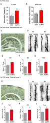
Oral administration of ALWPs to 5x FAD mice significantly enhances long-term memory and dendritic spine number. (A,B) 5x FAD mice were orally administered ALWPs (200 mg/kg, p.o.) or PBS (control) daily for 2 weeks. After 12 days, Y-maze (5x FAD CON, n = 8 mice; 5x FAD ALWPs, n = 8 mice) and NOR (5x FAD CON, n = 8 mice; 5x FAD ALWPs, n = 8 mice) tests were performed. (C,D) Representative AO and BS dendrites from hippocampal CA1 neurons of 5x FAD mice treated with PBS or ALWPs (200 mg/kg, p.o.) as indicated. (E–G) Dendritic spine density in hippocampal AO dendrites (E) and BS dendrites (F) and total average spine density (G) in hippocampal dendrites of 5x FAD mice (n = 8 mice/group, CA1: con, n = 58–72/neurons; ALWPs, n = 68–83/neurons). (H,I) Representative AO and BS dendrites from cortical layer V neurons of 5x FAD mice treated with PBS or ALWPs (200 mg/kg, p.o.) as indicated. (J–L) Dendritic spine density in cortical layer V AO dendrites (J) and BS dendrites (K) and total average spine density (L) in cortical dendrites of 5x FAD mice (n = 8 mice/group, con, n = 36–42/neurons ; ALWPs, n = 36–37/neurons). *p < 0.05, **p < 0.01, ***p < 0.0001.
Figure 6
Figure 6
ALWPs significantly promote spinogenesis in primary hippocampal neurons. (A) Primary hippocampal neurons were transfected with GFP plasmid DNA, followed by treatment with ALWPs (500 μg/ml) or PBS for 24 h and measurement of dendritic spine density. (B) Quantification of data from (A; CON, n = 29; ALWPS, n = 27). (C,D) The cumulative distribution percentage of spine head width and spine length in primary hippocampal neurons treated with ALWPs (500 μg/ml) or PBS for 24 h (Kolmogorov–Smirnov test). (E) Primary hippocampal neurons were transfected with GFP plasmid DNA, followed by treatment with ALWPs (750 μg/ml) or PBS for 24 h and measurement of dendritic spine density. (F) Quantification of data from (F; CON, n = 34; ALWPS, n = 26). (G,H) The cumulative distribution percentage of spine head width and spine length in primary hippocampal neurons treated with ALWPs (750 μg/ml) or PBS for 24 h (Kolmogorov–Smirnov test). *p < 0.05, **p < 0.01.
Figure 7
Figure 7
Oral administration of ALWPs to WT mice significantly promotes dendritic spine formation. (A,B) Representative AO and BS dendrites from hippocampal CA1 neurons of WT mice treated with the control or ALWPs (200 mg/kg, p.o.) as indicated. (C–E) Dendritic spine density in hippocampal AO dendrites (C) and BS dendrites (D) and total average spine density (E) in hippocampal dendrites of WT mice (n = 4 mice/group, con, n = 20–37/neurons ; ALWPs, n = 27–36/neurons). (F,G) The cumulative distribution percentage of spine length (F) and spine head width (G) in hippocampal CA1 AO dendrites of WT mice (Kolmogorov–Smirnov test). (H,I) The cumulative distribution percentage of spine length (H) and spine head width (I) in hippocampal CA1 BS dendrites of WT mice (Kolmogorov–Smirnov test). (J–K) Representative AO and BS dendrites from cortical layer V neurons of WT mice treated with the control or ALWPs (200 mg/kg, p.o.), as indicated. (L–N) Dendritic spine density in cortical layer V AO dendrites (L) and BS dendrites (M) and total average spine density (N) in cortical dendrites of WT mice (n = 4 mice/group, con, n = 42–44/neurons ; ALWPs, n = 42–43/neurons). (O,P) The cumulative distribution percentage of spine length (O) and spine head width (P) in cortical layer V AO dendrites of WT mice (Kolmogorov–Smirnov test). (Q–R) The cumulative distribution percentage of spine length (Q) and spine head width (R) in cortical layer V BS dendrites of WT mice (Kolmogorov–Smirnov test). *p < 0.05, **p < 0.01, ***p < 0.0001.
Figure 8
Figure 8
ALWPs require extracellular signal-regulated kinase (ERK) signaling to modulate dendritic spine formation. (A) Primary hippocampal neurons were transfected with GFP plasmid DNA for 24 h, treated with ALWPs (500 μg/ml) or PBS for 24 h, and immunostained with an anti-p-ERK antibody. (B) Quantification of data from (A; con, n = 88 dendrites; ALWPs, n = 100 dendrites). (C) Primary cortical neurons were treated with ALWPs (500 μg/ml) or PBS for 24 h and immunoblotted with anti-p-ERK or anti-ERK antibodies. (D,E) Quantification of data from (C; p-ERK, n = 8; ERK, n = 4). (F) Primary hippocampal neurons were transfected with GFP plasmid DNA for 24 h, pretreated with an ERK inhibitor (PD98059, 10 μM) or vehicle (1% DMSO) for 1 h, and treated with ALWPs (500 μg/ml) or PBS for 24 h, followed by measurement of dendritic spine density. (G) Quantification of data from (F; con, n = 86; ALWPs, n = 71; PD98059, n = 84; ALWPS+PD98059, n = 91). **p < 0.01, ***p < 0.0001.

Similar articles

Cited by

References

    1. Alonso A. D., Di Clerico J., Li B., Corbo C. P., Alaniz M. E., Grundke-Iqbal I., et al. . (2010). Phosphorylation of tau at Thr212, Thr231 and Ser262 combined causes neurodegeneration. J. Biol. Chem. 285, 30851–30860. 10.1074/jbc.m110.110957 - DOI - PMC - PubMed
    1. Asai M., Hattori C., Szabo B., Sasagawa N., Maruyama K., Tanuma S., et al. . (2003). Putative function of ADAM9, ADAM10, and ADAM17 as APP α-secretase. Biochem. Biophys. Res. Commun. 301, 231–235. 10.1016/s0006-291x(02)02999-6 - DOI - PubMed
    1. Asai H., Ikezu S., Tsunoda S., Medalla M., Luebke J., Haydar T., et al. . (2015). Depletion of microglia and inhibition of exosome synthesis halt tau propagation. Nat. Neurosci. 18, 1584–1593. 10.1038/nn.4132 - DOI - PMC - PubMed
    1. Augustinack J. C., Schneider A., Mandelkow E. M., Hyman B. T. (2002). Specific tau phosphorylation sites correlate with severity of neuronal cytopathology in Alzheimer’s disease. Acta Neuropathol. 103, 26–35. 10.1007/s004010100423 - DOI - PubMed
    1. Brewer G. J., Torricelli J. R., Evege E. K., Price P. J. (1993). Optimized survival of hippocampal neurons in B27-supplemented Neurobasal, a new serum-free medium combination. J. Neurosci. Res. 35, 567–576. 10.1002/jnr.490350513 - DOI - PubMed

LinkOut - more resources