. 2019 Aug 13:10:1877.
doi: 10.3389/fmicb.2019.01877. eCollection 2019.

Inhibition of HTLV-1 Infection by HIV-1 First- and Second-Generation Integrase Strand Transfer Inhibitors

Affiliations

Inhibition of HTLV-1 Infection by HIV-1 First- and Second-Generation Integrase Strand Transfer Inhibitors

Michał S Barski et al. Front Microbiol. .

Abstract

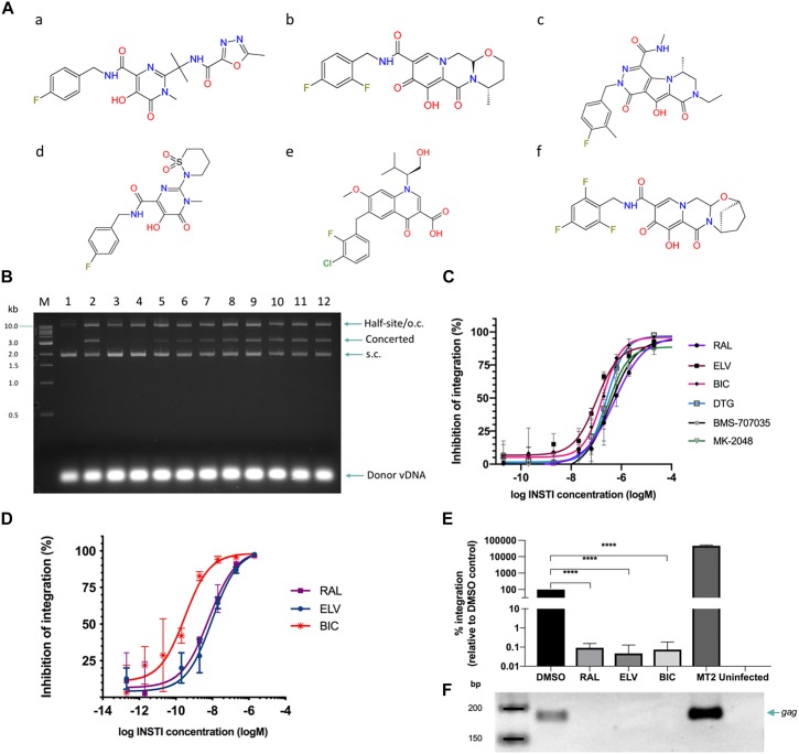
More than 10 million people worldwide are infected with the retrovirus human T-cell lymphotropic virus type 1 (HTLV-1). Infection phenotypes can range from asymptomatic to severe adult T-cell leukemia/lymphoma (ATLL) and HTLV-1-associated myelopathy. HTLV-1, like human immunodeficiency virus type 1 (HIV-1), is a blood-borne pathogen and viral infection happens in a similar fashion, with the major mode of transmission through breastfeeding. There is a strong correlation between time of infection and disease development, with a higher incidence of ATLL in patients infected during childhood. There is no successful therapeutic or preventative regimen for HTLV-1. It is therefore essential to develop therapies to inhibit transmission or block the onset/development of HTLV-1 associated diseases. Recently, we have seen the overwhelming success of integrase strand transfer inhibitors (INSTIs) in the treatment of HIV-1. Previously, raltegravir was shown to inhibit HTLV-1 infection. Here, we tested FDA-approved and two Phase II HIV-1 INSTIs in vitro and in a cell-to-cell infection model and show that they are highly active in blocking HTLV-1 infection, with bictegravir (EC50 = 0.30 ± 0.17 nM) performing best overall. INSTIs, in particular bictegravir, are more potent in blocking HTLV-1 transmission than tenofovir disproxil fumarate (TDF), an RT inhibitor. Our data suggest that HIV-1 INSTIs could present a good clinical strategy in HTLV-1 management and justifies the inclusion of INSTIs in clinical trials.

Keywords: HTLV-1; INSTI; PVL; bictegravir; elvitegravir; integrase; raltegravir.


Figures

FIGURE 1
FIGURE 1
Strand transfer activity of recombinant HTLV-1. (A) The formation of two types of product, concerted and half-site, is shown in the schematic. (B) The products are resolved on a 1.5% agarose gel and visualized with standard ethidum bromide treatment. M denotes the marker ladder (NEB, 1 kB), vDNA (U5_S18 3′-pre-processed, 18 nucleotide-long, double-stranded oligonucleotide donor mimicking the viral U5 LTR region), tDNA (supercoiled plasmid pGEM9Zf). (C) Activity of IN for U5- and U3-derived donors of different lengths is assayed. Sx: substrate of x nt in length. (D) HTLV-1 IN is sensitive to the use of different buffering species. When performing the strand transfer reaction in Hepes pH 7, B′γ strongly stimulates the concerted integration activity of HTLV-1 IN. In contrast, under optimal conditions (Pipes pH 7), no B′γ is required for high strand transfer activity. (E) Under conditions of optimal activity, IN remains active in the presence of 200 mM NaCl. Migration of DNA species in the gel is indicated on the right of the gel. S.c., supercoiled; o.c., open circular. The 1 kb DNA ladder (NEB, indicated on the left of the gel) was used as a reference. Presence of IN, B′γ, vDNA, tDNA, and buffer conditions are indicated above each gel.
FIGURE 2
FIGURE 2
INSTIs efficiently block HTLV-1 integration in vitro and HTLV-1 infection by cell-to-cell transmission. (A) Chemical structures of the INSTIs used in this study. (a) raltegravir, (b) dolutegravir, (c) MK-2048, (d) BMS-707035, (e) elvitegravir, and (f) bictegravir. Strand transfer is significantly inhibited by the addition of raltegravir (B) or other tested INSTIs (C). (B) Lane 1, negative control: no IN; lane 2: positive control, IN in the absence of drug. Lanes 3–12: in the presence of raltegravir; lane 3: 200 μM, lane 4: 20 μM, lane 5: 2 μM, lane 6: 634 nM, lane 7: 200 nM, lane 8: 63.4 nM, lane 9: 20 nM, lane 10: 2 nM, lane 11: 200 pM, and lane 12: 20 pM. Migration of DNA species in the gel is indicated on the right of the gel. S.c., supercoiled; o.c., open circular. The 1 kb DNA ladder (NEB, indicated on the left of the gel) was used as a reference. For each of the six compounds tested, the concerted integration bands were quantified by densitometry and values plotted as dose-response curves (C). (D–F) INSTIs efficiently block HTLV-1 infection in Jurkat cells. Jurkat cells were pre-treated with INSTIs (2 μM down to 0.2 pM serially diluted 1/10) and infected with HTLV-1 by co-culture with gamma-irrradiated MT-2 cells. Following depletion of MT-2 cells and expansion of the infected Jurkat cells, genomic DNA was isolated. (D) Infection was measured by determining the relative PVL of INSTI treated cells compared to DMSO treated control cells. The vertical axis shows the percentage of inhibition of HTLV-1 IN defined as the percentage of (1 – relative PVL). (E) Integrated provirus was quantified by Alu-qPCR and normalized to GAPDH numbers. DMSO treated Jurkat cells infected with HTLV-1 were arbitrarily set to 100%. Averages and standard deviations from three independent experiments are shown. RAL, raltegravir; ELV, elvitegravir; BIC, bictegravir; Uninfected stands for uninfected Jurkat cells (negative control). Data shown here is for the samples treated with 2 μM of the indicated drug. ****: p-value = 0.0001 (F) Representative gel illustrating products of the nested gag PCR (23 cycles) following Alu-PCR. Lanes correspond to the bar graphs in panel (E).
FIGURE 3
FIGURE 3
The active sites of HIV-1 and HTLV-1 IN are highly conserved. A homology model of HTLV-1 CCD is presented (A), overlaid on a previously reported model of HIV-1 intasome, based on the crystal structure of PFV intasome with raltegravir present in the active site (Krishnan et al., 2010). (C) A pairwise sequence similarity between the HIV-1 and HTLV-1 INs was conducted with MAFFT (Katoh and Standley, 2013), and the resulting similarity score for each residue was overlaid on the structure, represented in a color scale (red for highest conservation through to blue for lowest/no conservation), using the Alebrije script (B). The catalytic DDE triad as well as residues known to be involved in INSTI resistance are shown in stick representation (B) and indicated in the alignment with black squares (C). The residues of the DDE motif are also marked with asterisks. Catalytically important magnesium atoms are represented in purple (B). The predominance of residues highly conserved between the two INs is apparent within the active site. Pairwise alignment represented using ESPRIPT (Gouet et al., 1999), the model in panel (B) was produced using PyMOL (Schrodinger, 2015).

Similar articles

Cited by

References

    1. Afonso P. V., Mekaouche M., Mortreux F., Toulza F., Moriceau A., Wattel E., et al. (2010). Highly active antiretroviral treatment against STLV-1 infection combining reverse transcriptase and HDAC inhibitors. Blood 116 3802–3808. 10.1182/blood-2010-02-270751 - DOI - PubMed
    1. Alais S., Mahieux R., Dutartre H. (2015). Viral source-independent high susceptibility of dendritic cells to human T-cell leukemia virus type 1 infection compared to that of T lymphocytes. J. Virol. 89 10580–10590. 10.1128/JVI.01799-15 - DOI - PMC - PubMed
    1. Azran I., Schavinsky-Khrapunsky Y., Aboud M. (2004). Role of tax protein in human T-cell leukemia virus type-I leukemogenicity. Retrovirology 1 20. - PMC - PubMed
    1. Balakrishnan M., Jonsson C. B. (1997). Functional identification of nucleotides conferring substrate specificity to retroviral integrase reactions. J. Virol. 71 1025–1035. - PMC - PubMed
    1. Bar-Magen T., Sloan R. D., Donahue D. A., Kuhl B. D., Zabeida A., Xu H., et al. (2010). Identification of novel mutations responsible for resistance to MK-2048, a second-generation HIV-1 integrase inhibitor. J. Virol. 84 9210–9216. 10.1128/JVI.01164-10 - DOI - PMC - PubMed