

MALT1 Proteolytic Activity Suppresses Autoimmunity in a T Cell Intrinsic Manner
- PMID: 31474984
- PMCID: PMC6702287
- DOI: 10.3389/fimmu.2019.01898
MALT1 Proteolytic Activity Suppresses Autoimmunity in a T Cell Intrinsic Manner
Abstract
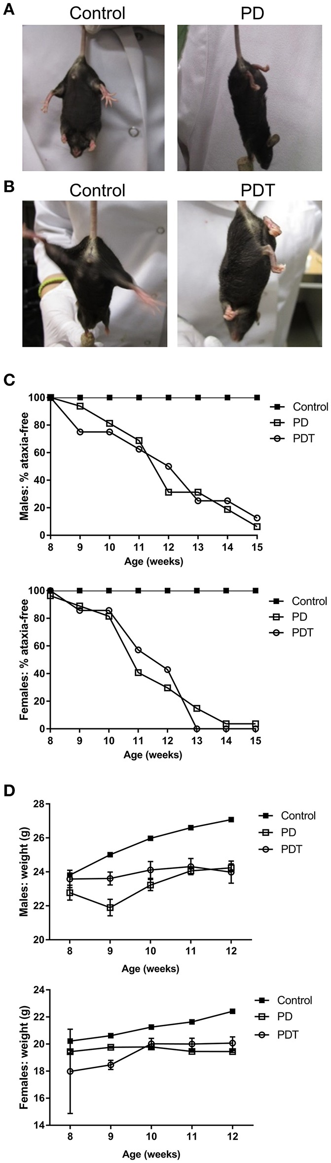
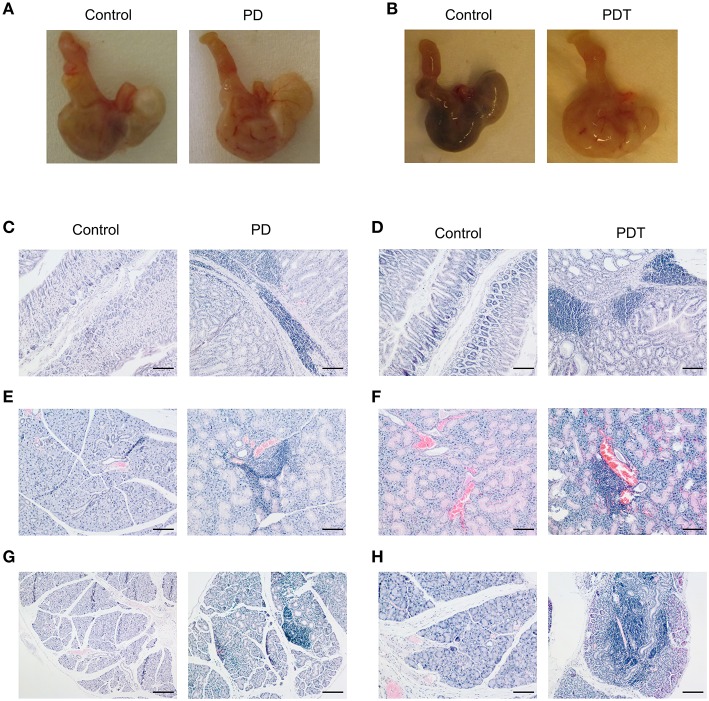
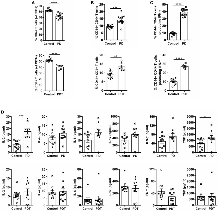
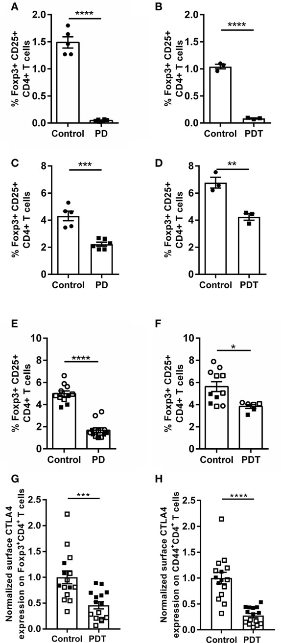
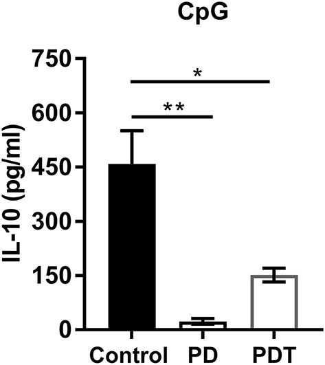
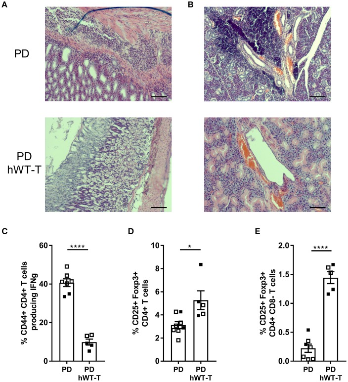
MALT1 is a central signaling component in innate and adaptive immunity by regulating NF-κB and other key signaling pathways in different cell types. Activities of MALT1 are mediated by its scaffold and protease functions. Because of its role in lymphocyte activation and proliferation, inhibition of MALT1 proteolytic activity is of high interest for therapeutic targeting in autoimmunity and certain lymphomas. However, recent studies showing that Malt1 protease-dead knock-in (Malt1-PD) mice suffer from autoimmune disease have somewhat tempered the initial enthusiasm. Although it has been proposed that an imbalance between immune suppressive regulatory T cells (Tregs) and activated effector CD4+ T cells plays a key role in the autoimmune phenotype of Malt1-PD mice, the specific contribution of MALT1 proteolytic activity in T cells remains unclear. Using T cell-conditional Malt1 protease-dead knock-in (Malt1-PDT) mice, we here demonstrate that MALT1 has a T cell-intrinsic role in regulating the homeostasis and function of thymic and peripheral T cells. T cell-specific ablation of MALT1 proteolytic activity phenocopies mice in which MALT1 proteolytic activity has been genetically inactivated in all cell types. The Malt1-PDT mice have a reduced number of Tregs in the thymus and periphery, although the effect in the periphery is less pronounced compared to full-body Malt1-PD mice, indicating that also other cell types may promote Treg induction in a MALT1 protease-dependent manner. Despite the difference in peripheral Treg number, both T cell-specific and full-body Malt1-PD mice develop ataxia and multi-organ inflammation to a similar extent. Furthermore, reconstitution of the full-body Malt1-PD mice with T cell-specific expression of wild-type human MALT1 eliminated all signs of autoimmunity. Together, these findings establish an important T cell-intrinsic role of MALT1 proteolytic activity in the suppression of autoimmune responses.
Keywords: Breg; CTLA-4; Treg; autoimmunity; inflammation; lymphocyte; paracaspase; protease.
Figures
Similar articles
-
Malt1 self-cleavage is critical for regulatory T cell homeostasis and anti-tumor immunity in mice.Eur J Immunol. 2018 Oct;48(10):1728-1738. doi: 10.1002/eji.201847597. Epub 2018 Aug 7. Eur J Immunol. 2018. PMID: 30025160 Free PMC article.
-
A MALT1 inhibitor suppresses human myeloid DC, effector T-cell and B-cell responses and retains Th1/regulatory T-cell homeostasis.PLoS One. 2020 Sep 1;15(9):e0222548. doi: 10.1371/journal.pone.0222548. eCollection 2020. PLoS One. 2020. PMID: 32870913 Free PMC article.
-
MALT1 protease function in regulatory T cells induces MYC activity to promote mitochondrial function and cellular expansion.Eur J Immunol. 2022 Jan;52(1):85-95. doi: 10.1002/eji.202149355. Epub 2021 Nov 2. Eur J Immunol. 2022. PMID: 34668583
-
Targeting MALT1 Proteolytic Activity in Immunity, Inflammation and Disease: Good or Bad?Trends Mol Med. 2016 Feb;22(2):135-150. doi: 10.1016/j.molmed.2015.12.004. Epub 2016 Jan 17. Trends Mol Med. 2016. PMID: 26787500 Review.
-
MALT1--a universal soldier: multiple strategies to ensure NF-κB activation and target gene expression.FEBS J. 2015 Sep;282(17):3286-97. doi: 10.1111/febs.13325. Epub 2015 Jun 10. FEBS J. 2015. PMID: 25996250 Review.
Cited by
-
CARD11 signaling in regulatory T cell development and function.Adv Biol Regul. 2022 May;84:100890. doi: 10.1016/j.jbior.2022.100890. Epub 2022 Feb 26. Adv Biol Regul. 2022. PMID: 35255409 Free PMC article. Review.
-
Potential role of MALT1 as a candidate biomarker of disease surveillance and treatment response prediction in inflammatory bowel disease patients.J Clin Lab Anal. 2022 Feb;36(2):e24130. doi: 10.1002/jcla.24130. Epub 2022 Jan 8. J Clin Lab Anal. 2022. PMID: 34997981 Free PMC article.
-
Proteolytic Activity of the Paracaspase MALT1 Is Involved in Epithelial Restitution and Mucosal Healing.Int J Mol Sci. 2023 Apr 17;24(8):7402. doi: 10.3390/ijms24087402. Int J Mol Sci. 2023. PMID: 37108564 Free PMC article.
-
Porcine Reproductive and Respiratory Syndrome Virus Adapts Antiviral Innate Immunity via Manipulating MALT1.mBio. 2022 Jun 28;13(3):e0066422. doi: 10.1128/mbio.00664-22. Epub 2022 Apr 25. mBio. 2022. PMID: 35467421 Free PMC article.
-
The role of the CBM complex in allergic inflammation and disease.J Allergy Clin Immunol. 2022 Nov;150(5):1011-1030. doi: 10.1016/j.jaci.2022.06.023. Epub 2022 Aug 16. J Allergy Clin Immunol. 2022. PMID: 35981904 Free PMC article. Review.
References
-
- Ruland J, Duncan GS, Wakeham A, Mak TW. Differential requirement for Malt1 in T and B cell antigen receptor signaling. Immunity. (2003) 19:749–58. - PubMed
Publication types
MeSH terms
Substances
LinkOut - more resources
Full Text Sources
Other Literature Sources
Molecular Biology Databases
Research Materials