. 2019 Aug 16:10:1954.
doi: 10.3389/fimmu.2019.01954. eCollection 2019.

Individual Effector/Regulator T Cell Ratios Impact Bone Regeneration

Affiliations

Individual Effector/Regulator T Cell Ratios Impact Bone Regeneration

Claudia Schlundt et al. Front Immunol. .

Abstract

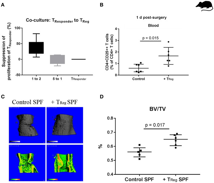
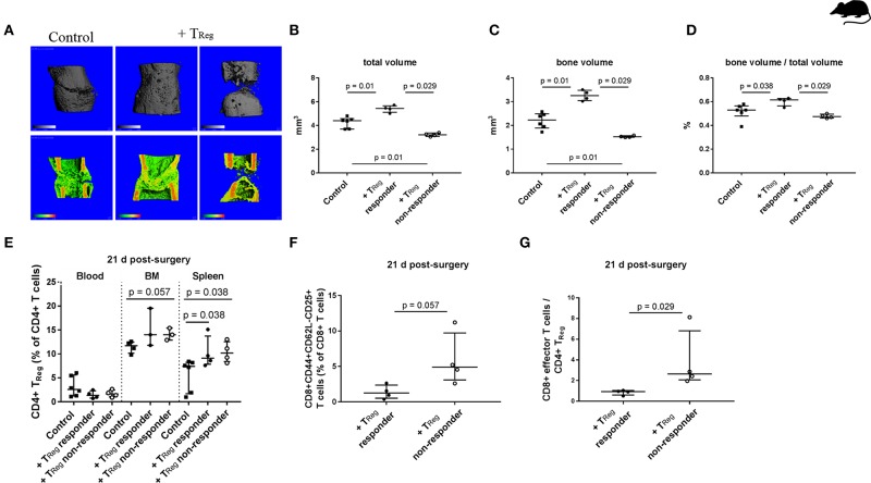
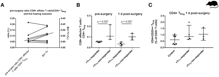
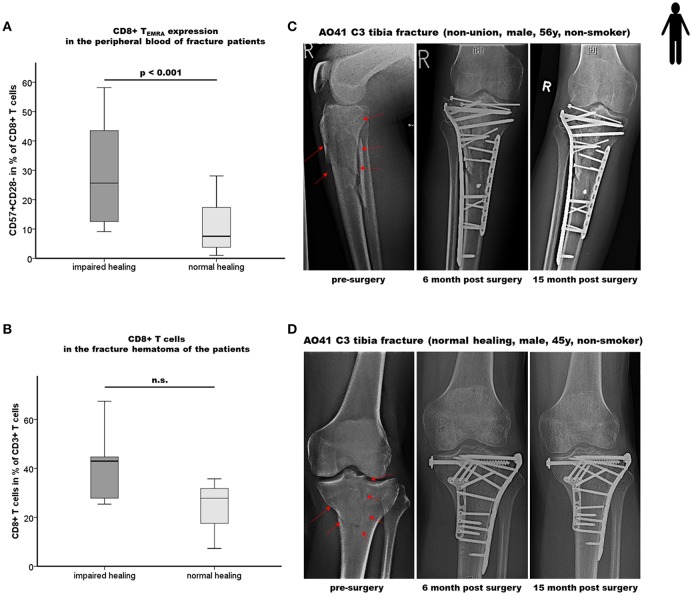
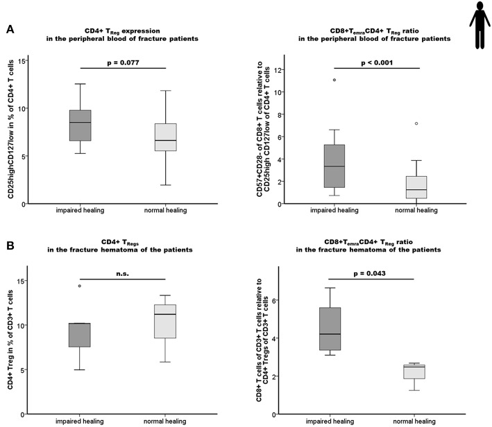
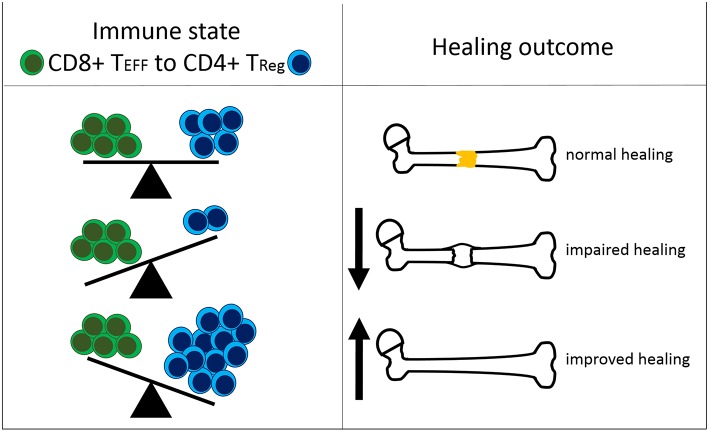
There is increasing evidence that T lymphocytes play a key role in controlling endogenous regeneration. Regeneration appears to be impaired in case of local accumulation of CD8+ effector T cells (TEFF), impairing endogenous regeneration by increasing a primary "useful" inflammation toward a damaging level. Thus, rescuing regeneration by regulating the heightened pro-inflammatory reaction employing regulatory CD4+ T (TReg) cells could represent an immunomodulatory option to enhance healing. Hypothesis was that CD4+ TReg might counteract undesired effects of CD8+ TEFF. Using adoptive TReg transfer, bone healing was consistently improved in mice possessing an inexperienced immune system with low amounts of CD8+ TEFF. In contrast, mice with an experienced immune system (high amounts of CD8+ TEFF) showed heterogeneous bone repair with regeneration being dependent upon the individual TEFF/TReg ratio. Thus, the healing outcome can only be improved by an adoptive TReg therapy, if an unfavorable TEFF/TReg ratio can be reshaped; if the individual CD8+ TEFF percentage, which is dependent on the individual immune experience can be changed toward a favorable ratio by the TReg transfer. Remarkably, also in patients with impaired fracture healing the TEFF/TReg ratio was higher compared to uneventful healers, validating our finding in the mouse osteotomy model. Our data demonstrate for the first time the key-role of a balanced TEFF/TReg response following injury needed to reach successful regeneration using bone as a model system. Considering this strategy, novel opportunities for immunotherapy in patients, which are at risk for impaired healing by targeting TEFF cells and supporting TReg cells to enhance healing are possible.

Keywords: bone healing; effector T cell; mouse model; regeneration; regulatory T (Treg) cell.


Figures

Figure 1
Figure 1
Enhancing bone healing by adoptive transfer of murine CD4+ TReg in SPF mice. (A) Demonstration of the functionality of isolated murine CD4+ TReg in a suppression assay, illustrated is the suppression of the proliferation of CD4+CD25- TResponder cells by using freshly isolated CD4+ TReg (n = 6). (B) Status of CD4+ TReg in the peripheral blood of the experimental mice 1 d after adoptive transfer, control (n = 6) vs. CD4+ TReg enriched mice (n = 6). (C,D) Improved bone healing by adoptive CD4+ TReg transfer as demonstrated by μCT evaluation after 21 d, control (C, left, control SPF) vs. TReg enriched mice (C, right, +TReg SPF). Quantification of the μCT data revealed a significantly higher BV/TV in the mice that received a CD4+ TReg transfer (+TReg) (n = 6) compared to the control SPF mice (n = 5) (D). BV, bone volume; TV, total callus volume; BV/TV, ratio of BV to TV; Mann-Whitney U-test.
Figure 2
Figure 2
Heterogeneous impact of adoptive transfer of CD4+ TReg on bone healing outcome in mice kept under non-SPF housing conditions. (A–D) μCT evaluation of the healing outcome after adoptive CD4+ TReg transfer after 21 d. (A) Representative images of the analyzed volume of interest of the healing bones are shown. +TReg treated mice clustered into responder (+TReg left, n = 4) and non-responder (+TReg right, n = 4). Control mice showed no complete bridging after 21 d (left, control, n = 6). In contrast, +TReg responder showed complete bridging, whereas non-responder showed no bridging. (B–D) Quantification of the μCT analysis, + TReg responder vs. + TReg non-responder vs. control mice. (E–G) Evaluation of the immune cell composition of +TReg responder vs. non-responder 21 d post-surgery. (E) Confirmation of CD4+ TReg engraftment in both, +TReg responder and non-responder compared to the control. (F,G) +TReg non-responder showed an elevated percentage of CD8+ TEFF (F) as well as a significantly higher ratio of CD8+ TEFF /CD4+ TReg (G) in the peripheral blood in comparison to the +TReg responder. TV, total volume; BV, bone volume; BV/TV, ratio of BV to TV; Mann–Whitney U-test, Bonferroni correction (B–E).
Figure 3
Figure 3
Individual effector/regulatory T cell ratio pre-osteotomy determines the healing outcome in mice. (A) Mice having a lower pre-surgery CD8+ TEFF to CD4+ TReg ratio (left y-axis) showed a higher BV/TV (right y-axis, axis is reversed for a clearer depiction). Dashed line indicates the cut off between the +TReg responder (below the dashed line) and the +TReg non-responder (above the dashed line). (B) Adoptive CD4+ TReg transfer did not reshape the unfavorable CD8+ TEFF/CD4+ TReg ratio. (C) Engraftment of CD4+CD25++ T cells 1 d post-surgery is elevated in both, the +TReg responder (n = 4) and +TReg non-responder (n = 3) in comparison to the control group (n = 6). 45BV, total bone volume; TV, total callus volume; BV/TV, ratio of BV to TV; Mann–Whitney U-test.
Figure 4
Figure 4
Impaired bone healing correlates with an elevated CD8+ TEMRA level in both the peripheral blood as well as in the local fracture hematoma of patients. (A) Results of the flow cytometry analyses of terminally differentiated CD8+ effector T cells (CD8+TEMRA) in the peripheral blood of fracture patients at the time of operation are shown. Impaired fracture healing patients showed a significantly higher frequency of CD45+CD3+CD8+CD57+CD28- T cells (CD57+CD28-+ in % of CD45+CD3+CD8+ T cells) compared to the normal healing patients indicates a dramatic increase of CD8+ TEMRA: n = 12 for the impaired healing group, n = 23 for the normal healing group. (B) Quantification of CD8+ T cells in the hematoma region of fracture healing patients by epigenetic qPCR analysis. Impaired fracture healing patients showed an elevated level of CD8+ T cells in the local fracture area: n = 5 for the impaired healing group, n = 3 for the normal healing group (C,D) Representative x-ray images from the study cohort over a period of 15 months including the pre-surgery as well as the 6 and 15 months post-surgery states of fracture healing of an impaired healing (C) and a normal healing (D) patient, respectively. Student's t-test.
Figure 5
Figure 5
Impaired fracture healing correlates with an elevated level of CD4+ TReg and a higher ratio of CD8+ TEMRA/CD4+ TReg. (A) Results of the flow cytometry analyses of the CD4+ TReg and the ratio CD8+TEMRA/CD4+TReg in the peripheral blood of fracture patients at the time of operation are shown. Impaired fracture healing patients showed slightly elevated levels of CD45+CD3+CD4+CD25highCD127low TReg (CD25highCD127low in % of CD45+CD3+CD4+) compared to the normal healing patients indicates a partial, but not sufficient compensatory effect. The ratio of CD8+TEMRA/CD4+TReg was significantly higher in patients with a delayed healing: n = 12 for the impaired healing group, n = 23 for the normal healing group. (B) Impaired fracture healing patients showed a significantly higher ratio of CD8+T cells/CD4+ TReg in the hematoma region demonstrating an insufficient compensatory effect of the CD4+ TReg in the impaired fracture healing patients: n = 5 for the impaired healing group, n = 3 for the normal healing group. Student's t-test.
Figure 6
Figure 6
Interdependency of the immune status and the healing outcome after bone fracture. The ratio of CD8+ effector T cells (CD8+ TEFF) to CD4+ regulatory T cells (CD4+ TReg) affects the healing outcome after bone fracture. Under normal healing conditions, the T cell ratio is balanced. An elevated amount of CD8+ TEFF relative to CD4+ TReg is unfavorable for successful healing. A shifted balance in the CD4+ TRegsb direction improves bone fracture healing.

References

    1. Einhorn TA, Gerstenfeld LC. Fracture healing: mechanisms and interventions. Nat Rev Rheumatol. (2015) 11:45–54. 10.1038/nrrheum.2014.164 - DOI - PMC - PubMed
    1. Dimitriou R, Tsiridis E, Giannoudis PV. Current concepts of molecular aspects of bone healing. Injury. (2005) 36:1392–404. 10.1016/j.injury.2005.07.019 - DOI - PubMed
    1. Schlundt C, Bucher CH, Tsitsilonis S, Schell H, Duda GN, Schmidt-Bleek K. Clinical and research approaches to treat non-union fracture. Curr Osteoporos Rep. (2018) 16:155–68. 10.1007/s11914-018-0432-1 - DOI - PubMed
    1. Bucher CH, Lei H, Duda GN, Volk HD, Schmidt-Bleek K. The role of immune Reactivity in bone regeneration. In: Zorzi AR, editor. Advanced Techniques in Bone Regeneration. IntechOpen. (2016). Available online at: https://www.intechopen.com/books/advanced-techniques-in-bone-regeneratio...
    1. Shapouri-Moghaddam A, Mohammadian S, Vazini H, Taghadosi M, Esmaeili SA, Mardani F, et al. . Macrophage plasticity, polarization, and function in health and disease. J Cell Physiol. (2018) 233:6425–40. 10.1002/jcp.26429 - DOI - PubMed

Publication types