Case Reports
. 2019 Aug 14:9:734.
doi: 10.3389/fonc.2019.00734. eCollection 2019.

Pembrolizumab in a Patient With a Metastatic CASTLE Tumor of the Parotid

Affiliations
Case Reports

Pembrolizumab in a Patient With a Metastatic CASTLE Tumor of the Parotid

Lisa Lorenz et al. Front Oncol. .

Abstract

Carcinoma showing thymus-like elements (CASTLE) is a rare tumor, most commonly found in the thyroid gland. Here we report a case of CASTLE tumor localized to the parotid gland, recognized in retrospect after a late manifestation of symptomatic pleural carcinomatosis. The original tumor in the parotid gland was treated by surgery followed by radiotherapy. Ten years later, a metastatic disease with recurrent pleural effusions occurred. Pleural carcinomatosis was strongly positive for CD5, CD117, and p63 as was the original tumor of the parotid, which allowed the diagnosis of a CASTLE tumor. Additionally, the pleural tumor expressed high levels of programmed death ligand 1 (PD-L1), and the patient underwent treatment with the monoclonal PD-L1 inhibitor pembrolizumab achieving a partial remission. To the best of our knowledge, this is the first patient with a metastatic CASTLE tumor treated with a PD-L1 inhibitor.

Keywords: CASTLE; PD-L1; carcinoma; checkpoint inhibitor; extrathyroidal; immunotherapy; parotid; thymus-like.


Figures
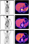
Figure 1
Figure 1
Initial and follow-up FDG PET/CT: (A) Initial FDG-MIP with multiple hypermetabolic lesions in the upper lobe of the right lung, in mediastinal, hilar and upper mesenteric lymph nodes and at the left pleura. Increased turnover of the bone marrow. (B) Initial fusion FDG-PET/CT axial slice with pathologic high FDG-uptake in the left pleura, upper mesenteric lymph nodes, and bone marrow. Normal presentation of liver, spleen, and stomach. (C) Follow-up FDG-MIP after 4 months on pembrolizumab shows a generally reduced metabolism at all tumor locations. Normalization of the bone marrow turnover. Due to newly incipient traumatic injury at the left shoulder the patient could not lift up the left arm. (D) Fusion FDG-PET/CT axial slice after 4 months on pembrolizumab with residual FDG-uptake at the left pleura and in upper mesenteric lymph nodes. Normal presentation of the bone marrow. (E) Follow-up FDG-MIP after 11 months on pembrolizumab shows further regression of lesions in the upper lobe of the right lung as well as in mediastinal, hilar, and upper mesenteric lymph nodes (the focal lesions in the left mediastinum and supraclavicular right represent the central venous catheter). Residual focal FDG-Uptake in the left pleura. Normal turnover of the bone marrow. (F) Fusion FDG-PET/CT axial slice after 11 months on pembrolizumab with only little elevated FDG-uptake in the left pleura. The formerly upper mesenteric lymph node is healed up. Normal presentation of liver, spleen, stomach, and bone marrow.
Figure 2
Figure 2
Histomorphological and immunohistochemical analysis of parietal pleura, biopsy from 2018: (A) H & E staining, (B) Immunostaining for CD5, (C) Immunostaining for CD117, and (D) Immunostaining for PD-L1.
Figure 3
Figure 3
Histomorphological and immunohistochemical analysis of parotid, tumor excisate from 2008: (A) H & E staining, (B) Immunostaining for CD5, and (C) Immunostaining for CD117.

References

    1. Youens KE, Bean SM, Dodd LG, Jones CK. Thyroid carcinoma showing thymus-like differentiation (CASTLE): case report with cytomorphology and review of the literature. Diagn Cytopathol. (2011) 39:204–9. 10.1002/dc.21399 - DOI - PubMed
    1. Miyauchi A, Kuma K, Matsuzuka F, Matsubayashi S, Kobayashi A, Tamai H, et al. . Intrathyroidal epithelial thymoma - an entity distinct from squamous-cell carcinoma of the thyroid. World J Surg. (1985) 9:128–34. 10.1007/BF01656263 - DOI - PubMed
    1. Chan JK, Rosai J. Tumors of the neck showing thymic or related branchial pouch differentiation: a unifying concept. Hum Pathol. (1991) 22:349–67. 10.1016/0046-8177(91)90083-2 - DOI - PubMed
    1. Marx A, Chan JK, Coindre JM, Detterbeck F, Girard N, Harris NL, et al. . The 2015 world health organization classification of tumors of the thymus: continuity and changes. J Thoracic Oncol. (2015) 10:1383–95. 10.1097/JTO.0000000000000654 - DOI - PMC - PubMed
    1. Ge W, Yao YZ, Chen G, Ding YT. Clinical analysis of 82 cases of carcinoma showing thymus-like differentiation of the thyroid. Oncol Lett. (2016) 11:1321–6. 10.3892/ol.2015.4055 - DOI - PMC - PubMed

Publication types