. 2019 Sep 27:19:715-727.
doi: 10.1016/j.isci.2019.08.008. Epub 2019 Aug 8.

Pim1 Impacts Enterovirus A71 Replication and Represents a Potential Target in Antiviral Therapy

Affiliations

Pim1 Impacts Enterovirus A71 Replication and Represents a Potential Target in Antiviral Therapy

Fanghang Zhou et al. iScience. .

Abstract

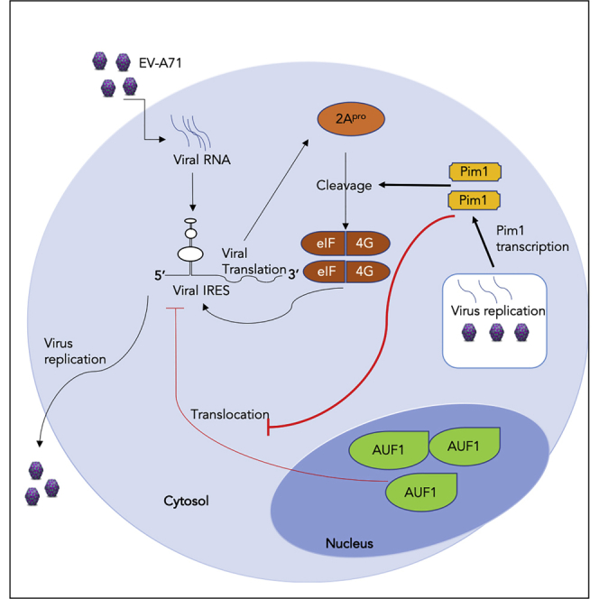
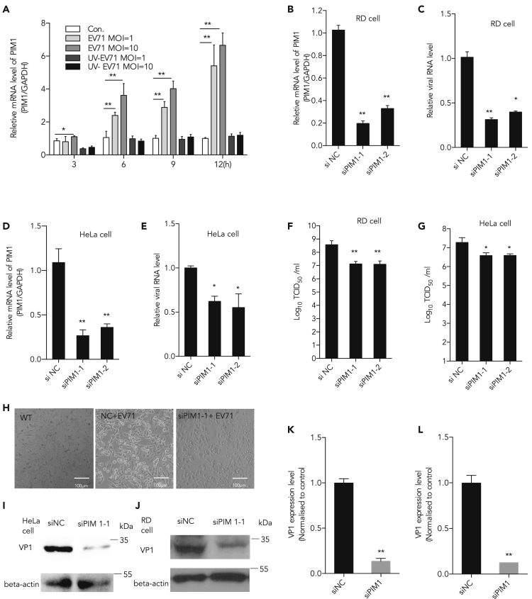
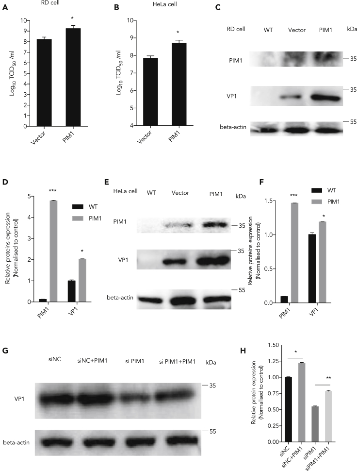
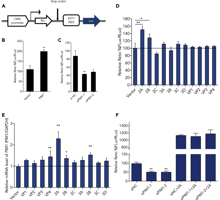
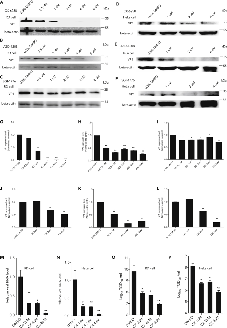
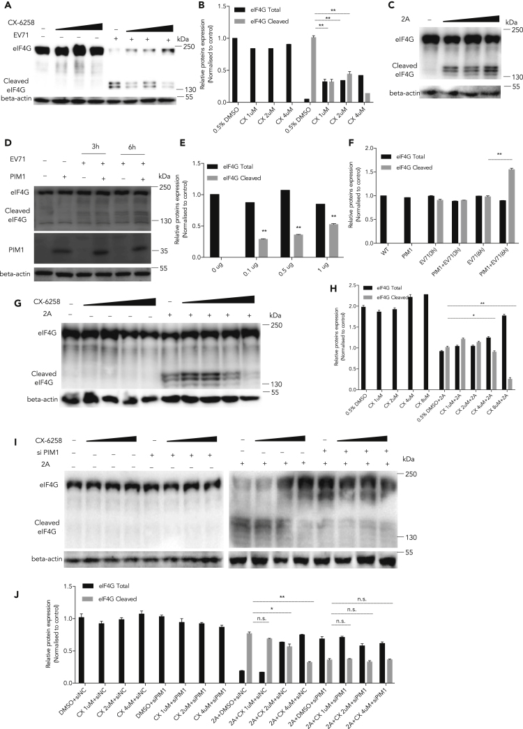
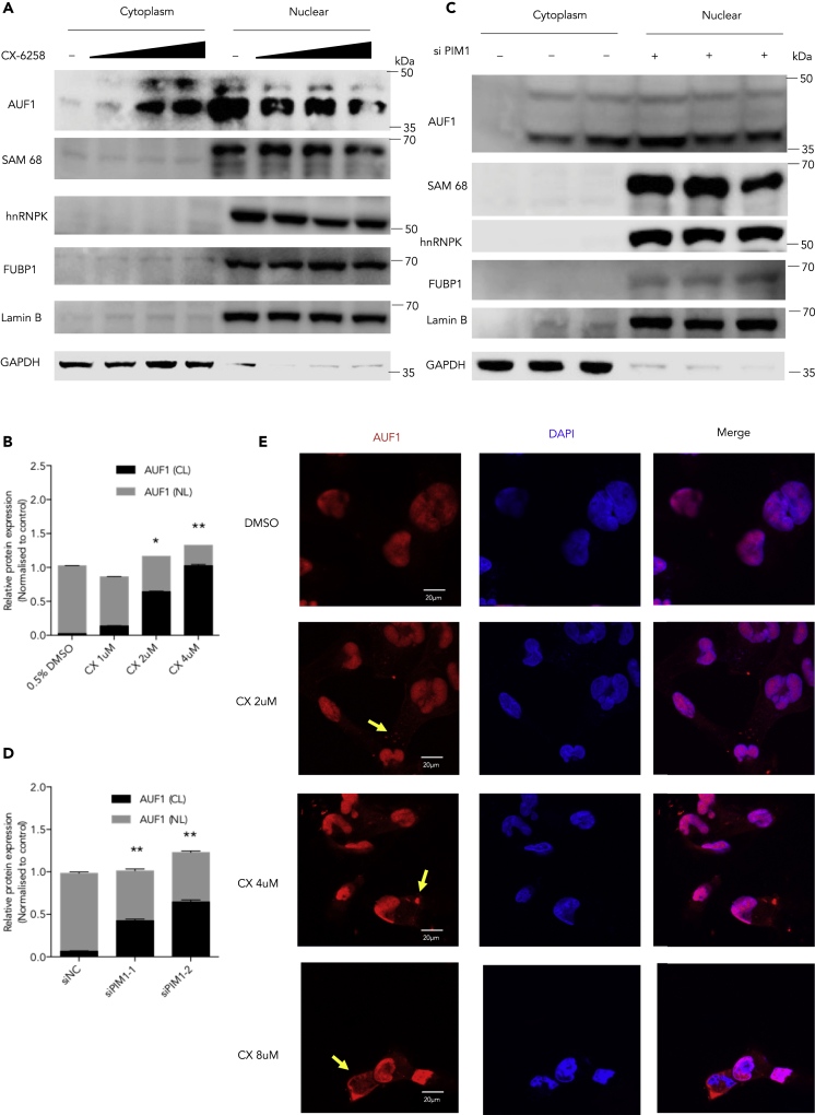
Enterovirus A71 (EV-A71) infection causes hand-foot-and-mouth disease (HFMD) and fatal neurological diseases, and there are no effective treatments. Host factors play key roles in establishing viral infection and determining the disease progression and outcome of antiviral therapies. In this study, we found that the expression of Pim1 was significantly upregulated in EV-A71 infection. Ectopic expression or silencing of Pim1 promoted or inhibited EV-A71 replication through two distinct mechanisms. Pim1 enhanced viral IRES activity by increasing viral 2A protease-mediated eIF4G cleavage and blocked AUF1, a suppressor of IRES, translocation from the nucleus to cytosol. More importantly, we discovered that Pim1 inhibitors (SGI-1776, AZD-1208, and CX-6258) reduced EV-A71 reproduction. Particularly, CX-6258 remarkably reduced EV-A71 reproduction more than 1,000 times, providing a potential therapeutic agent for EV-A71 treatment.

Keywords: Biochemistry; Biological Sciences; Cell Biology; Microbiology; Molecular Microbiology; Viral Microbiology; Virology.


Conflict of interest statement

The authors declare no competing financial interests.

Figures

None
Graphical abstract
Figure 1
Figure 1
Inhibiting EV-A71 Replication by Knockdown of Pim1 (A) RD cells were infected with EV-A71 or inactive EV-A71 (UV light-inactive virus, negative control) at MOI of 10, and cellular mRNA was extracted at different time points p.i. Pim1 mRNA level was determined by qRT-PCR. (B and D) Pim1 was silenced by two individual siRNA duplexes si-Pim1-1 and si-Pim1-2. The relative cellular Pim1 mRNA level was quantified by qRT-PCR at 48 h after transfection in both (B) RD and (D) HeLa cells. GAPDH was used as the internal control. (C and E) (C) RD and (E) HeLa cells treated with Pim1-specific siRNA and infected with EV-A71 at MOI 0.01. The extracellular viral RNA level was calculated at 48 h. (F and G) Viral titer was further determined in both (F) RD and (G) HeLa cells. (H) Cell survival image was taken in RD cells. (I and J) Pim1 was silenced in EV-A71-infected (J) RD and (I) HeLa cells. (K) The quantification results of (I). (L) The quantification results of (J). Protein levels of viral VP1 protein were detected and quantitated. The density of VP1 to β-actin was set as 1, and the relative fold change is indicated. Data are represented as mean ± SD (n = 3). Student's t test, *p < 0.05, compared with mock group; **p < 0.01, compared with mock group.
Figure 2
Figure 2
Promoting EV-A71 Replication by Ectopic Expression of Pim1 (A and B) After ectopic expression of Pim1, viral titer was determined following the below-mentioned methods in (A) RD and (B) HeLa cells. (C and E) Pim1 was ectopically expressed in EV-A71-infected cells at MOI 1 for 24 h in (C) RD and (E) HeLa cells. Protein levels of Pim1 and viral protein VP1 were detected. β-Actin was used as the internal control. (D) The quantification results of (C). (F) The quantification results of (E). (G) siRNA targeting Pim1 3′ UTR at 40 nM was co-transfected with the Pim1 expression plasmid for 48 h in RD cells and then infected with EV71 at MOI 1 for 24 h. (H) The quantification results of (G). The cell lysates were collected for western blot assay. Protein levels were detected and quantitated. The density of individual protein to β-actin in the control was set as 1, and the relative fold change is indicated. Data are represented as mean ± SD (n = 3). *p < 0.05, compared with mock group; **p < 0.01, compared with mock group.
Figure 3
Figure 3
Enhancing Viral IRES Activity by Pim1 (A) A diagram of the dicistronic reporter plasmid, which was used to evaluate EV-A71 IRES activity when Pim1 was modulated. (B and C) (B) The relative IRES activity after ectopic expression of Pim1 or (C) knockdown of Pim1 in 293T cells. (D) After co-transfection of the reporter plasmid and individual viral protein expression plasmid into 293T cells for 48 h, the RLuc and FLuc activities were analyzed. (E) The relative mRNA level of Pim1 was determined by qRT-PCR assay after transfection of the individual viral protein expression plasmid into 293T cells for 48 h. (F) IRES activities after ectopic expression of 2Apro, with or without silencing of Pim1. Results are represented as mean ± SD from three independent experiments. *p < 0.05, **p < 0.01.
Figure 4
Figure 4
Suppressing EV-A71 Infection by Pim1 Inhibitors (A–F) Different Pim1 inhibitors CX-6258 (A and D), AZD-1208 (B and E), and SGI-1776 (C and F) were added in the culture media at the indicated concentration for 2 h with 10% fetal bovine serum and then cells were infected with EV-A71 at MOI 1 for 24 h; 0.5% DMSO was loaded in the control group. The VP1 protein level was determined, and beta-actin was loaded as internal control. (G and J) The quantification results of (A) and (D), respectively. (H and K) The quantification results of (B) and (E), respectively. (I and L) The quantification results of (C) and (F), respectively. (M and N) RD and HeLa Cells were firstly treated with CX-6258 at the indicated concentrations for 2 h, then infected with EV-A71 at MOI 0.01 for 72 h. The extracellular viral RNA levels in the supernatant were determined by qRT-PCR. (O and P) RD and HeLa Cells were firstly treated with CX-6258 at the indicated concentration for 2 h, then infected EV-A71 at MOI 0.01 for 72 h. The viral titer in the supernatant was measured by TCID50 assay. Protein levels were detected and quantitated by image density. The density of individual protein to β-actin in the control was set as 1, and the relative fold change is indicated. Results are represented as mean ± SD from three independent experiments. *p < 0.05, **p < 0.01, ***p < 0.001. See also Figure S1.
Figure 5
Figure 5
Inhibiting 2Apro-mediated eIF4G Cleavage by CX-6258 (A) RD cells were treated with different concentrations of CX-6258 and then infected with EV-A71 at MOI 10. After culture for 9 h, cell lysates were collected and the cleavage of eIF4G was determined. (B) The quantification results of (A). (C) HEK293T cells were transfected with p2Apro (0.1, 0.5, and 1 μg) for 36 h, and the cleavage of eIF4G was determined. (D) HEK293T cells were first treated with different concentrations of CX-6258 for 2 h and transfected with 1 μg 2Apro-expressing plasmid for 36 h. The cleavage of eIF4G was determined. (E and F) The quantification results of (C) and (D), respectively. (G) 293T cells were transfected with a Pim1-expressing plasmid for 48 h and then infected with EV-A71 at MOI 10 for the indicated time. The cleavage of eIF4G was determined. (H) The quantification results of (G). (I) 293T cells were first transfected with Pim1-specific siRNA for 48 h and then treated with CX-6258 at the indicated concentrations for 2 h, followed by transfection of a 2Apro-expressing plasmid at 48 h. Cell lysates were collected, and the status of eIF4G cleavage was examined. (J) The quantification results of (I). Protein levels were detected and quantitated by image density. The density of individual protein to β-actin or GAPDH in the control was set as 1, and the relative fold change is indicated. Results are represented as mean ± SD from three independent experiments. *p < 0.05, **p < 0.01.
Figure 6
Figure 6
Inducing AUF1 Cytoplasmic Accumulation by CX-6258 (A) RD cells were treated with CX-6258 at the indicated concentrations for 2 h, and then the cytoplasmic and nuclear proteins were extracted. The cytoplasm and nuclear distribution of AUF1, Sam68, hnRNP K and FUBP1 were determined by western blot assay. (B) The quantification results of (A). (C) Pim1-specific siRNAs were transfected into RD cells for 48 h. The cytoplasmic and nuclear proteins were extracted. The protein levels of AUF1, Sam68, hnRNP K, and FUBP1 were examined by western blot assays. (D) The quantification results of (C). (E) The cytoplasmic and nuclear distribution of AUF1 was detected in RD cells after treating with CX-6258 at indicated concentrations for 2 h under a fluorescence microscope. The yellow arrows indicated the representative cells of AUF1 translocated in the cytosole. Results are represented as mean ± SD from three independent experiments. *p < 0.05, **p < 0.01.
Figure 7
Figure 7
Diagram of Pim1 Function in EV-A71 Infection

Similar articles

Cited by

References

    1. Braso-Maristany F., Filosto S., Catchpole S., Marlow R., Quist J., Francesch-Domenech E., Plumb D.A., Zakka L., Gazinska P., Liccardi G. PIM1 kinase regulates cell death, tumor growth and chemotherapy response in triple-negative breast cancer. Nat. Med. 2016;22:1303–1313. - PMC - PubMed
    1. Chen L.S., Redkar S., Bearss D., Wierda W.G., Gandhi V. Pim kinase inhibitor, SGI-1776, induces apoptosis in chronic lymphocytic leukemia cells. Blood. 2009;114:4150–4157. - PMC - PubMed
    1. Dan X., Wan Q., Yi L., Lu J., Jiao Y., Li H., Song D., Chen Y., Xu H., He M.L. Hsp27 responds to and facilitates enterovirus A71 replication by enhancing viral internal ribosome entry site-mediated translation. J. Virol. 2019;93 e02322–18. - PMC - PubMed
    1. De Vries M., Smithers N.P., Howarth P.H., Nawijn M.C., Davies D.E. Inhibition of Pim1 kinase reduces viral replication in primary bronchial epithelial cells. Eur. Respir. J. 2015;45:1745–1748. - PubMed
    1. Dong Q., Men R., Dan X., Chen Y., Li H., Chen G., Zee B., Wang M.H.T., He M.L. Hsc70 regulates the IRES activity and serves as an antiviral target of enterovirus A71 infection. Antiviral. Res. 2018;150:39–46. - PubMed