. 2019 Sep 2;19(1):77.
doi: 10.1186/s12880-019-0376-7.

Combination of CEUS and MRI for the diagnosis of periampullary space-occupying lesions: a retrospective analysis

Affiliations

Combination of CEUS and MRI for the diagnosis of periampullary space-occupying lesions: a retrospective analysis

Xin-Pei Chen et al. BMC Med Imaging. .

Abstract

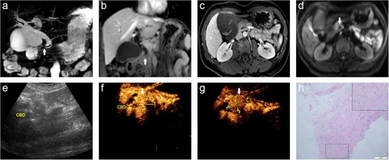
Background: The value of magnetic resonance imaging (MRI), contrast-enhanced ultrasound (CEUS), and the combination of CEUS and MRI (CCWM) for the diagnosis of periampullary space-occupying lesions (PSOL) was investigated.

Methods: A total of 102 patients diagnosed with PSOLs by surgery or biopsy were recruited retrospectively. The sensitivity, specificity, positive predictive value (PPV), negative predictive value (NPV), and accuracy of MRI, CEUS, and CCWM were analyzed.

Results: MRI, CEUS, and CCWM allowed for the accurate detection of 91.17, 92.15, and 99.01% of PSOLs, respectively. The specificity, PPV, and accuracy of CCWM were significantly different from MRI and CEUS (p < 0.05). However, there the sensitivity and NPV were not significantly different among the three diagnostic technologies. In addition, the specificity, PPV, and accuracy were not significantly different between MRI and CEUS (all p > 0.05).

Conclusions: CCWM is valuable for differentiating benign and malignant PSOL, which provides important guiding significances for the clinic.

Keywords: CCMW; Contrast-enhanced ultrasound (CEUS); Magnetic resonance imaging (MRI); Periampullary cancer; Periampullary space-occupying lesions.


Conflict of interest statement

The authors declare that they have no competing interests.

Figures

Fig. 1
Fig. 1
A 58-year-old male was misdiagnosed as having a common bile duct stone by MRI, which was pathologically-confirmed as choledochal adenocarcinoma; (a) MRCP image displays the narrowing (arrow) of the distal CBD suddenly and the expansion of the biliary tract; (b) Axial T1-weighted images and (c) T1-weighted DCE-MRI show high signal nodules in the CBD, and the possibility of stones was considered (arrow), yet no enhancement is seen in the structure of bile duct wall; (d) CUS shows dilated CBD; (e, f) CEUS shows a cauliflower-like mass with uneven enhancement (arrow), which should be considered for common bile duct carcinomas; (g) Histopathological examination reveals that some malignant glands infiltrated into the bile duct wall. CBD: common bile duct; PA: pancreas
Fig. 2
Fig. 2
A 56-year-old female with common bile duct inflammation stenosis was misdiagnosed as having duodenal papillary carcinoma by CEUS; (a) MRCP image shows dilation of the intrahepatic and extrahepatic bile ducts with the lower part of the CBD tapering gradually (arrow) and the gallbladder increasing significantly. b Coronary contrast-enhanced T1WI shows the lower part of the CBD gradually becoming thin and occlusive (arrow) with no definite sign of mass-occupying lesions in the travel area of the intrahepatic and extrahepatic bile ducts; (c) Axial T1-weighted DCE-MR image shows the obstruction plane is in the lower part of the CBD (arrow), and the possibility of inflammatory stenosis and occlusion is considered. d DWI shows no abnormal signal is found; (e) CUS displays the choledochoectasia; (f, g) Uneven and highly intensified duodenal papilla region (arrow), and duodenal papillary carcinoma is considered; (h) Histopathological examination shows proliferative fibrous connective tissue of the bile duct wall, and the hyperplastic bile duct epithelium can be seen in the deep layer of the bile duct wall. CBD: common bile duct

Similar articles

Cited by

References

    1. Berberat PO, Kunzli BM, Gulbinas A, Ramanauskas T, Kleeff J, Muller MW, et al. An audit of outcomes of a series of periampullary carcinomas. Eur J Surg Oncol. 2009;35:187–191. doi: 10.1016/j.ejso.2008.01.030. - DOI - PubMed
    1. Baghmar S, Agrawal N, Kumar G, Bihari C, Patidar Y, Kumar S, et al. Prognostic factors and the role of adjuvant treatment in Periampullary carcinoma: a single-Centre experience of 95 patients. J Gastrointest Cancer. 2018. - PubMed
    1. Hashemzadeh Shahryar, Mehrafsa Behzad, Kakaei Farzad, Javadrashid Reza, Golshan Rosa, Seifar Fatemeh, Hajibonabi Farid, Salmannezhad Khorami Farzad. Diagnostic Accuracy of a 64-Slice Multi-Detector CT Scan in the Preoperative Evaluation of Periampullary Neoplasms. Journal of Clinical Medicine. 2018;7(5):91. doi: 10.3390/jcm7050091. - DOI - PMC - PubMed
    1. Puli SR, Singh S, Hagedorn CH, Reddy J, Olyaee M. Diagnostic accuracy of EUS for vascular invasion in pancreatic and periampullary cancers: a meta-analysis and systematic review. Gastrointest Endosc. 2007;65:788–797. doi: 10.1016/j.gie.2006.08.028. - DOI - PubMed
    1. Chang S, Lim JH, Choi D, Kim SK, Lee WJ. Differentiation of ampullary tumor from benign papillary stricture by thin-section multidetector CT. Abdom Imaging. 2008;33:457–462. doi: 10.1007/s00261-007-9295-0. - DOI - PubMed