. 2019 Sep 2;7(1):142.
doi: 10.1186/s40478-019-0787-2.

Unique α-synuclein pathology within the amygdala in Lewy body dementia: implications for disease initiation and progression

Affiliations

Unique α-synuclein pathology within the amygdala in Lewy body dementia: implications for disease initiation and progression

Zachary A Sorrentino et al. Acta Neuropathol Commun. .

Abstract

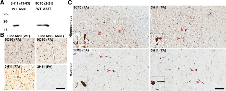
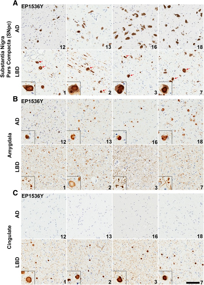
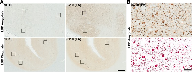
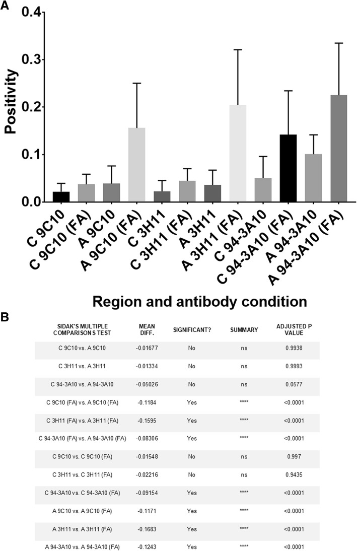
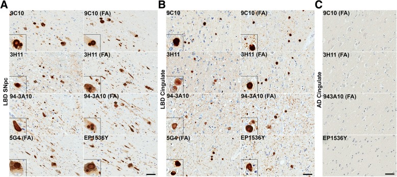
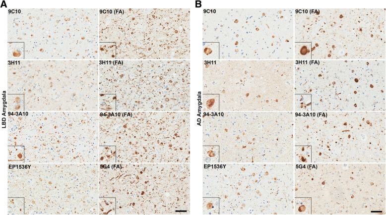
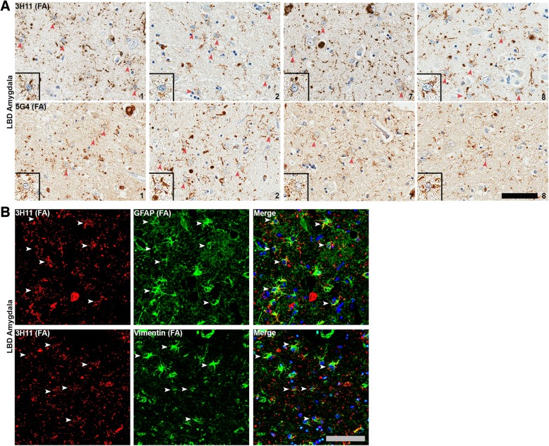
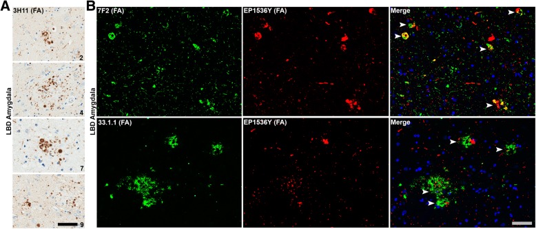
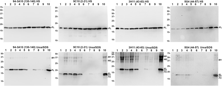
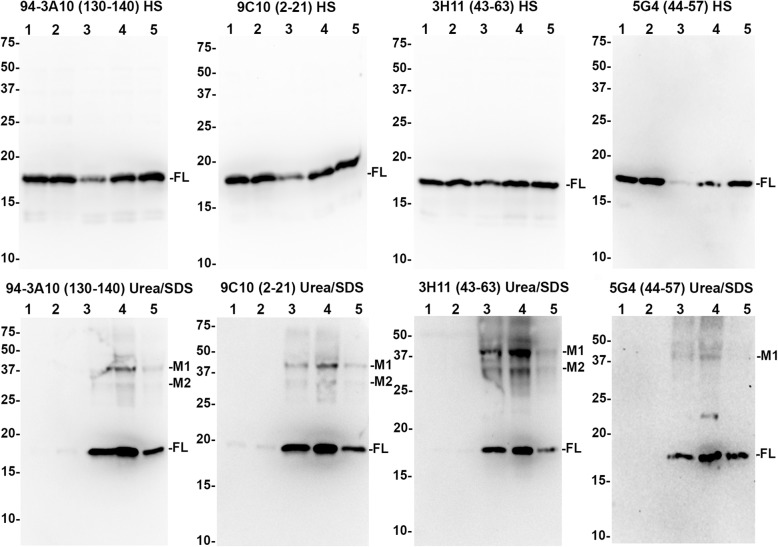
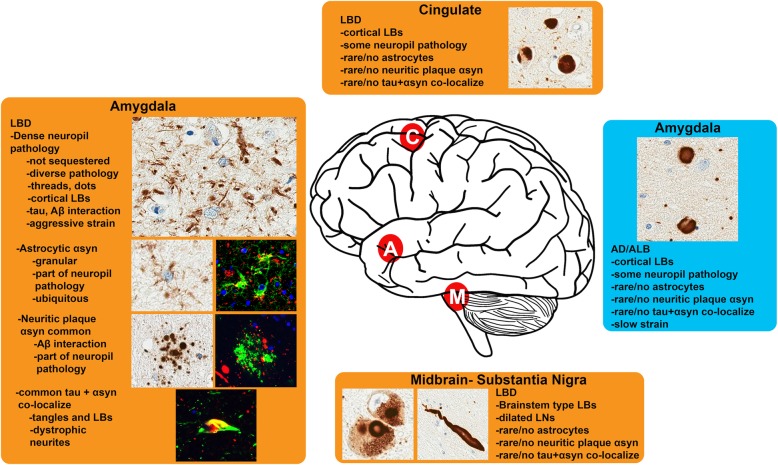
The protein α-synuclein (αsyn) forms pathologic aggregates in a number of neurodegenerative diseases including Lewy body dementia (LBD) and Parkinson's disease (PD). It is unclear why diseases such as LBD may develop widespread αsyn pathology, while in Alzheimer's disease with amygdala restricted Lewy bodies (AD/ALB) the αsyn aggregates remain localized. The amygdala contains αsyn aggregates in both LBD and in AD/ALB; to understand why αsyn pathology continues to progress in LBD but not in AD/ALB, tissue from the amygdala and other regions were obtained from 14 cases of LBD, 9 cases of AD/ALB, and 4 controls for immunohistochemical and biochemical characterization. Utilizing a panel of previously characterized αsyn antibodies, numerous unique pathologies differentiating LBD and AD/ALB were revealed; particularly the presence of dense neuropil αsyn aggregates, astrocytic αsyn, and αsyn-containing dystrophic neurites within senile plaques. Within LBD, these unique pathologies were predominantly present within the amygdala. Biochemically, the amygdala in LBD prominently contained specific carboxy-truncated forms of αsyn which are highly prone to aggregate, suggesting that the amygdala may be prone to initiate development of αsyn pathology. Similar to carboxy-truncated αsyn, it was demonstrated herein that the presence of aggregation prone A53T αsyn is sufficient to drive misfolding of wild-type αsyn in human disease. Overall, this study identifies within the amygdala in LBD the presence of unique strain-like variation in αsyn pathology that may be a determinant of disease progression.

Keywords: Amygdala; Astrocyte; Inclusion formation; Lewy body; Lewy body dementia; Neurodegeneration; Parkinson’s disease; Truncation; α-Synuclein.


Conflict of interest statement

The authors declare that they have no competing interests.

Figures

Fig. 1
Fig. 1
LRP comprised of WT αsyn detected by selective antibody 3H11. a Western blot of 200 ng recombinant WT or A53T human αsyn protein probed with antibody 9C10 (residues 2–21) or antibody 3H11 (residues 43–63); A53T αsyn does not react with antibody 3H11. b Immunohistochemical staining with antibody 3H11 or 9C10 in αsyn transgenic mice. Using antibody 3H11, αsyn aggregates are extensively detected within line M20 mice overexpressing WT human αsyn but not in line M83 mice overexpressing A53T human αsyn demonstrating the histochemical specificity of this antibody. Labeling with antibody 9C10 depicts αsyn pathology in both types of αsyn transgenic mice. Scale bar 50 μm. c Immunohistochemical staining of tissue from the midbrain and hippocampus of a familial case of PD/LBD due to a heterozygous A53T mutation in SNCA. Antibody 9C10 detects both WT and A53T αsyn and detects abundant pathology in both regions; Antibody 3H11 only detects WT αsyn but also labels many pathologic inclusions, indicating that WT αsyn is recruited to aggregate by the presence of the A53T αsyn mutation. Arrowheads indicate LRP. All the sections depicted were treated with FA. Scale bar 50 μm
Fig. 2
Fig. 2
Comparison of LRP between LBD and AD/ALB. a Representative sections of the SNpc from 4 AD/ALB and 4 LBD cases stained with pSer129 αsyn antibody EP1536Y. Abundant LNs and LBs (red arrows) are seen within pigmented neurons in all LBD cases, however AD/ALB cases display only rare examples of thread like neurites. Insets display LBs in pigmented neurons. b Representative sections of the amygdala from AD/ALB versus LBD cases labeled with antibody EP1536Y. Cortical-type LBs are apparent in dense patches within amygdalas of either disease type, however abundant thread-like neurites are more apparent in LBD cases compared with AD/ALB. Insets highlight cortical LBs or neurites. c Representative sections of the cingulate cortex from AD/ALB compared with LBD using antibody EP1536Y. LRP is entirely absent in all examined AD/ALB cases, whereas LBD cases display extensive LBs in deeper cortical layers and dot like inclusions in more superficial layers. Case numbers are indicated in lower right corner. Scale bar 100 μm
Fig. 3
Fig. 3
Significant brain regional difference in formic acid antigen retrieval of LRP. a Low magnification sections from the amygdala and cingulate cortex of an LBD case labeled with antibody 9C10 (residues 2–21 αsyn) without or with FA retrieval as indicated. FA retrieval had a major impact in increasing staining of αsyn aggregates in the amygdala compared with the cingulate cortex. Scale bar 1 mm. b A region of dense Lewy pathology within the amygdala was used to optimize threshold values for positive pixel count analysis. All antibodies were tested to ensure similar detection of pathology with minimal background as shown in bottom panel where red colored pixels are positive. Scale bar 50 μm
Fig. 4
Fig. 4
Quantitation of LRP in LBD brain regions across a panel of antibodies. a Three areas of dense pathology within the cingulate (C) or amygdala (A) of LBD (N = 9) cases stained with 3 different antibodies without or with (FA) retrieval were subject to positive pixel analysis; average positivity and error bars (std) are displayed for each region and antibody without or with FA. All cases were averaged for each region and antibody for comparison. Without FA retrieval, all antibodies detect similar amounts of LRP within the amygdala versus the cingulate cortex; with FA retrieval a large increase in labeled amygdala pathology is evident with all antibodies whereas a lesser increase in pathology is seen in the cingulate cortex and only with antibody 94-3A10. With FA retrieval, the average amygdala pathology burden is significantly greater than the cingulate cortex for all antibodies. b A statistical summary of positivity comparisons for data presented in A. The average positivity for each antibody between and within regions along with presence or absence of FA retrieval were tested for significant differences using one-way ANOVA and the Sidak post-hoc multiple comparisons test. Mean difference is the absolute difference in positivity values
Fig. 5
Fig. 5
LRP in LBD SNpc and cingulate cortex is similarly detected across a panel of αsyn antibodies. a Representative sections of the SNpc from an LBD case labeled with 5 different αsyn antibodies as indicted in the top, left corners without or with FA as indicated. Insets display LBs in pigmented neurons. Within the SNpc, abundance of pathology is similar regardless of antibody used or antigen retrieval albeit with minor differences. Scale bar 50 μm. b Representative staining of the cingulate cortex from an LBD case labeled with 5 different αsyn antibodies without or with FA as indicated. Insets display cortical LBs. Within the cingulate, pathology is similar for most antibody and antigen retrieval conditions. αsyn-positive neurites are slightly more apparent with FA retrieval or C-terminal antibodies, particularly 94-3A10 with FA which is reflected in the quantitative positivity analysis. Scale bar 50 μm. c Sections from the cingulate of an AD/ALB case with no LRP were stained with 4 αsyn antibodies without or with FA, as indicated. Little to no positive staining was detected in the AD/ALB cingulate cortex with any of these antibodies. Scale bar 50 μm
Fig. 6
Fig. 6
Comparison of LRP in the amygdala of LBD versus AD/ALB across a panel of αsyn antibodies. a Representative sections of dense amygdala pathology from a LBD case labeled with 5 different αsyn antibodies without or with FA retrieval, as indicated. Insets display cortical LBs and αsyn-positive neurites. In the LBD amygdala, extensive neuritic pathology is detected for multiple antibodies when FA retrieval was used. The robust increase in apparent Lewy pathology with FA treatment is mainly seen in neuritic and possible glial processes; the amount of cortical LBs per visual field remain the same regardless of antibody or FA treatment. This immunohistochemical staining profile contrasts with the only modest increase in detected pathology seen in the LBD SNpc and cingulate cortex. Scale bar 50 μm. b Stained sections of amygdala pathology from an AD/ALB case labeled with 5 different αsyn antibodies without or with FA retrieval, as indicated. Insets display cortical LBs and αsyn-positive neurites. In the AD/ALB amygdala, neuritic pathology is modestly enhanced for multiple antibodies when FA retrieval is used; however, the density of neuropil αsyn staining pathology in LBD is not apparent in AD/ALB. Scale bar 50 μm
Fig. 7
Fig. 7
αsyn astrocytic pathology is common in LBD amygdala. a Amygdala sections from 4 different LBD cases (1, 2, 7 and 8) were labeled with central αsyn antibodies either 3H11 or 5G4 along with FA retrieval. Typically, more than 5 cells with astrocytic morphology (depicted with arrowheads) stained with these antibodies was readily observed per field; insets show examples for each case and each antibody. Although astrocytic αsyn inclusions may be present in other brain regions, they are only this densely abundant within the MTL, particularly the amygdala. Cases are indicated in lower right corner. Scale bar 50 μm. b Double labeling immunofluorescent analysis of LBD amygdala sections using central αsyn antibody 3H11 in conjunction with astrocytic markers GFAP or vimentin. Astrocytic αsyn often appears as vesicular granules outlining 2–3 astrocytic processes as opposed to the more uniform αsyn staining seen within nearby cortical LBs, suggesting a differing subcellular co-localization or form of aggregates. Scale bar 30 μm
Fig. 8
Fig. 8
αsyn aggregates within the LBD amygdala often co-localizes with Alzheimer’s disease-type inclusion pathology. a Amygdala sections from 4 different LBD cases (2, 4, 7 and 9) stained with central αsyn antibody 3H11 along with FA retrieval reveal abundant αsyn dystrophic neurites within Aβ senile plaques. These αsyn-positive dystrophic neurites within Aβ plaques were common within the LBD amygdalas but rare in the AD/ALB amygdalas examined. Cases are indicated in lower right corner. Scale bar 50 μm. b Double labeled immunofluorescence microscopy of LBD amygdala sections from 2 different LBD cases using rabbit monoclonal anti-pSer129 αsyn antibody EP1536Y and mouse monoclonal anti-pThr205 tau antibody 7F2 or mouse monoclonal anti-Aβ antibody 33.1.1. Co-localization of tau and αsyn aggregates was common in the LBD amygdala both in neuronal cell bodies and processes. Scale bar 50 μm
Fig. 9
Fig. 9
Immunoblotting comparison of αsyn species in LBD and AD/ALB amygdala. High salt (HS) and SDS/urea fractions were obtained from the MTL of 5 LBD cases (lanes 1–5), 2 cases of AD/ALB (lanes 6–7), 2 non-synucleinopathy controls (lanes 8–9), and for one LBD case the amygdala was specifically isolated (lane 10). 20 μg of lysate for each case and fraction were subject to western blot analysis using a panel of 4 antibodies which are indicated. In the HS fraction, all antibodies predominantly revealed full-length (FL) αsyn in all cases. In the SDS/Urea fractions, monomeric FL αsyn is present in high amounts for 4/5 LBD cases (lanes 2, 3, 4, 5, and 10); 1 LBD and 1 AD/ALB case (lanes 1 and 7, respectively) have an intermediate amount of FL αsyn in this fraction and 1 AD/ALB case along with 2 controls (lanes 6, 8, and 9) have very little FL αsyn in this fraction. For the LBD cases, 2 prominent truncation bands are present for all antibodies except for C-terminal antibody 94-3A10 suggesting these are carboxy-truncated forms of αsyn (T1 and T2) in the SDS/urea fractions from the MTL in LBD but not controls or AD/ALB; the truncation bands are strongest in the LBD amygdala. Additional higher molecular mass bands are prominent in LBD cases (M1 and M2); these bands are less visible in AD/ALB or controls. The relative mobilities of molecular mass protein markers are identified on the left of the blots
Fig. 10
Fig. 10
Immunoblotting comparison of αsyn species in LBD cingulate cortex. High salt (HS) and SDS/urea fractions were obtained from the cingulate cortex of 2 control cases (lanes 1–2) and 3 LBD cases (lanes 3–5). 20 μg of lysate for each case and fraction were subject to western blot analysis using a panel of 4 antibodies, as indicated. In the HS fraction, all antibodies demonstrate similar amounts of monomeric FL αsyn except for one of the LBD cases which had a diminished level. In the SDS/Urea fractions, monomeric FL αsyn is present in high amounts for all LBD cases while the 2 controls have almost no αsyn in this fraction. Although higher molecular mass M1 and M2 bands are present in the cingulate SDS/urea fraction, truncation bands T1 and T2 are not as apparent which represents a biochemical difference between αsyn in the amygdala and MTL compared with cingulate cortex. The relative mobilities of molecular mass protein markers are identified on the left of the blots
Fig. 11
Fig. 11
Diagram of pathologic determinants associated with further progression of αsyn pathology. The diversity of αsyn pathologies across different brain regions in LBD is shown in orange while the more limited αsyn pathology in AD/ALB is in blue (C = cingulate cortex, A = amygdala, M = midbrain-substantia nigra pars compacta). In LBD, diverse αsyn pathologies are most abundant within the amygdala where common co-localization with tau and Aβ are seen along with dense αsyn-positive neuropil aggregates including in glial processes that may represent a less easily sequestered strain of αsyn. In AD/ALB, αsyn pathology is largely limited to large LB type inclusions that may represented a more sequestered and less pathogenic strain of αsyn

References

    1. Abdelmotilib H, Maltbie T, Delic V, Liu Z, Hu X, Fraser KB, Moehle MS, Stoyka L, Anabtawi N, Krendelchtchikova V, Volpicelli-Daley LA, West A. α-Synuclein fibril-induced inclusion spread in rats and mice correlates with dopaminergic neurodegeneration. Neurobiol Dis. 2017;105:84–98. doi: 10.1016/j.nbd.2017.05.014. - DOI - PMC - PubMed
    1. Anderson JP, Walker DE, Goldstein JM, de Laat R, Banducci K, Caccavello RJ, Barbour R, Huang J, Kling K, Lee M, Diep L, Keim PS, Shen X, Chataway T, Schlossmacher MG, Seubert P, Schenk D, Sinha S, Gai WP, Chilcote TJ. Phosphorylation of Ser-129 is the dominant pathological modification of α-Synuclein in familial and sporadic Lewy body disease. J Biol Chem. 2006;281:29739–29752. doi: 10.1074/jbc.M600933200. - DOI - PubMed
    1. Arai Y, Yamazaki M, Mori O, Muramatsu H, Asano G, Katayama Y. Alpha-synuclein-positive structures in cases with sporadic Alzheimer’s disease: morphology and its relationship to tau aggregation. Brain Res. 2001;888:287–296. doi: 10.1016/s0006-8993(00)03082-1. - DOI - PubMed
    1. Baba M, Nakajo S, Tu PH, Tomita T, Nakaya K, Lee VM, Trojanowski JQ, Iwatsubo T. Aggregation of alpha-synuclein in Lewy bodies of sporadic Parkinson’s disease and dementia with Lewy bodies. Am J Pathol. 1998;152:879–884. - PMC - PubMed
    1. Beach TG, White CL, Hamilton RL, Duda JE, Iwatsubo T, Dickson DW, Leverenz JB, Roncaroli F, Buttini M, Hladik CL, Sue LI, Noorigian JV, Adler CH. Evaluation of α-synuclein immunohistochemical methods used by invited experts. Acta Neuropathol. 2008;116:277–288. doi: 10.1007/s00401-008-0409-8. - DOI - PMC - PubMed

Publication types

MeSH terms

Substances