. 2019 Sep 2;7(1):141.
doi: 10.1186/s40478-019-0791-6.

Aβ43 in human Alzheimer's disease: effects of active Aβ42 immunization

Affiliations

Aβ43 in human Alzheimer's disease: effects of active Aβ42 immunization

Lieke Jäkel et al. Acta Neuropathol Commun. .

Abstract

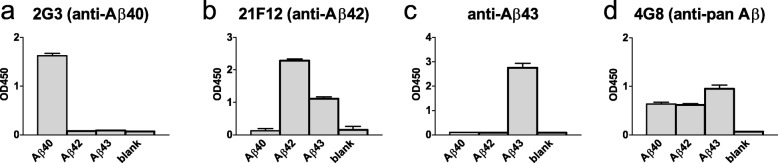
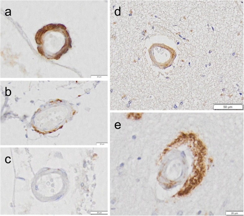
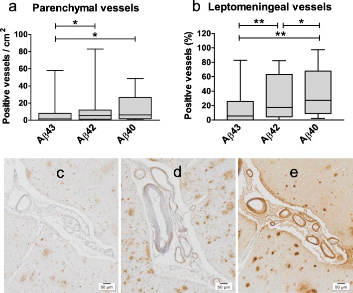
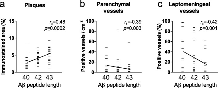
Neuropathological follow-up of patients with Alzheimer's disease (AD) who participated in the first clinical trial of Amyloid-β 42 (Aβ42) immunization (AN1792, Elan Pharmaceuticals) has shown that immunization can induce removal of Aβ42 and Aβ40 from plaques, whereas analysis of the cerebral vessels has shown increased levels of these Aβ peptides in cerebral amyloid angiopathy (CAA). Aβ43 has been less frequently studied in AD, but its aggregation propensity and neurotoxic properties suggest it may have an important pathogenic role. In the current study we show by using immunohistochemistry that in unimmunized AD patients Aβ43 is a frequent constituent of plaques (6.0% immunostained area), similar to Aβ42 (3.9% immunostained area). Aβ43 immunostained area was significantly higher than that of Aβ40 (2.3%, p = 0.006). In addition, we show that Aβ43 is only a minor component of CAA in both parenchymal vessels (1.5 Aβ43-positive vessels per cm2 cortex vs. 5.3 Aβ42-positive vessels, p = 0.03, and 6.2 Aβ40-positive vessels, p = 0.045) and leptomeningeal vessels (5.6% Aβ43-positive vessels vs. 17.3% Aβ42-positive vessels, p = 0.007, and 27.4% Aβ40-positive vessels, p = 0.003). Furthermore, we have shown that Aβ43 is cleared from plaques after Aβ immunotherapy, similar to Aβ42 and Aβ40. Cerebrovascular Aβ43 levels did not change after immunotherapy.

Keywords: Alzheimer’s disease; Amyloid-β; Aβ immunotherapy; Aβ43; Cerebral amyloid angiopathy; Human study; Immunohistochemistry.


Conflict of interest statement

The authors declare that they have no competing interests.

Figures

Fig. 1
Fig. 1
Specificity of anti-Aβ antibodies. The anti-Aβ43 antibody specifically recognizes Aβ43 (a). The 21F12 antibody also recognizes Aβ43, in addition to Aβ42 (b). The 2G3 antibody is specific for Aβ40 (c). Detection with the 4G8 antibody shows that the different Aβ isoforms are present in comparable amounts and bind to the plate with similar affinity (d). Data are presented as mean (standard deviation)
Fig. 2
Fig. 2
Parenchymal Aβ load in AD cases. Representative images of parenchymal tissue stained by antibodies directed against Aβ43 (a), Aβ42 (b), and Aβ40 (c) at 20x objective magnification. Indicated areas are shown at higher magnification (d-f). A significantly larger portion of the cortex is immunostained by the Aβ43- and Aβ42-specific antibodies compared to the Aβ40-specific antibody (g). The cases shown in a-f are representative of the median immunostained area as plotted in g. Box plot shows median values with the 25th and 75th percentile as boundaries and whiskers indicating minimum and maximum values. Scale bar = 50 μm (upper panel) or 20 μm (lower panel). *p ≤ 0.05; **p ≤ 0.01
Fig. 3
Fig. 3
Representative examples of Aβ43 staining in cerebral small vessels. Aβ43-immunostaining was detected in both leptomeningeal (a, b) and parenchymal (d, e) vessels. Some immunostained vessels were partially stained (b, e). In others, Aβ43 staining was detected in the full circumference and thickness of the vessel wall (a, d). Many vessels were devoid of Aβ43 (c). Scale bar = 20 μm (a, b, c, e) or 50 μm (d)
Fig. 4
Fig. 4
Aβ peptides in the cerebrovasculature of AD cases. Analysis of the numbers of parenchymal (a) and leptomeningeal (b) vessels affected by Aβ43, Aβ42, and Aβ40 in AD cases shows significantly lower numbers of vessels affected by Aβ43 compared to the other peptides. Images of cerebrovascular immunostaining of Aβ43 (c), Aβ42 (d), and Aβ40 (e) in the same cortical area of an AD case. Box plots show median values with the 25th and 75th percentile as boundaries and whiskers indicating minimum and maximum values. Scale bar = 50 μm. *p ≤ 0.05; **p ≤ 0.01
Fig. 5
Fig. 5
Plaque and CAA severity correlate to Aβ peptide length. Analysis of plaque and CAA severity in brain tissue of AD patients immunostained for Aβ43, Aβ42, and Aβ40 shows that Aβ peptide length correlates to plaque load (a). An inverse correlation between Aβ peptide length and both the number of Aβ-affected parenchymal vessels (b) and leptomeningeal vessels (c) is observed
Fig. 6
Fig. 6
Effect of immunotherapy on parenchymal Aβ load. Aβ43 (a), Aβ42 (b), and Aβ40 (c) loads are significantly lower in iAD cases compared to non-immunized AD cases. Representative images of Aβ43 (d, g), Aβ42 (e, h), and Aβ40 (f, i) load in AD (d-f) and immunized AD cases (g-i). Box plots show median values with the 25th and 75th percentile as boundaries and whiskers indicating minimum and maximum values. Scale bar = 100 μm. **p ≤ 0.01; ***p ≤ 0.001
Fig. 7
Fig. 7
Effect of immunotherapy on cerebrovascular Aβ load. The numbers of parenchymal (a) and leptomeningeal (b) vessels affected by Aβ43, Aβ42, or Aβ40 did not differ between AD and iAD cases. Images of cerebrovascular immunostaining of Aβ43 (c), Aβ42 (d), and Aβ40 (e) in the same cortical area of an immunized AD case. Box plots show median values with the 25th and 75th percentile as boundaries and whiskers indicating minimum and maximum values. Scale bar = 100 μm
Fig. 8
Fig. 8
The numbers of vessels affected by the different Aβ isoforms strongly correlate to each other. Parv = parenchymal vessels; Lepv = leptomeningeal vessels

References

    1. Attems J. Sporadic cerebral amyloid angiopathy: pathology, clinical implications, and possible pathomechanisms. Acta Neuropathol. 2005;110(4):345–359. doi: 10.1007/s00401-005-1074-9. - DOI - PubMed
    1. Attems J, Jellinger KA, Lintner F. Alzheimer's disease pathology influences severity and topographical distribution of cerebral amyloid angiopathy. Acta Neuropathol. 2005;110(3):222–231. doi: 10.1007/s00401-005-1064-y. - DOI - PubMed
    1. Bales KR, et al. Passive immunotherapy targeting amyloid-beta reduces cerebral amyloid angiopathy and improves vascular reactivity. Brain. 2016;139(Pt 2):563–577. doi: 10.1093/brain/awv313. - DOI - PubMed
    1. Bayer AJ, et al. Evaluation of the safety and immunogenicity of synthetic Abeta42 (AN1792) in patients with AD. Neurology. 2005;64(1):94–101. doi: 10.1212/01.WNL.0000148604.77591.67. - DOI - PubMed
    1. Boche D, et al. Consequence of Abeta immunization on the vasculature of human Alzheimer's disease brain. Brain. 2008;131(Pt 12):3299–3310. doi: 10.1093/brain/awn261. - DOI - PubMed

Publication types

MeSH terms