Multicenter Study
. 2019 Sep 2;10(1):3935.
doi: 10.1038/s41467-019-11862-x.

The molecular origin and taxonomy of mucinous ovarian carcinoma

Dane Cheasley  1 Matthew J Wakefield  2   3 Georgina L Ryland  1 Prue E Allan  1 Kathryn Alsop  1   3 Kaushalya C Amarasinghe  1 Sumitra Ananda  1   4 Michael S Anglesio  5 George Au-Yeung  1   3 Maret Böhm  6 David D L Bowtell  1   3 Alison Brand  7 Georgia Chenevix-Trench  8 Michael Christie  3   9 Yoke-Eng Chiew  7 Michael Churchman  10 Anna DeFazio  7 Renee Demeo  1 Rhiannon Dudley  11 Nicole Fairweather  11 Clare G Fedele  1   3 Sian Fereday  1   3 Stephen B Fox  1   3 C Blake Gilks  5 Charlie Gourley  10 Neville F Hacker  12 Alison M Hadley  13 Joy Hendley  1 Gwo-Yaw Ho  2 Siobhan Hughes  1 David G Hunstman  5 Sally M Hunter  1 Tom W Jobling  14 Kimberly R Kalli  15 Scott H Kaufmann  15 Catherine J Kennedy  7 Martin Köbel  16 Cecile Le Page  17 Jason Li  1 Richard Lupat  1 Orla M McNally  3   18 Jessica N McAlpine  5 Anne-Marie Mes-Masson  17   19 Linda Mileshkin  1 Diane M Provencher  17   20 Jan Pyman  18   21 Kurosh Rahimi  17   20 Simone M Rowley  1 Carolina Salazar  1 Goli Samimi  6 Hugo Saunders  1 Timothy Semple  1 Ragwha Sharma  7   22 Alice J Sharpe  23 Andrew N Stephens  11 Niko Thio  1 Michelle C Torres  1 Nadia Traficante  1   3 Zhongyue Xing  1 Magnus Zethoven  1 Yoland C Antill  24   25 Clare L Scott  1   2   3   9 Ian G Campbell  1   3 Kylie L Gorringe  26   27
Affiliations
Multicenter Study

The molecular origin and taxonomy of mucinous ovarian carcinoma

Dane Cheasley et al. Nat Commun. .

Abstract

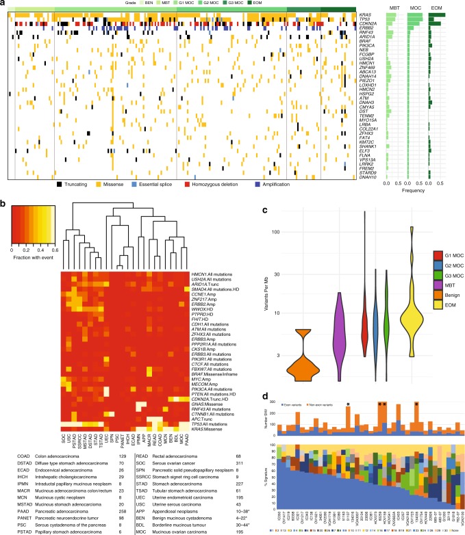
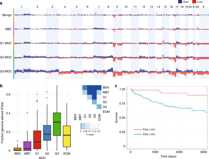
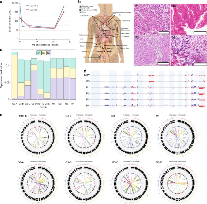
Mucinous ovarian carcinoma (MOC) is a unique subtype of ovarian cancer with an uncertain etiology, including whether it genuinely arises at the ovary or is metastatic disease from other organs. In addition, the molecular drivers of invasive progression, high-grade and metastatic disease are poorly defined. We perform genetic analysis of MOC across all histological grades, including benign and borderline mucinous ovarian tumors, and compare these to tumors from other potential extra-ovarian sites of origin. Here we show that MOC is distinct from tumors from other sites and supports a progressive model of evolution from borderline precursors to high-grade invasive MOC. Key drivers of progression identified are TP53 mutation and copy number aberrations, including a notable amplicon on 9p13. High copy number aberration burden is associated with worse prognosis in MOC. Our data conclusively demonstrate that MOC arise from benign and borderline precursors at the ovary and are not extra-ovarian metastases.


Conflict of interest statement

The authors declare no competing interests.

Figures

Fig. 1
Fig. 1
Variant analysis. a Summary of variants across the cohort for genes mutated in > 5% of MOC; also includes copy number alterations for CDKN2A and ERBB2. BEN, benign mucinous; MBT, borderline mucinous; MOC, mucinous ovarian carcinoma; EOM, extra-ovarian metastases. b Comparison of copy number alterations and mutations with other tumor types, summarised by frequency for each. Number of cases shown below. *Higher number is for selected genes tested by Sanger sequencing/SNP arrays (see Methods); lower number from exome analysis. c Number of variants per Mb by group (ANOVA, two-sided, F = 1.55, df = 5, p = 0.18), combining exome and targeted sequencing cohorts. d Top: number of single nucleotide variants (SNV) input to signature detection from whole exome and whole genome sequencing, with each column an MOC case. Note whole genome samples truncated at 300 (asterisk). Bottom: COSMIC mutation signatures
Fig. 2
Fig. 2
Copy number analysis. a Comparison of copy number frequency across the genome, comparing benign (BEN), borderline (MBT), MOC grade 1 (G1), grade 2 (G2) and grade 3 (G3). b Fraction of the genome altered (FGA) by group including extra-ovarian (EOM) mucinous tumors (ANOVA, two-sided p < 0.001, F = 17.0, df = 5). Tukey post-test comparison p-values shown at right (two-sided). Error bars are “Tukey” (geom-boxplot in ggplot2). c MOC disease-specific survival by FGA. Score (logrank) test = 11.98 on 1 df, p = 0.002
Fig. 3
Fig. 3
9p13 amplification. a WGS case HOV159 showing complex amplification with the majority of breakpoints rejoining internally to chromosome 9. b WGS case 5950 showing simpler amplification with a mixture of internal and external breakpoint fusions. c Overview of all amplified samples (gains in blue, losses in red) showing minimal region of overlap at 33.785–35.159 Mb and association with 9p loss. d All MOC with 9p13 amplification have TP53 mutation and 16/17 also have have CDKN2A loss/loss of heterozygosity (LOH). HD, homozygous deletion. For TP53: No: no mutation detected, Yes: mutation detected. e RNAseq analysis showing genes significantly differentially expressed between amplified and non-amplified (FDR < 0.05). Blue genes = within 9p amplicon, *genes connected in STRING network related to chromosome condensation
Fig. 4
Fig. 4
Molecular evolution of Grade 3 MOC and metastatic disease. a Serum markers of rapid autopsy case. Dashed line indicates maximum normal level. b Schematic of location of metastatic tissues taken at autopsy and haematoxylin and eosin stained sections of metastatic sites (M1 and M3) as well as two areas of the primary tumor, the majority borderline morphology (MBT) and the small area of high-grade invasive tumor identified in the frozen section (G3). Scale bars are 200 µm. c Mutation signatures (S1 - S3) identified by de novo analysis of 5 Grade 3 cases (A-E) showing the shift in mutation signature profile from primary (MBT-E, G3-E) to metastatic sites at autopsy (M1 - M4). d Copy number of primary and metastatic sites showing dramatic increase in structural and copy number alterations in the metastatic sites. Blue, gain; red, loss. e Circos plots illustrating structural variants in primary (MBT-E, G3-E) and metastatic sites (M1–M4) in the rapid autopsy case, and in four independent Grade 3 MOC cases (G3-A, G3-B, G3-C, G3-D). G3-A and G3-B have chromosome 9p amplicons

References

    1. Ledermann JA, et al. Gynecologic Cancer InterGroup (GCIG) consensus review for mucinous ovarian carcinoma. Int J. Gynecol. Cancer. 2014;24:S14–S19. doi: 10.1097/IGC.0000000000000296. - DOI - PubMed
    1. Perren TJ. Mucinous epithelial ovarian carcinoma. Ann. Oncol. 2016;27:i53–i57. doi: 10.1093/annonc/mdw087. - DOI - PubMed
    1. Vaughan S, et al. Rethinking ovarian cancer: recommendations for improving outcomes. Nat. Rev. Cancer. 2011;11:719–725. doi: 10.1038/nrc3144. - DOI - PMC - PubMed
    1. Simons M, et al. A novel algorithm for better distinction of primary mucinous ovarian carcinomas and mucinous carcinomas metastatic to the ovary. Virchows Arch. 2019;474:289–296. doi: 10.1007/s00428-018-2504-0. - DOI - PMC - PubMed
    1. Ovarian cancer: recognition and initial management (NICE Clinical guideline No. 122). National Institute for Health and Care Excellence (2011). - PubMed

Publication types

MeSH terms