. 2020 Aug;27(7-8):585-598.
doi: 10.1038/s41417-019-0129-3. Epub 2019 Sep 3.

Oncolytic activity of HF10 in head and neck squamous cell carcinomas

Affiliations

Oncolytic activity of HF10 in head and neck squamous cell carcinomas

Shinichi Esaki et al. Cancer Gene Ther. 2020 Aug.

Abstract

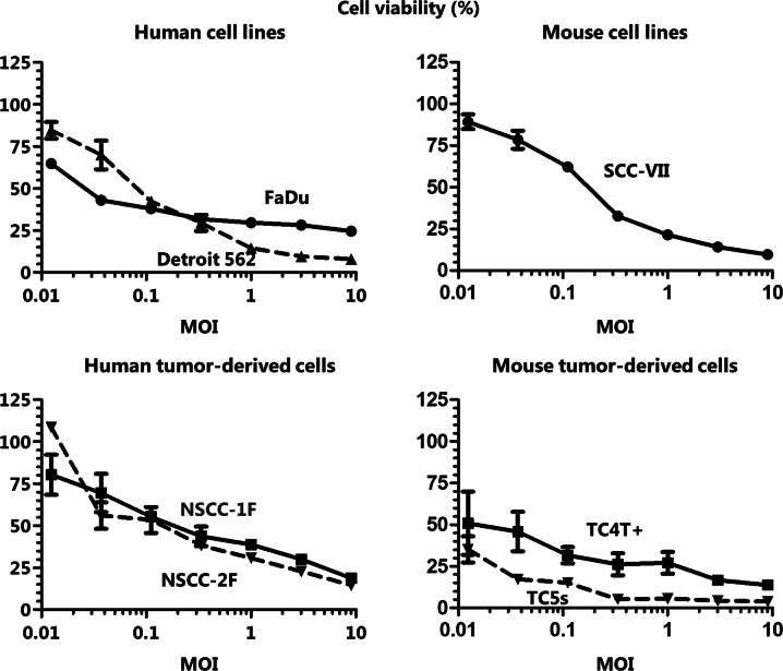
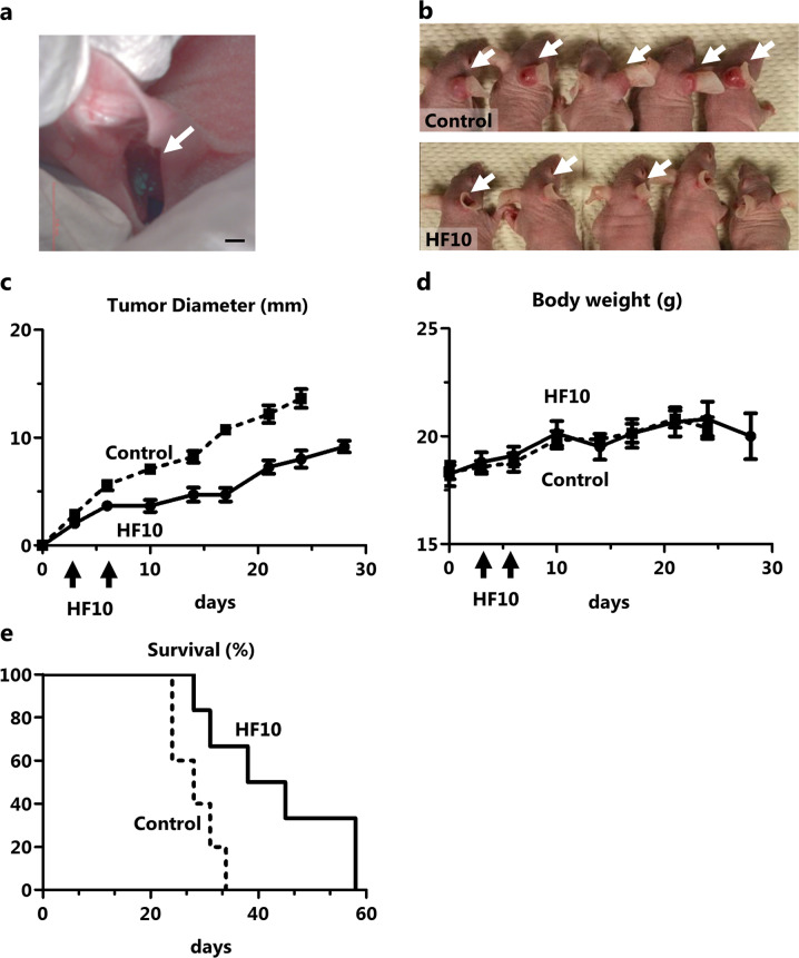
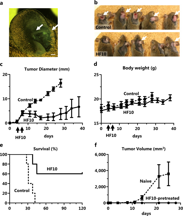
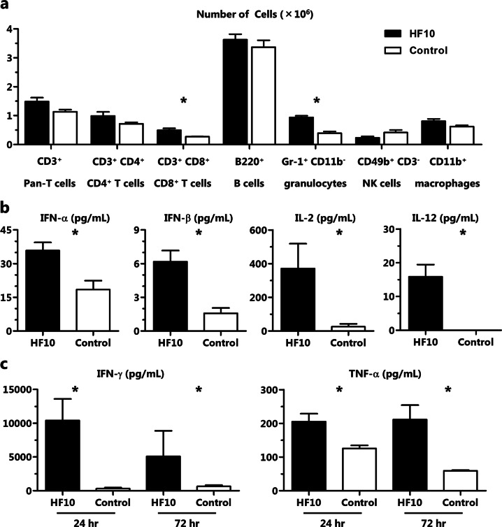
Recent developments in therapeutic strategies have improved the prognosis of head and neck squamous cell carcinoma (HNSCC). Nevertheless, 5-year survival rate remains only 40%, necessitating new therapeutic agents. Oncolytic virotherapy entails use of replication-competent viruses to selectively kill cancer cells. We aimed to explore the potential of HF10 as an oncolytic virus against human or mouse HNSCC cell lines, and primary-cultured HNSCC cells. HF10 replicated well in all the HNSCC cells, in which it induced cytopathic effects and cell killing. Next, we investigated the oncolytic effects of HF10 in ear tumor models with human or mouse tumor cells. We detected HF10-infected cells within the ear tumors based on their expression of green fluorescent protein. HF10 injection suppressed ear tumor growth and prolonged overall survival. In the syngeneic model, HF10 infection induced tumor necrosis with infiltration of CD8-positive cells. Moreover, the splenocytes of HF10-treated mice released antitumor cytokines, IL-2, IL-12, IFN-alpha, IFN-beta, IFN-gamma, and TNF-alpha, after stimulation with tumor cells in vitro. The HF10-treated mice that survived their original tumor burdens rejected tumor cells upon re-challenge. These results suggested that HF10 killed HNSCC cells and induced antitumoral immunity, thereby establishing it as a promising agent for the treatment of HNSCC patients.


Conflict of interest statement

The authors declare that they have no conflict of interest.

Figures

Fig. 1
Fig. 1
HF10 killed human and mouse HNSCC cells in vitro. The indicated cells were infected with HF10 at various MOIs and viability was assessed at 72 h post infection. Cell viability, calculated from the mean of samples tested in triplicate, is presented as the percent of live cells in the test group relative to in the uninfected control
Fig. 2
Fig. 2
HF10 replicated in human and mouse HNSCC cells and induced CPEs. a One-step (MOI = 3) and multistep (MOI = 0.03) growth curves of HF10 in the indicated cells. b Representative images of FaDu, SCC-VII, and Vero cells after HF10 infection (MOI = 3 or 0.03). Scale bars, 50 μm
Fig. 3
Fig. 3
HF10 suppressed the growth of FaDu ear tumors. a HF10-GFP was injected into FaDu ear tumors. GFP-positive cells were observed at 24 h after injection. The tumor is indicated by the arrow. Scale bars, 1 mm. b Photographs of the mice on day 14 after control or HF10 treatment. The arrows indicate the ear tumors. ce HF10 or PBS was intratumorally injected on days 3 and 6 after tumor inoculation (n = 5). HF10 injection suppressed tumor growth (p < 0.001) and significantly prolonged survival (p < 0.01)
Fig. 4
Fig. 4
HF10-suppressed SCC-VII ear tumor growth and induced antitumor immunity. a HF10-GFP was injected into SCC-VII ear tumors. GFP-positive cells were observed at 24 h after injection. The tumor is indicated by the arrow. Scale bars, 1 mm. b Photographs of the mice after each treatment on day 17. The arrows indicate the ear tumors. ce HF10 or PBS was intratumorally injected on days 4 and 7 after tumor inoculation (n = 5). HF10 injection significantly suppressed tumor growth (p < 0.001) and significantly prolonged survival (p < 0.05). After HF10 treatment, 3 of 5 mice rejected their tumors and survived long-term. f SCC-VII cells were subcutaneously injected into naïve mice (n = 3) or the 3 mice that rejected their tumors after HF10 injection
Fig. 5
Fig. 5
HF10 injection induced tumor necrosis, CD8+ T cell accumulation in and around the tumor, and antitumor and immune-stimulatory cytokine production. a After HF10 injection into SCC-VII tumors, we performed H&E, anti-HSV, and anti-CD8 staining. Scale bars, 100 μm. b The SCC-VII tumors were removed at 3 and 24 h after the second injections of HF10 or PBS. We analyzed IL-2, IFN-γ, and TNF-α in the tumors (*p < 0.05)
Fig. 6
Fig. 6
HF10 treatment-induced CD8+ T cell and granulocyte accumulation in the spleen and the production of antitumor and immune-stimulatory cytokines. a CD45+ leukocytes in the spleen were analyzed by flow cytometry for CD3+CD8+ T cells and Gr-1+CD11b granulocytes in the spleen. b, c The splenocytes were co-cultured with the SCC-VII cells for 24 and 72 h. We analyzed the release of IFN-α, IFN-β, IFN-γ, TNF-α, IL-2, and IL-12 in the supernatants after 24 h (*p < 0.05) and IFN-γ and TNF-α after 72 h (*p < 0.05)

References

    1. Leemans CR, Braakhuis BJ, Brakenhoff RH. The molecular biology of head and neck cancer. Nat Rev. 2011;11:9–22. - PubMed
    1. Siegel RL, Miller KD, Jemal A. Cancer Statistics, 2017. CA Cancer J Clin. 2017;67:7–30. - PubMed
    1. Hori M, Matsuda T, Shibata A, Katanoda K, Sobue T, Nishimoto H, et al. Cancer incidence and incidence rates in Japan in 2009: a study of 32 population-based cancer registries for the Monitoring of Cancer Incidence in Japan (MCIJ) project. Jpn J Clin Oncol. 2015;45:884–91. - PubMed
    1. Cattaneo R, Miest T, Shashkova EV, Barry MA. Reprogrammed viruses as cancer therapeutics: targeted, armed and shielded. Nat Rev Microbiol. 2008;6:529–40. - PMC - PubMed
    1. Pol J, Kroemer G, Galluzzi L. First oncolytic virus approved for melanoma immunotherapy. Oncoimmunology. 2016;5:e1115641. - PMC - PubMed

Publication types