Review
. 2019 Dec;20(12):721-737.
doi: 10.1038/s41580-019-0162-y. Epub 2019 Sep 2.

Control of DNA replication timing in the 3D genome

Affiliations
Review

Control of DNA replication timing in the 3D genome

Claire Marchal et al. Nat Rev Mol Cell Biol. 2019 Dec.

Abstract

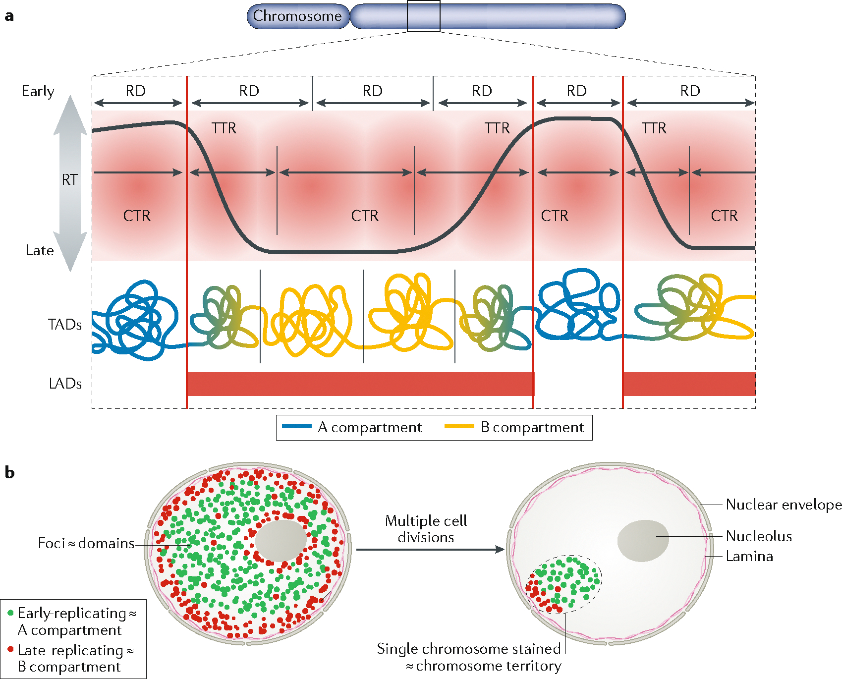
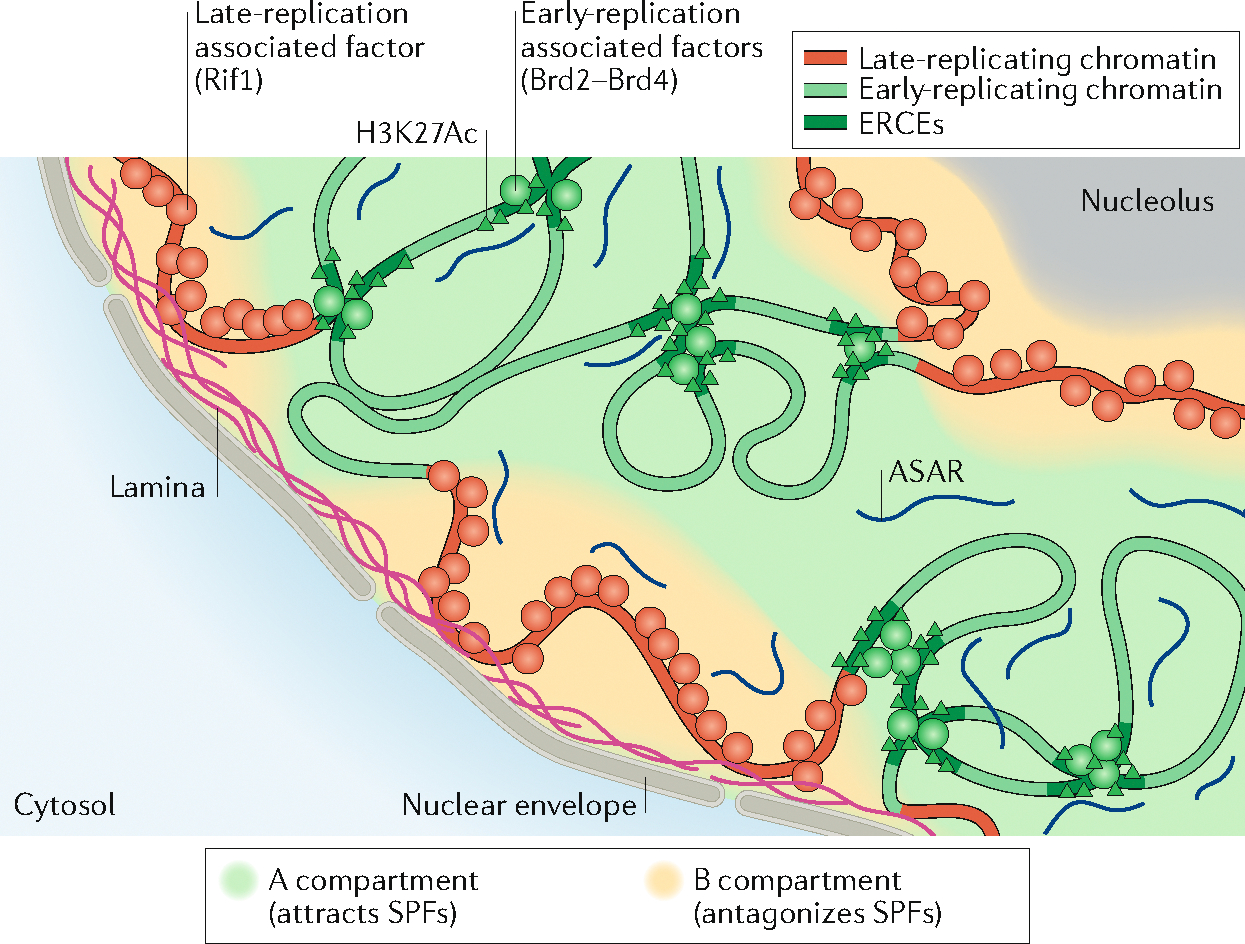
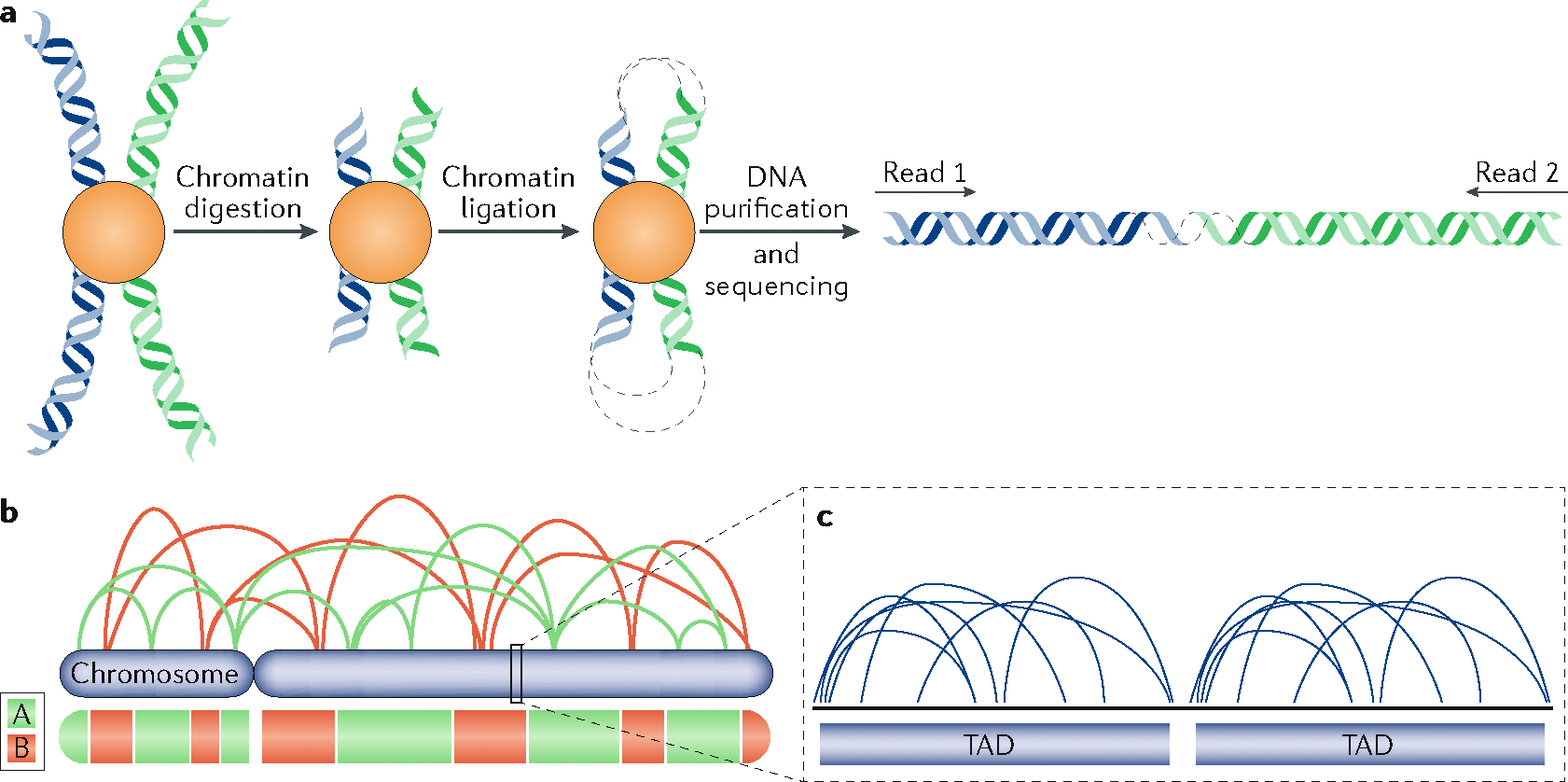
The 3D organization of mammalian chromatin was described more than 30 years ago by visualizing sites of DNA synthesis at different times during the S phase of the cell cycle. These early cytogenetic studies revealed structurally stable chromosome domains organized into subnuclear compartments. Active-gene-rich domains in the nuclear interior replicate early, whereas more condensed chromatin domains that are largely at the nuclear and nucleolar periphery replicate later. During the past decade, this spatiotemporal DNA replication programme has been mapped along the genome and found to correlate with epigenetic marks, transcriptional activity and features of 3D genome architecture such as chromosome compartments and topologically associated domains. But the causal relationship between these features and DNA replication timing and the regulatory mechanisms involved have remained an enigma. The recent identification of cis-acting elements regulating the replication time and 3D architecture of individual replication domains and of long non-coding RNAs that coordinate whole chromosome replication provide insights into such mechanisms.


Conflict of interest statement

Competing interests

The authors declare no competing interests.

Figures

Fig. 1 |
Fig. 1 |. Replication timing relationship to 3D chromatin structure.
a | Current model of the relationship between replication timing (RT) and chromatin structure. Early and late constant timing regions (CTRs) are 1–5 Mb regions separated by timing transition regions (TTRs) as demarcated within the red shaded area. These CTRs consist of one to several replication domains (RDs), defined as chromatin segments that coordinately switch RT during cell fate changes (that is, between different cell types; see FIG. 2a). RDs share the properties and approximate boundaries of a subset of topologically associated domains (TADs), aligning most closely with TADs that are at compartment boundaries. Early CTRs correspond to the A compartment, but late CTRs correspond to the B compartment and TTRs correspond to the transitions between compartments. Both TTRs and late CTRs correspond to lamina-associated domains (LADs). b | RT illuminates genome architecture: nuclei after an early S pulse label (green) followed by several hours of a chase period and then a late S pulse label (red). In this model, observable foci of DNA synthesis correspond to the replication domains in panel a and early/late-replicating chromatin corresponds to A/B compartments. After multiple passages, only one chromosome per cell remains labelled, marking the chromosome territory,. However, the foci retain their label intensity and genetic continuity, demonstrating that the DNA that is synthesized during one cell cycle remains clustered together as a structural unit of chromosomes for many generations.
Fig. 2 |
Fig. 2 |. 3D chromatin structure and replication timing are dynamic during cell differentiation and during the cell cycle.
a | Replication timing (RT) is regulated during differentiation. Here, RT is shown for a region of chromosome 8, in human embryonic stem cells H9 (H9 ESC) and two differentiated H9 cells. Some regions switch RT during differentiation (black to grey, or grey to black), whereas others remain constant. RT data are available at www.replicationdomain.com. b | Both a defined RT programme and interphase chromatin architecture are set up coincidently at the timing decision point (TDP) during the G1 phase,. The information that defines RT is lost during the G2 phase. In nuclei that are artificially forced to replicate their DNA before the TDP or after the S phase, DNA replication does not follow any specific RT,. The early- and late-replicating 3D compartments illuminated by replication labelling in the prior S phase are re-established at the TDP and persist through the remainder of interphase into the G2 phase, demonstrating that this spatial organization is not sufficient to dictate an RT programme. 3D chromatin interactions — both the separation between large-scale spatial compartments and the distinction between topologically associated domains (TADs) — are dismantled during mitosis and re-formed at the TDP, coincident with the establishment of RT. Whereas compartments and TADs become slightly more or less distinct, respectively, during the course of the S phase, the major architectural changes in genome architecture occur during entry into and exit from mitosis.
Fig. 3 |
Fig. 3 |. Epigenetic versus sequence-specific regulation of replication timing.
a | The cluster of ribosomal DNA consists of repeated genes coding for ribosomal RNAs. Correlated with the methylation status of each copy of these genes, they will replicate late (highly methylated) or early (low methylation level),. b | The β-globin locus is an example of developmental regulation of replication timing (RT): in erythroid cells, the locus is activated, rich in histone acetylation and early replicating. In non-erythroid cells, the locus is silenced, without histone acetylation and late replicating. Inducing histone acetylation in non-erythroid cells with trichostatin A (TSA) treatment promotes early replication of the locus, whereas depleting the locus for histone acetylation by targeting histone deacetylases (HDACs) to a β-globin transgene in erythroid cells induces late replication of the locus. c | Sequence-specific RT can be observed using subspecies F1 hybrid mouse embryonic stem cells (ESCs). Here, some non-imprinted regions have different RT, depending on the allele. Data from GEO series GSE95091 (REF.).
Fig. 4 |
Fig. 4 |. Cis- and trans-acting elements regulating replication timing.
a | Long non-coding RNA asynchronous replication and autosomal RNAs (ASARs) are expressed from one locus specific to each homologous chromosome, and coat the whole chromosome from which they are expressed. This coating is necessary to define the temporal window during which DNA replication can occur on each chromosome. b | Early-replication control elements (ERCEs) are sequences that are found to interact with each other within a megabase-sized domain, forming a cluster or ‘hub’ of ERCEs. They are large domains of acetylated histones and are bound by master transcription factors. ERCEs are necessary for the early replication of the chromosome domain in which they reside. c | Replication origins are activated by recruitment of S-phase promoting factors (SPFs) such as the kinases Dbf4-dependent kinase (DDK) and cyclin-dependent kinase (CDK), and the protein Treslin, and inhibited by factors such as Rif1 and the deacetylase Sirt1 (see also BOX 2) that inhibit SPFs. All known trans-acting regulators of replication timing act on DDK, CDK or Treslin but potentially other such regulators may exist that act upon other SPFs such as TopBP1, Cdc45, Mcm10 and GINS.
Fig. 5 |
Fig. 5 |. Proposed model of organization within the nucleus.
Model for the compartmentalization of the nucleus into and early- and late-replicating chromatin regions. In this model, early-replicating regions, within the A compartment, contain elements rich in H3K27Ac, bound by factors such as Brd2 and Brd4 that recruit S-phase-promoting factors (SPFs; see FIG. 4) such as Treslin. These elements, called early-replication control elements (ERCEs), form a platform for recruiting SPFs within the domain in which they belong. In this model, the replication of late-replicating regions, within the B compartment and associated with the nuclear lamina and the nucleolus, would be delayed by factors inhibiting origin firing (that is, antagonizing SPFs), such as Rif1 (which antagonizes DDK). At the chromosome level, coating of each chromosome by long non-coding RNAs known as asynchronous replication and autosomal RNAs (ASARs) could ensure the presence of replication machinery factors within each chromosome territory.
None
None

References

    1. Taylor JH Asynchronous duplication of chromosomes in cultured cells of Chinese hamster. J. Biophys. Biochem. Cytol. 7, 455–464 (1960). - PMC - PubMed
    1. Desprat R et al. Predictable dynamic program of timing of DNA replication in human cells. Genome Res. 19, 2288–2299 (2009). - PMC - PubMed
    1. Farkash-Amar S et al. Global organization of replication time zones of the mouse genome. Genome Res. 18, 1562–1570 (2008). - PMC - PubMed
    1. Hiratani I et al. Global reorganization of replication domains during embryonic stem cell differentiation. PLOS Biol. 6, e245 (2008). - PMC - PubMed
    1. MacAlpine DM, Rodríguez HK & Bell SP Coordination of replication and transcription along a Drosophila chromosome. Genes Dev. 18, 3094–3105 (2004). - PMC - PubMed

Publication types

MeSH terms

Substances

LinkOut - more resources