. 2019 Dec;4(12):2226-2236.
doi: 10.1038/s41564-019-0539-x. Epub 2019 Sep 2.

Life cycle progression and sexual development of the apicomplexan parasite Cryptosporidium parvum

Affiliations

Life cycle progression and sexual development of the apicomplexan parasite Cryptosporidium parvum

Jayesh Tandel et al. Nat Microbiol. 2019 Dec.

Abstract

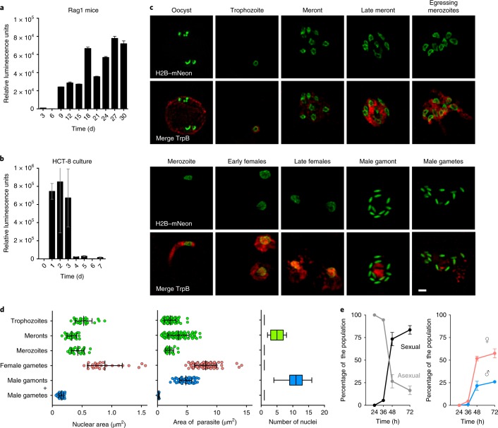
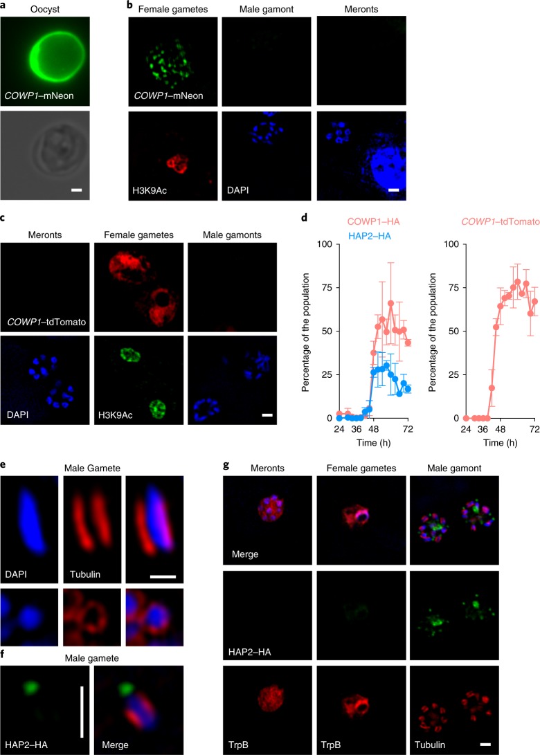
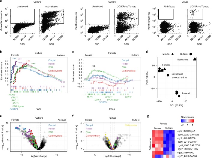
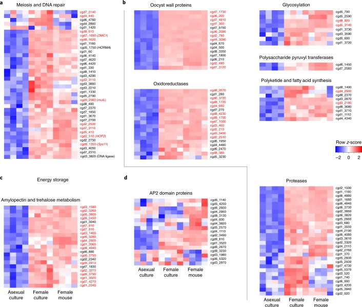
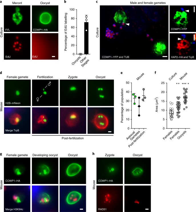
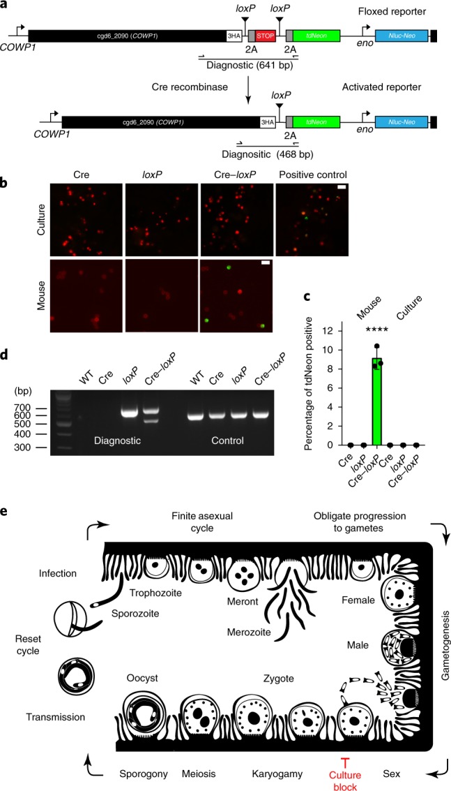
The apicomplexan parasite Cryptosporidium is a leading global cause of severe diarrhoeal disease and an important contributor to early childhood mortality. Currently, there are no fully effective treatments or vaccines available. Parasite transmission occurs through ingestion of oocysts, through either direct contact or consumption of contaminated water or food. Oocysts are meiotic spores and the product of parasite sex. Cryptosporidium has a single-host life cycle in which both asexual and sexual processes occur in the intestine of infected hosts. Here, we genetically engineered strains of Cryptosporidium to make life cycle progression and parasite sex tractable. We derive reporter strains to follow parasite development in culture and in infected mice and define the genes that orchestrate sex and oocyst formation through mRNA sequencing of sorted cells. After 2 d, parasites in cell culture show pronounced sexualization, but productive fertilization does not occur and infection falters. By contrast, in infected mice, male gametes successfully fertilize female parasites, which leads to meiotic division and sporulation. To rigorously test for fertilization, we devised a two-component genetic-crossing assay using a reporter that is activated by Cre recombinase. Our findings suggest obligate developmental progression towards sex in Cryptosporidium, which has important implications for the treatment and prevention of the infection.


Conflict of interest statement

The authors declare no competing interests.

Figures

Fig. 1
Fig. 1. Cryptosporidium life cycle stages revealed by the H2B–mNeon transgene.
a,b, C. parvum infection was monitored by luciferase activity in mice lacking mature T and B cells (a; faeces were measured every 3 d) and HCT-8 cultures (b). Data are mean ± s.d. from three independent biological replicates. c, HCT-8 cultures were infected with H2B–mNeon transgenic parasites and fixed at 24 h (‘Oocyst’, ‘Trophozoite’, ‘Meront’, ‘Late meront’ and ‘Egressing merozoites’), 36 h (‘Merozoite’) and 48 h (‘Early females’, ‘Late females’, ‘Male gamont’ and ‘Male gametes’) time intervals. Green, nuclei; red, cytoplasm (antibody against tryptophan synthase B (TrpB), cgd5_4560). This experiment was performed three times with similar results. Scale bar, 1 µm. d, Morphometric analyses of the size (n= 25) and number (n = 100) of nuclei and the area for each stage (n= 75) on the basis of the markers shown in c. The nuclear area (left) and total area (middle) of parasites stages are shown as mean ± s.d. of individual values represented as dots. The number of nuclei at particular parasite stages are represented as box plots (right). The box shows median and quartile range and whiskers represent extreme values. e, A time-course experiment in which stages were scored using the parameters defined in d revealed abrupt sexualization of cultures at 48 h into culture. Data are mean ± s.d. from three independent biological replicates.
Fig. 2
Fig. 2. Exclusive molecular markers for the sexual stages of C. parvum.
ad, C. parvum were engineered to express COWP1–mNeon (a,b) and COWP1–HA (Supplementary Fig. 3) from the native locus or COWP1-promoter-driven tdTomato from the ectopic TK locus (c). Note the mNeon labelling of the wall in oocysts purified from infected mice and punctate labelling in female gametes observed in infected HCT-8 cells. Labelling becomes apparent after 42 h of culture and is never observed in asexual meronts or male gametes (b,d). The COWP1 promoter alone is sufficient to confer female-specific expression to a reporter protein (c,d). Anti-H3K9Ac antibodies were used to label the nuclei of females because they stain poorly with 4,6-diamidino-2-phenylindole (DAPI). For the time intervals in d, cultures were infected with the indicated transgenic strains and triplicate coverslips were fixed and processed for immunofluorescence assays. Parasite stages were scored for HA staining, the mean ± s.d. percentage of HA-positive stages among all of the parasites is shown for three independent biological replicates. e, Male gametes show a characteristic array of microtubules around the nucleus after staining with anti-tubulin antibodies. f,g, When parasites were engineered to express HAP2–HA from the native locus, antibody staining revealed exclusive labelling of free gametes (f) and male gamonts (g). HAP2 labels a single pole per mature gamete. This staining becomes apparent after 42 h of culture (d, blue). All of the microscopy experiments shown in this figure were performed independently three times. Scale bars in ac,f,g, 1 µm; scale bar in e, 0.5 µm.
Fig. 3
Fig. 3. Isolation of parasite stages by cell sorting and RNA sequencing.
a, Flow cytometry of infected cells with the indicated markers and origins. Gates used for sorting are shown as boxes. This experiment was performed twice. SSC, side scatter. b,c, Gene set enrichment analysis (GSEA) with multiple testing correction comparing cultured asexual and female parasites (b) or females sorted from mice or culture (c). Custom gene signatures were generated using Gene Ontology or community datasets available at CryptoDB (b; carbohydrate (GO:0005975; normalized enrichment score (NES) = 1.67, FDR-adjusted P = 0.004), DNA (GO:0006259; NES = 1.68, FDR-adjusted P = 0.005), redox (GO:0055114; NES = 1.99, FDR-adjusted P = 0) and oocyst wall proteome dataset (NES = 1.76, FDR-adjusted P = 0.001)). ***P ≤ 0.005. Enrichment analysis comparing between females was not significant (NS). n = 4 biological replicates per group. d, Principal component analysis of all RNA-sequencing datasets generated during this study (Supplementary Fig. 7). e,f, Volcano plots showing C. parvum genes that were differentially expressed between asexual and female parasites from culture (e) or between in vitro and in vivo female parasites (f). n = 4 biological replicates per group. Each symbol represents a C. parvum gene, those genes representing the leading edge from b are indicated by the colour according to the pathway that they act in. The horizontal dashed line shows an FDR-adjusted P value of 0.01; the vertical dashed lines indicate a log2-transformed fold change of −1 and 1, respectively. g, A heat map of glideosome components, which are indicated in yellow in f. n = 4 biological replicates per group.
Fig. 4
Fig. 4. Female gametes express genes that are required for genetic recombination and oocyst formation.
ac, Heat maps illustrating expression of genes associated with specific molecular functions that are upregulated in females (generally results from in vivo and in vitro females concur, although there are some exceptions). Genetic recombination (a), oocyst environmental resilience (b) and energy storage (c); n = 4 biological replicates per group. d, Expression heat map for all C. parvum AP2 DNA-binding proteins. Note the pronounced difference identifying four genes upregulated in all females and two only in in vivo females. As we were unable to sequence males, we cannot formally exclude that some genes that show high female-specific expression may be upregulated in all sexual stages. Expression values are given as row z-scores and annotated genes list are provided as Supplementary Files. Genes from the leading edge in Fig. 3b are highlighted in red text.
Fig. 5
Fig. 5. Cryptosporidium males locate females in culture, but fertilization and meiosis only occur in vivo.
a,b, HCT-8 cells were infected with COWP1–HA C. parvum and after 36 h of infection, the nucleotide analogue EdU was added to the medium. Then, 12 h later, cells were click labelled and counterstained with anti-HA antibodies or Vicia villosa lectin (VVL; a). Cells were scored for nuclear EdU labelling (b); 100 stages were quantified for three biological replicates, and the experiment was performed twice. Data are mean ± s.d. c, Representative images of encounters between male and female gametes in culture; gametes were identified using the indicated transgenes or antibodies, and attached males are highlighted by arrowheads. YFP, yellow fluorescent protein. dh, Ifng−/− mice were infected with H2B–mNeon-expressing (d) or COWP1–HA-expressing (g,h) parasites, and intestines were sectioned and prepared for immunofluorescence assays and counterstained with anti-TrpB, anti-H3K9Ac or anti-RAD51 antibodies. Representative micrographs show progression of events following fertilization. Post-fertilization stages are abundant in vivo (e) and these stages were significantly larger than those found in vitro (f); each symbol represents a parasite, n = 25. For e, data are mean ± s.d. from three independent mice. For f, data are mean ± s.d.; the statistical analysis was performed using a two-sided Student’s t-test comparing cultured females with in vivo females (**P = 0.0023) or with in vivo oocysts (****P = 0.0001). All of the microscopy experiments shown in dh were performed twice with similar results. Scale bars, 1 µm.
Fig. 6
Fig. 6. A genetic fusion assay demonstrates fertilization in vivo but not in vitro.
a, To detect gamete fusion, we engineered two C. parvum strains, one that constitutively expresses Cre recombinase (Supplementary Fig. 11) and a second that carries a tdNeon reporter flanked by loxP at the COWP1 locus. b, Cre-mediated excision of a terminator results in reporter expression. HCT-8 cultures and Ifng−/− mice were infected with each strain individually or in combination. This experiment was performed twice. Cultured parasites were counterstained with anti-TrpB antibodies, oocysts with Macula pomifera agglutinin (both red) and scored for tdNeon expression. Scale bars, 10 µm. c, Three replicates were quantified for green fluorescence and 1,000 cells were counted for each replicate. Data are mean ± s.d. Green fluorescence was only observed after in vivo infection and only when both strains were present (****P= 0.0002, two-sided Student’s t-test). As a positive control, cells were infected with parasites that were crossed in vivo (indicated in b). d, PCR mapping of the floxed (diagnostic) and α-tubulin (control) loci using the primer pair shown in a. Genomic DNA was isolated from wild-type parasites as well as oocysts from the mouse infection experiments. Crossing resulted in a new amplicon that was consistent with precise Cre excision. This experiment was performed twice with similar results. e, Schematic model of the C. parvum life cycle that highlights the model of obligate progression to sex and the fertilization block in HCT-8 culture. We do not show type II meronts here, which are often depicted as an obligate step towards gametes. Although we observed meronts with four and eight nuclei, we did not find a quantitative link between the meronts with four nuclei and gametes (Supplementary Fig. 12).

References

    1. Liu L, et al. Global, regional, and national causes of under-5 mortality in 2000-15: an updated systematic analysis with implications for the Sustainable Development Goals. Lancet. 2016;388:3027–3035. - PMC - PubMed
    1. Kotloff KL, et al. Burden and aetiology of diarrhoeal disease in infants and young children in developing countries (the Global Enteric Multicenter Study, GEMS): a prospective, case-control study. Lancet. 2013;382:209–222. - PubMed
    1. Checkley W, et al. A review of the global burden, novel diagnostics, therapeutics, and vaccine targets for Cryptosporidium. Lancet Infect. Dis. 2015;15:85–94. - PMC - PubMed
    1. Platts-Mills JA, et al. Pathogen-specific burdens of community diarrhoea in developing countries: a multisite birth cohort study (MAL-ED) Lancet Glob. Health. 2015;3:E564–E575. - PMC - PubMed
    1. Korpe PS, Petri WA., Jr. Environmental enteropathy: critical implications of a poorly understood condition. Trends Mol. Med. 2012;18:328–336. - PMC - PubMed

Publication types