. 2019 Oct;15(10):966-974.
doi: 10.1038/s41589-019-0353-z. Epub 2019 Sep 2.

mRNA structure determines modification by pseudouridine synthase 1

Affiliations

mRNA structure determines modification by pseudouridine synthase 1

Thomas M Carlile et al. Nat Chem Biol. 2019 Oct.

Abstract

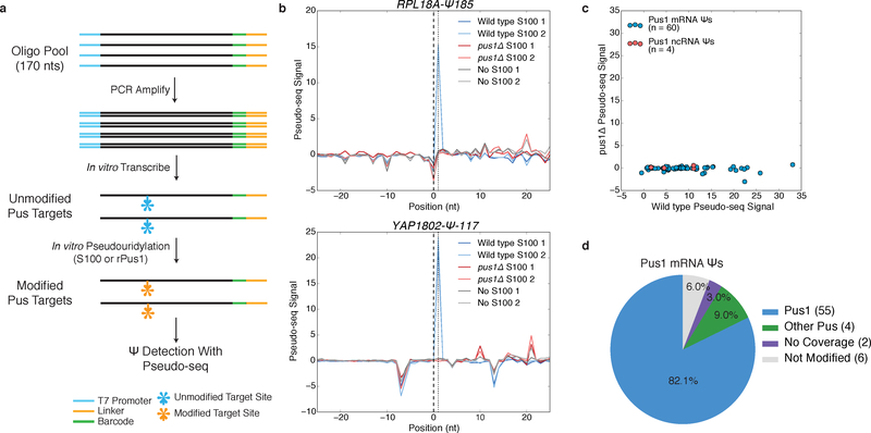
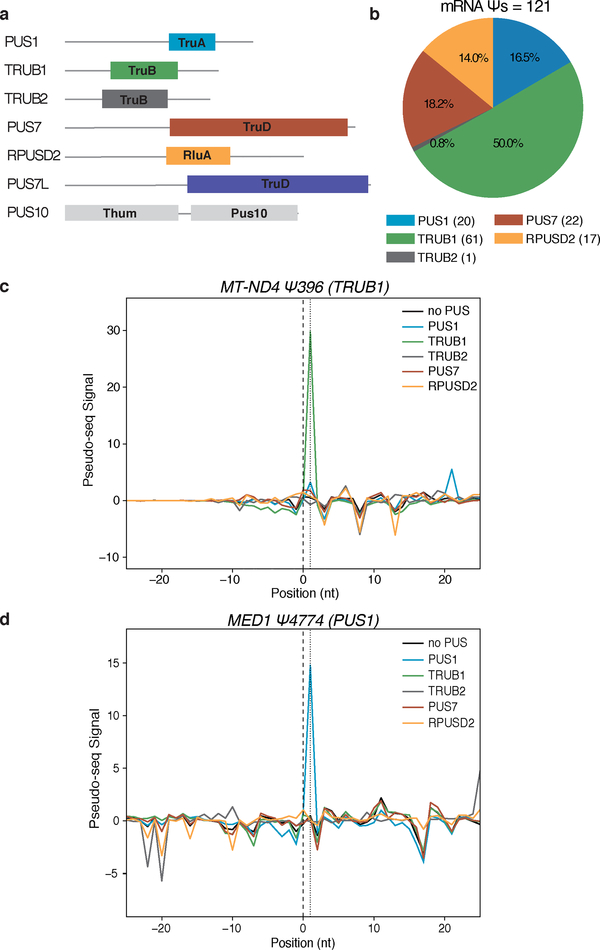
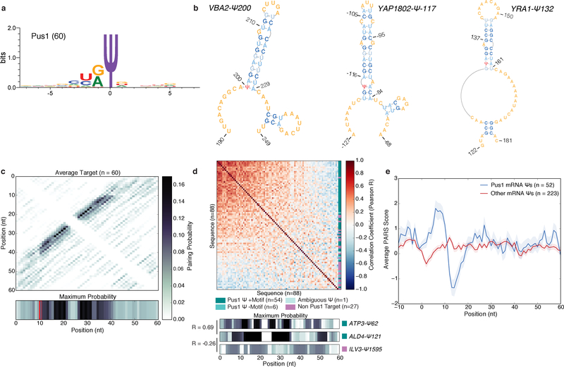
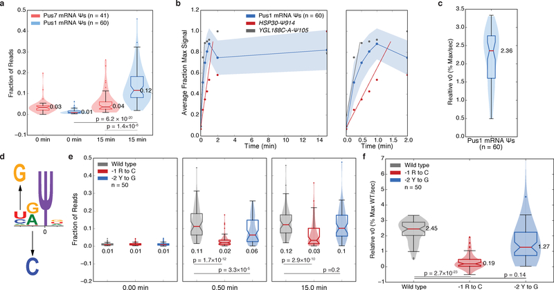
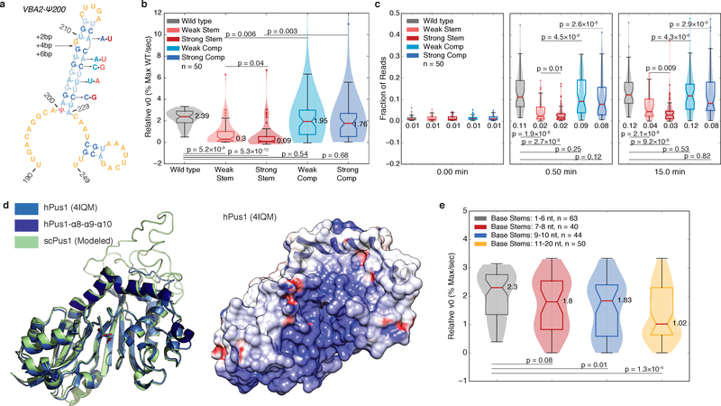
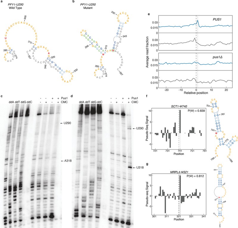
Pseudouridine (Ψ) is a post-transcriptional RNA modification that alters RNA-RNA and RNA-protein interactions that affect gene expression. Messenger RNA pseudouridylation was recently discovered as a widespread and conserved phenomenon, but the mechanisms responsible for selective, regulated pseudouridylation of specific sequences within mRNAs were unknown. Here, we have revealed mRNA targets for five pseudouridine synthases and probed the determinants of mRNA target recognition by the predominant mRNA pseudouridylating enzyme, Pus1, by developing high-throughput kinetic analysis of pseudouridylation in vitro. Combining computational prediction and rational mutational analysis revealed an RNA structural motif that is both necessary and sufficient for mRNA pseudouridylation. Applying this structural context information predicted hundreds of additional mRNA targets that were pseudouridylated in vivo. These results demonstrate a structure-dependent mode of mRNA target recognition by a conserved pseudouridine synthase and implicate modulation of RNA structure as the probable mechanism to regulate mRNA pseudouridylation.


Conflict of interest statement

The authors declare no competing financial interests.

Figures

Figure 1:
Figure 1:. A High-Throughput In vitro Pseudouridylation Assay
a) A schematic of in vitro pseudouridylation of oligo pool-derived RNAs and Ψ detection with Pseudo-seq. b) Pseudo-seq signal for mRNA substrates incubated with wild type S100 (blue), pus1Δ S100 (red), or no extract (gray). RPL18A-Ψ185 (upper), YAP1802-Ψ-117 (lower). c) A scatter plot of Pseudo-seq signal for pools incubated with wild type or pus1Δ S100 extracts. Sequences correspond to Pus1 ncRNA (red, n=4 sequences) and mRNA (blue, n=60 sequences) substrates. Values represent an average of n=2 replicates. d) Summary of in vitro pseudouridylation of PUS1-dependent mRNA Ψs.
Figure 2:
Figure 2:. Identification of Human PUS mRNA Substrates In vitro
a) Schematic of the pseudouridine synthase domain structures of 7 human PUS proteins. b-d) RNA pools of sequences of mRNA Ψs from H. sapiens were pseudouridylated with recombinant PUS proteins: PUS1 (blue), TRUB1 (green), TRUB2 (gray), PUS7 (red), RPUSD2 (yellow), no PUS (black). b) A summary of mRNA Ψs assigned to hPUS proteins. c,d) Pseudo-seq signal for (c) a TRUB1 mRNA target: MT-ND4-Ψ396, and (d) a PUS1 mRNA target: MED1-Ψ4774.
Figure 3:
Figure 3:. A Structural Motif Associated with Pus1 mRNA Targets in Yeast
a) The sequence motif surrounding n=60 high confidence PUS1-dependent mRNA Ψs, generated with WebLogo 3.5. b) MFE structures for VBA2-Ψ200 (left), YAP1802-Ψ-117 (middle) YRA1-Ψ132 (right). c) A heatmap of the average pairing probability matrix from RNAfold (upper), and the average maximum pairing probability for each base (lower) for n=60 high-confidence Pus1 targets. d) A heatmap of pairwise correlation coefficients (Pearson R) between the arrays of maximum pairing probabilities for each mRNA site (n=88 Ψs with genetic evidence for Pus1-dependence in vivo). In vitro modified mRNA Ψs with a stem-loop motif (dark teal, n=54 seqeunces), in vitro modified without a stem-loop motif (medium teal, n=6 sequences), with ambiguous in vitro data (light teal, n=1 sequence), and not modified in vitro (purple, n=27 sequences). Rows and columns are ordered by the sum of R values across the row/column. Indicated on the right is the classification of each sequence (upper). The maximum pairing probability and correlation values for 3 sequences (lower). e) Average PARS score ±SEM for our high confidence Pus1 mRNA substrates (blue, n=52 targets), and all other mRNA Ψs (red, n=223 targets) identified in log phase and high density for which there is PARS data.
Figure 4:
Figure 4:. Kinetic Analysis Reveals Sequence Features Important for mRNA Pseudouridylation by Pus1
a) Violin plots (center lines, medians; notches, 95% confidence intervals; boxes, 25th to 75th percentiles; whiskers, 1.5X inter-quartile range; dots, values outside of the 1.5X IQR) of the distributions of the fraction of reads mapping to the expected RT stop positions for Pus7 mRNA targets (pink, n=41 sequences) and Pus1 mRNA targets (blue, n=61 sequences). Medians, and p-values (unpaired t-test, two-tailed) are indicated. b) The average fraction of maximum signal for n=60 high confidence Pus1 mRNA targets is shown ± standard deviation (blue), for HSP30-Ψ914 (red), YGL188C-A-Ψ105 (gray) on a 0–15 min timescale (left) or a 0–2 min timescale (right). Lines indicating the slope (v0,rel) of the fit are indicated. c) A violin plot (elements as above) of the v0,rel values for n=60 high confidence Pus1 mRNA targets. Median indicated. d) A schematic of sequence motif mutations. e-f) Violin plots (elements as above) of the kinetics of pseudouridylation as indicated by (e) the fraction of reads at expected RT stop positions at indicated timepoints, or (f) v0,rel values for wild type (gray, n=50 sequences), −1 R to C mutants (red, n=50 sequences), and −2 H to G mutants (blue, n=50 sequences). Medians, and p-values (paired t-test, two-tailed) are indicated.
Figure 5:
Figure 5:. The Rate of mRNA Pseudouridylation Depends on Stem Length and Stability
a) A schematic of stem disrupting and compensatory mutations for VBA2-Ψ200. See b-c) for description of color scheme. b-c) Violin plots (center lines, medians; notches, 95% confidence intervals; boxes, 25th to 75th percentiles; whiskers, 1.5X inter-quartile range; dots, values outside of the 1.5X IQR) of (b) v0,rel values, or (c) the fraction of reads at the expected RT stop positions at indicated timepoints for wild type (gray, n=50 sequences), weak stem disrupting mutations (pink, n=50 sequences), strong stem disrupting mutations (red, n=50 sequences), weak compensatory mutations (light blue, n=50 sequences) and strong compensatory mutations (dark blue, n=50 sequences). Medians, and p-values (paired t-test, two-tailed) are indicated. d) The structure of hPus1 (light blue, 4IQM, Czudnochowski et al. 2013) with the three-helical RNA binding channel cap (dark blue) with modelled yeast Pus1 (left). An electrostatic surface map of hPus1, with the helical cap in ribbon form (right). e) Violin plots (elements as in above) of v0,rel for wild type, and stem extension mutants binned by base stem length. 1–6 nt (gray, n=63 targets), 7–8 nt (red, n=40 targets), 9–10 nt (blue, n=44 targets), and 11–20 nt (yellow, n=50 targets) bins are shown. Medians, and p-values (unpaired t-test, two-tailed) are indicated.
Figure 6:
Figure 6:. The Pus1 Structural Motif is Sufficient for Pseudouridylation
a-b) MFE structures for a region surrounding (a) wild type, or (b) mutant PFY1-U290. Target Ψ nucleotides (red), adapter, padding, and T7 sequences (gray), and mutated nucleotides (green) are indicated. c-d) Primer extension gels of (c) PFY1-U290 wild type, and (d) PFY1-U290 mutant sequences. Positions of U290 and A/U318 are indicated. Gels are representative of n=4 replicates. Uncropped gel images can be found in Supplementary Figure 7a,b. e) Metaplot of RT stops for sites predicted to be Pus1 targets with P(Ψ) > 0.8, in +CMC libraries (blue) and −CMC libraries (grey) from high OD in vivo Pseudo-seq data. PUS1 panels show the aggregated reads from knockout libraries for pus2Δ,3Δ,4Δ,5Δ,6Δ,7Δ, and 9Δ. f-g) Pseudo-seq signal from the pooled PUS1 reads, and predicted secondary structure for a putative Pus1 target at (f) position SCT1-Ψ740 and (g) MRPL4-Ψ321.

References

    1. Boccaletto P et al. MODOMICS: A database of RNA modification pathways. 2017 update. Nucleic Acids Res. (2018). doi:10.1093/nar/gkx1030 - DOI - PMC - PubMed
    1. Gilbert WV, Bell TA & Schaening C Messenger RNA modifications: Form, distribution, and function. Science (80-. ). 352, 1408–1412 (2016). - PMC - PubMed
    1. Carlile TM et al. Pseudouridine profiling reveals regulated mRNA pseudouridylation in yeast and human cells. Nature 515, 143–6 (2014). - PMC - PubMed
    1. Schwartz S et al. Transcriptome-wide Mapping Reveals Widespread Dynamic-Regulated Pseudouridylation of ncRNA and mRNA. Cell 159, 148–62 (2014). - PMC - PubMed
    1. Lovejoy AF, Riordan DP & Brown PO Transcriptome-wide mapping of pseudouridines: pseudouridine synthases modify specific mRNAs in S. cerevisiae. PLoS One 9, e110799 (2014). - PMC - PubMed

Publication types

MeSH terms