. 2019 Sep 2;5(1):138.
doi: 10.1186/s40792-019-0696-4.

Primary thymic MALT lymphoma in a patient with Sjögren's syndrome and multiple lung cysts: a case report

Affiliations

Primary thymic MALT lymphoma in a patient with Sjögren's syndrome and multiple lung cysts: a case report

Yusuke Hirokawa et al. Surg Case Rep. .

Abstract

Background: Thymic mucosa-associated lymphoid tissue (MALT) lymphoma is rare and also known for its association with autoimmune diseases, especially Sjögren's syndrome (SjS), which could affect the systemic organs, and pulmonary involvement often reveals multiple lung cysts.

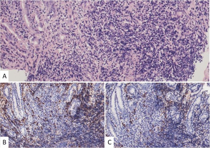
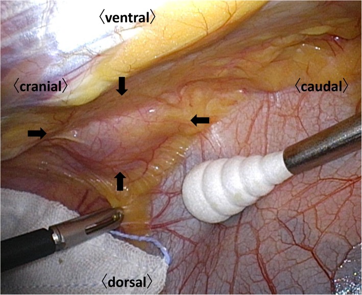
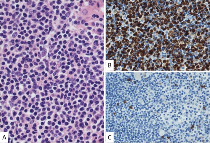
Case presentation: A 40-year-old woman presented with an anterior mediastinal mass and multiple lung cysts on computed tomography. We suspected thymoma concomitant with lymphangioleiomyomatosis and performed a total thymectomy and wedge resection of the lung as a surgical biopsy. The histopathological diagnosis of the mediastinal mass was a MALT lymphoma, and there were no specific findings in the lung specimen. She had a history of SjS, which had been overlooked during the initial work-up.

Conclusions: A history of SjS should raise suspicion of a MALT lymphoma for the differential diagnosis of an anterior mediastinal mass. A precise history taking is crucial for the correct diagnosis, and we could have avoided a lung resection in our case.

Keywords: Lung cysts; Sjögren’s syndrome; Thymic MALT lymphoma.


Conflict of interest statement

The authors declare that they have no competing interests.

Figures

Fig. 1
Fig. 1
a Computed tomographic scan of the chest showing a well-circumscribed mass with a heterogenous concentration measuring 49 × 22 mm in the anterior mediastinum. b Out-of-phase dynamic magnetic resonance imaging of the mediastinum showing the multilocular mass without invasion to the surrounding parenchyma
Fig. 2
Fig. 2
Computed tomographic scan of the chest showing multiple cysts in both lungs
Fig. 3
Fig. 3
Histopathological findings of the stomach lesion showing lymphocytic infiltration without any atypical cells in the hematoxylin and eosin staining (a) or any light chain restriction between the kappa (b) and lambda (c) chains in the immunostaining, consistent with inflammatory changes. We used antibodies with the product number n1510 (company, DAKO; dilutions, 5 times for the kappa chains) and product number ncl-lam (company, Leica; dilutions, 200 times for the lambda chains). The positive cells are stained with a brown color
Fig. 4
Fig. 4
A total thymectomy was performed by a bilateral approach via a video-assisted thoracoscopic surgery. The mass is enclosed by the arrows without any adhesions to the surrounding tissue
Fig. 5
Fig. 5
Histopathological findings of the mediastinal mass showing small- to medium-sized atypical lymphoid cells, and some of them exhibit plasmacytoid differentiation in the hematoxylin and eosin staining (a). A light chain restriction positive for kappa (b) and negative for lambda (c) chains is demonstrated. We used antibodies with the product number N1510 (company, DAKO; dilutions, 5 times for the kappa chains) and product number NCL-LAM (company, Leica; dilutions, 200 times for the lambda chains). The positive cells are stained with a brown color

References

    1. Isaacson P, Wright DH. Malignant lymphoma of mucosa-associated lymphoid tissue. A distinctive type of B-cell lymphoma. Cancer. 1983;52(8):1410–1416. doi: 10.1002/1097-0142(19831015)52:8<1410::AID-CNCR2820520813>3.0.CO;2-3. - DOI - PubMed
    1. Zucca E, Bertoni F, Roggero E, Cavalli F. The gastric marginal zone B-cell lymphoma of MALT type. Blood. 2000;96(2):410–419. - PubMed
    1. Servitje O, Gallardo F, Estrach T, Pujol RM, Blanco A, Fernandez-Sevilla A, et al. Primary cutaneous marginal zone B-cell lymphoma: a clinical, histopathological, immunophenotypic and molecular genetic study of 22 cases. Br J Dermatol. 2002;147(6):1147–1158. doi: 10.1046/j.1365-2133.2002.04961.x. - DOI - PubMed
    1. Zucca E, Conconi A, Pedrinis E, Cortelazzo S, Motta T, Gospodarowicz MK, et al. Nongastric marginal zone B-cell lymphoma of mucosa-associated lymphoid tissue. Blood. 2003;101(7):2489–2495. doi: 10.1182/blood-2002-04-1279. - DOI - PubMed
    1. Khalil MO, Morton LM, Devesa SS, Check DP, Curtis RE, Weisenburger DD, et al. Incidence of marginal zone lymphoma in the United States, 2001-2009 with a focus on primary anatomic site. Br J Haematol. 2014;165(1):67–77. doi: 10.1111/bjh.12730. - DOI - PMC - PubMed

LinkOut - more resources