. 2019 Oct;18(20):2800-2813.
doi: 10.1080/15384101.2019.1661174. Epub 2019 Sep 3.

Myotubularin related protein 7 is essential for the spermatogonial stem cell homeostasis via PI3K/AKT signaling

Affiliations

Myotubularin related protein 7 is essential for the spermatogonial stem cell homeostasis via PI3K/AKT signaling

Dan Zhao et al. Cell Cycle. 2019 Oct.

Abstract

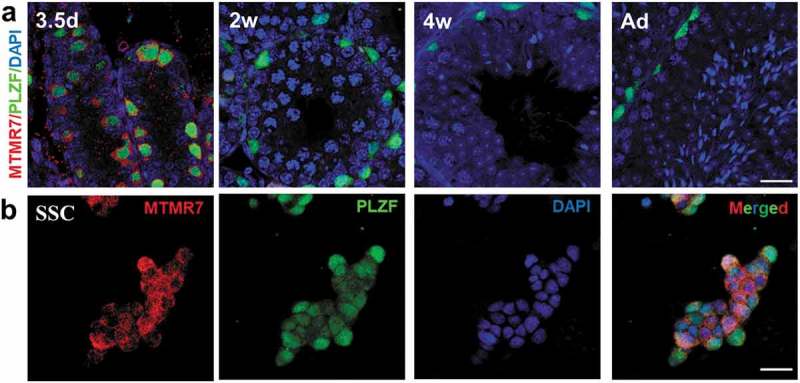
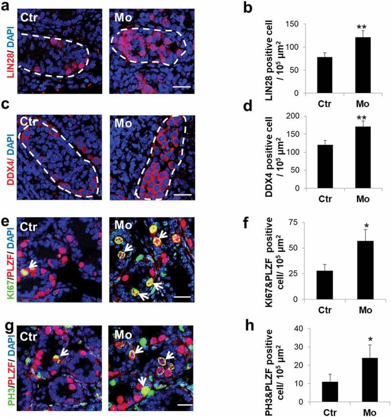
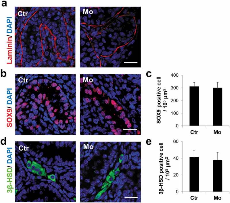
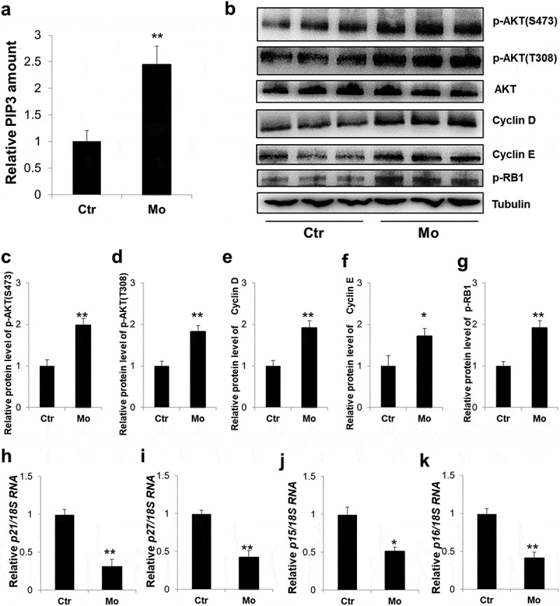
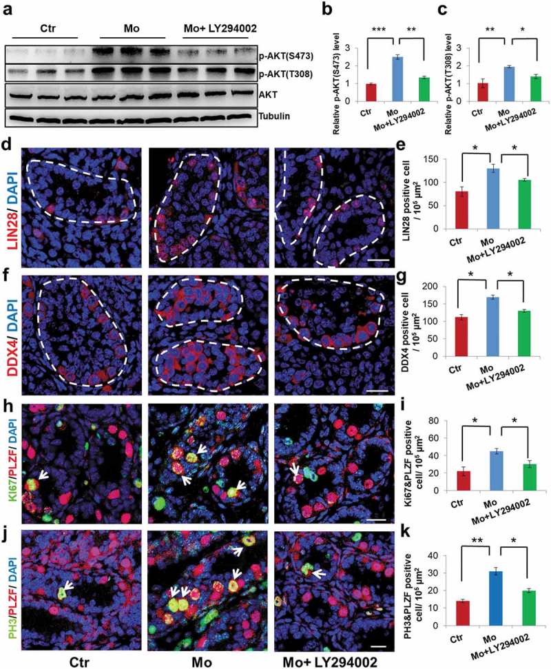
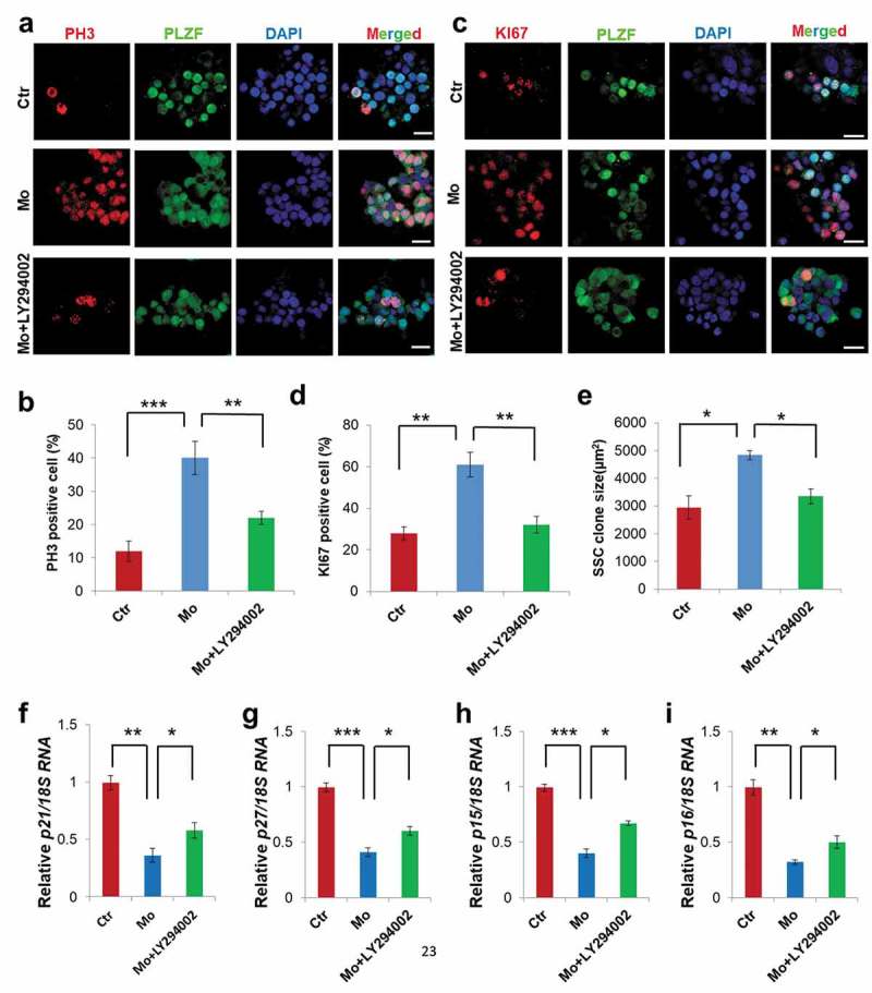
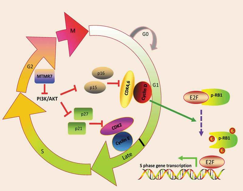
Myotubularin related protein 7 (MTMR7), a key member of the MTMR family, depicts phosphatase activity and is involved in myogenesis and tumor growth. We have previously identified MTMR7 in the proteomic profile of mouse spermatogonial stem cell (SSC) maturation and differentiation, implying that MTMR7 is associated with neonatal testicular development. In this study, to further explore the distribution and function of MTMR7 in mouse testis, we studied the effect of Mtmr7 knockdown on neonatal testicular development by testicular and SSC culture methods. Our results revealed that MTMR7 is exclusively located in early germ cells. Deficiency of MTMR7 by morpholino in neonatal testis caused excessive SSC proliferation, which was attributable to the aberrant PI3K/AKT signaling activation. Altogether, our study demonstrates that MTMR7 maintains SSC homeostasis by inhibiting PI3K/AKT signaling activation.

Keywords: Myotubularin related protein 7 (MTMR7); PI3K/AKT signaling; germ cells; spermatogonial stem cell (SSC).


Figures

Figure 1.
Figure 1.
Expression of MTMR7 in testis and SSC. (a) Co-immunostaining of MTMR7 and PLZF (undifferentiated spermatogonia marker) at 3.5-d, 2-wk, 4-wk and adult mouse testis. (b) Co-immunostaining of MTMR7 and PLZF in SSC. Scale bar: 20 μm.
Figure 2.
Figure 2.
Establishment of neonatal testis culture platform. (a) Schematic representation of neonatal testis culture. Neonatal testis was cut into pieces and placed on the medium/air interface of agarose gel stand. The fragments were cultured for 4 d either with 20 μM Mo or its Ctr. (b,c) Western blot analysis showed high knockdown efficiency of MTMR7 by Mo. (**p < 0.01, Student’s t-test, sample number = 3).
Figure 3.
Figure 3.
MTMR7 is required for germ cell development. (a,b) Representative images of LIN28 (undifferentiated spermatogonia marker) immunostaining in Ctr and Mo testes. (c,d) Representative images of DDX4 (germ cell marker) immunostaining in Ctr and Mo testes. (e,f) Co-immunostaining of KI67 (cellular proliferation marker) and PLZF in Ctr and Mo testes. (g,h) Co-immunostaining of PH3 (cellular proliferation marker) and PLZF in Ctr and Mo testes. Dotted lines indicate seminiferous tubule. White arrows indicate proliferating SSC. (*p < 0.05, **p < 0.01, Student’s t-test, sample number = 6). Scale bar: 20 μm.
Figure 4.
Figure 4.
MTMR7 knockdown does not disrupt the integrity of seminiferous tubules and somatic cells. (a) Representative images of Laminin immunostaining in Ctr and Mo testes. (b,c) Representative images of SOX9 (Sertoli marker) immunostaining in Ctr and Mo testes. (d,e) Representative images of 3β-HSD (Leydig marker) immunostaining in Ctr and Mo testes. (Student’s t-test, sample number = 6).Scale bar: 20 μm.
Figure 5.
Figure 5.
Aberrant activation of PI3K/AKT signaling in MTMR7-deficient testes. (a) ELISA assay of the PIP3level in Ctr and Mo testes. (b) Western blot analysis of the expression levels of p-AKT(S473), p-AKT(T308), total AKT, Cyclin D, Cyclin E and p-RB1 in Ctr and Mo testes. (c-g) Quantification of (b). Real-time PCR analysis of the expression levels of p21 (h), p27 (i), p15 (j) and p16 (k) in Ctr and Mo testes. (*p < 0.05, **p < 0.01, Student’s t-test. For (b-g) sample number = 3; for (h-k), sample number = 6).
Figure 6.
Figure 6.
PI3K inhibitor, LY294002, can partly rescue the altered phenotypes. (a,b,c) Western blot analysis showed p-AKT(S473), p-AKT(T308) and total AKT levels in Ctr, Mo and Mo+ LY294002 testes. (d,e) Representative images of LIN28 immunostaining in Ctr, Mo and Mo+ LY294002 testes. (f,g) Representative images of DDX4 immunostaining in Ctr, Mo and Mo+ LY294002 testes. (h,i) Co-immunostaining of KI67 and PLZF in Ctr, Mo and Mo+ LY294002 testes. (j,k) Co-immunostaining of PH3 and PLZF in Ctr, Mo and Mo+ LY294002 testes. Dotted lines indicate seminiferous tubule. White arrows indicate proliferating SSC. (*p < 0.05, **p < 0.01, ***p < 0.001, one-way ANOVA. For (a-c), sample number = 3; for (d-k), sample number = 6). Scale bar: 20 μm.
Figure 7.
Figure 7.
Influence of MTMR7 knockdown on SSC culture. (a,b) Co-immunostaining of PH3 and PLZF in Ctr, Mo and Mo+ LY294002 SSCs. (c,d) Co-immunostaining of KI67 and PLZF immunostaining in Ctr, Mo and Mo+ LY294002 SSCs. (e) Quantification of SSC clone. Real-time PCR analysis of the expression levels of p21 (f), p27 (g), p15 (h) and p16 (i) in Ctr, Mo and Mo+ LY294002 SSCs. (*p < 0.05, **p < 0.01, ***p < 0.001, one-way ANOVA, sample number = 4).Scale bar: 20 μm.
Figure 8.
Figure 8.
Schematic illustration of MTMR7 work model. MTMR7 acts as a negative regulator of cell cycle via depressing PI3K/AKT signaling. Knockdown of MTMR7 leads to PI3K/AKT activation, followed by the inactivated of CDKIs (p21, p27, p15 and p16). Then, cyclin D-CDK4,6 and cyclin E-CDK2 complex phosphorylate RB1, thus, contributing to E2F transcriptional activity and promoting G1-S phase transition.

References

    1. Hermann BP, Phillips BT, Orwig KE.. The elusive spermatogonial stem cell marker? Biol Reprod. 2011;85:221–223. - PMC - PubMed
    1. Phillips BT, Gassei K, Orwig KE.. Spermatogonial stem cell regulation and spermatogenesis. Philos Trans R Soc Lond B Biol Sci. 2010;365:1663–1678. - PMC - PubMed
    1. Hara K, Nakagawa T, Enomoto H, et al. Mouse spermatogenic stem cells continually interconvert between equipotent singly isolated and syncytial states. Cell Stem Cell. 2014;14:658–672. - PMC - PubMed
    1. Nakagawa T, Sharma M, Nabeshima Y, et al. Functional hierarchy and reversibility within the murine spermatogenic stem cell compartment. Science. 2010;328:62–67. - PMC - PubMed
    1. McLean DJ, Friel PJ, Johnston DS, et al. Characterization of spermatogonial stem cell maturation and differentiation in neonatal mice. Biol Reprod. 2003;69:2085–2091. - PubMed

MeSH terms

Substances